

Abstract
There are two proliferative populations in the developing cerebral wall: the pseudostratified ventricular epithelium (PVE) and the secondary proliferative population (SPP). The present experiments provide on embryonic day 14 (E14) in the mouse direct measures of the values for the proportions of daughter cells that continue to proliferate (proliferative, P fraction, or P) and for those that leave the cell cycle (quiescent, Q fraction, or Q; Q = 1 - P) for both of these populations. The range of values of P for the PVE, 0.62-0.66, would provide for the relatively low rate of neuronal output and for an expansion of the proliferative population appropriate for E14. The even higher values of P for the SPP, 0.73-1.0, would expand this population rapidly in preparation for the high glial cell output to occur later in cerebral histogenesis.
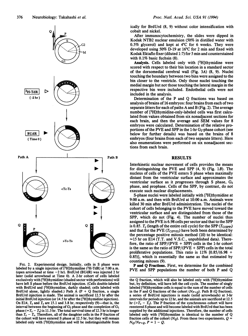
Full text
PDF


Images in this article


Selected References
These references are in PubMed. This may not be the complete list of references from this article.
- Caviness V. S., Jr Neocortical histogenesis in normal and reeler mice: a developmental study based upon [3H]thymidine autoradiography. Brain Res. 1982 Jul;256(3):293–302. doi: 10.1016/0165-3806(82)90141-9. [DOI] [PubMed] [Google Scholar]
- Fairén A., Cobas A., Fonseca M. Times of generation of glutamic acid decarboxylase immunoreactive neurons in mouse somatosensory cortex. J Comp Neurol. 1986 Sep 1;251(1):67–83. doi: 10.1002/cne.902510105. [DOI] [PubMed] [Google Scholar]
- Fishell G., Mason C. A., Hatten M. E. Dispersion of neural progenitors within the germinal zones of the forebrain. Nature. 1993 Apr 15;362(6421):636–638. doi: 10.1038/362636a0. [DOI] [PubMed] [Google Scholar]
- Grove E. A., Kirkwood T. B., Price J. Neuronal precursor cells in the rat hippocampal formation contribute to more than one cytoarchitectonic area. Neuron. 1992 Feb;8(2):217–229. doi: 10.1016/0896-6273(92)90289-p. [DOI] [PubMed] [Google Scholar]
- Hall P. A., Watt F. M. Stem cells: the generation and maintenance of cellular diversity. Development. 1989 Aug;106(4):619–633. doi: 10.1242/dev.106.4.619. [DOI] [PubMed] [Google Scholar]
- Levitt P., Cooper M. L., Rakic P. Coexistence of neuronal and glial precursor cells in the cerebral ventricular zone of the fetal monkey: an ultrastructural immunoperoxidase analysis. J Neurosci. 1981 Jan;1(1):27–39. doi: 10.1523/JNEUROSCI.01-01-00027.1981. [DOI] [PMC free article] [PubMed] [Google Scholar]
- McConnell S. K., Kaznowski C. E. Cell cycle dependence of laminar determination in developing neocortex. Science. 1991 Oct 11;254(5029):282–285. doi: 10.1126/science.254.5029.282. [DOI] [PubMed] [Google Scholar]
- Miller M. W. The migration and neurochemical differentiation of gamma-aminobutyric acid (GABA)-immunoreactive neurons in rat visual cortex as demonstrated by a combined immunocytochemical-autoradiographic technique. Brain Res. 1986 Jul;393(1):41–46. doi: 10.1016/0165-3806(86)90063-5. [DOI] [PubMed] [Google Scholar]
- Misson J. P., Edwards M. A., Yamamoto M., Caviness V. S., Jr Mitotic cycling of radial glial cells of the fetal murine cerebral wall: a combined autoradiographic and immunohistochemical study. Brain Res. 1988 Feb 1;466(2):183–190. doi: 10.1016/0165-3806(88)90043-0. [DOI] [PubMed] [Google Scholar]
- Murray A. W., Kirschner M. W. What controls the cell cycle? Sci Am. 1991 Mar;264(3):56–63. doi: 10.1038/scientificamerican0391-56. [DOI] [PubMed] [Google Scholar]
- O'Rourke N. A., Dailey M. E., Smith S. J., McConnell S. K. Diverse migratory pathways in the developing cerebral cortex. Science. 1992 Oct 9;258(5080):299–302. doi: 10.1126/science.1411527. [DOI] [PubMed] [Google Scholar]
- Pardee A. B. The Yang and Yin of cell proliferation: an overview. J Cell Physiol Suppl. 1987;Suppl 5:107–110. doi: 10.1002/jcp.1041330420. [DOI] [PubMed] [Google Scholar]
- Parnavelas J. G., Barfield J. A., Franke E., Luskin M. B. Separate progenitor cells give rise to pyramidal and nonpyramidal neurons in the rat telencephalon. Cereb Cortex. 1991 Nov-Dec;1(6):463–468. doi: 10.1093/cercor/1.6.463. [DOI] [PubMed] [Google Scholar]
- Potten C. S., Loeffler M. Stem cells: attributes, cycles, spirals, pitfalls and uncertainties. Lessons for and from the crypt. Development. 1990 Dec;110(4):1001–1020. doi: 10.1242/dev.110.4.1001. [DOI] [PubMed] [Google Scholar]
- Rakic P. Neurons in rhesus monkey visual cortex: systematic relation between time of origin and eventual disposition. Science. 1974 Feb 1;183(4123):425–427. doi: 10.1126/science.183.4123.425. [DOI] [PubMed] [Google Scholar]
- Rakic P. Specification of cerebral cortical areas. Science. 1988 Jul 8;241(4862):170–176. doi: 10.1126/science.3291116. [DOI] [PubMed] [Google Scholar]
- SAUER M. E., WALKER B. E. Radioautographic study of interkinetic nuclear migration in the neural tube. Proc Soc Exp Biol Med. 1959 Jul;101(3):557–560. doi: 10.3181/00379727-101-25014. [DOI] [PubMed] [Google Scholar]
- SIDMAN R. L., MIALE I. L., FEDER N. Cell proliferation and migration in the primitive ependymal zone: an autoradiographic study of histogenesis in the nervous system. Exp Neurol. 1959 Oct;1:322–333. doi: 10.1016/0014-4886(59)90024-x. [DOI] [PubMed] [Google Scholar]
- Schwartz M. L., Meinecke D. L. Early expression of GABA-containing neurons in the prefrontal and visual cortices of rhesus monkeys. Cereb Cortex. 1992 Jan-Feb;2(1):16–37. doi: 10.1093/cercor/2.1.16. [DOI] [PubMed] [Google Scholar]
- Schwartz M. L., Rakic P., Goldman-Rakic P. S. Early phenotype expression of cortical neurons: evidence that a subclass of migrating neurons have callosal axons. Proc Natl Acad Sci U S A. 1991 Feb 15;88(4):1354–1358. doi: 10.1073/pnas.88.4.1354. [DOI] [PMC free article] [PubMed] [Google Scholar]
- Sidman R. L., Rakic P. Neuronal migration, with special reference to developing human brain: a review. Brain Res. 1973 Nov 9;62(1):1–35. doi: 10.1016/0006-8993(73)90617-3. [DOI] [PubMed] [Google Scholar]
- Takahashi T., Nowakowski R. S., Caviness V. S., Jr BUdR as an S-phase marker for quantitative studies of cytokinetic behaviour in the murine cerebral ventricular zone. J Neurocytol. 1992 Mar;21(3):185–197. doi: 10.1007/BF01194977. [DOI] [PubMed] [Google Scholar]
- Takahashi T., Nowakowski R. S., Caviness V. S., Jr Cell cycle parameters and patterns of nuclear movement in the neocortical proliferative zone of the fetal mouse. J Neurosci. 1993 Feb;13(2):820–833. doi: 10.1523/JNEUROSCI.13-02-00820.1993. [DOI] [PMC free article] [PubMed] [Google Scholar]
- Walsh C., Cepko C. L. Cell lineage and cell migration in the developing cerebral cortex. Experientia. 1990 Sep 15;46(9):940–947. doi: 10.1007/BF01939387. [DOI] [PubMed] [Google Scholar]
- Walsh C., Cepko C. L. Clonal dispersion in proliferative layers of developing cerebral cortex. Nature. 1993 Apr 15;362(6421):632–635. doi: 10.1038/362632a0. [DOI] [PubMed] [Google Scholar]
- Walsh C., Cepko C. L. Widespread dispersion of neuronal clones across functional regions of the cerebral cortex. Science. 1992 Jan 24;255(5043):434–440. doi: 10.1126/science.1734520. [DOI] [PubMed] [Google Scholar]