Summary
A young woman with an occluded middle cerebral artery presented with a ruptured flow aneurysm distal on a Heubner artery as part of a moyamoya collateral network. Leptomeningeal collateral supply was tested by occlusion of the A1 origin of the Heubner artery. This test occlusion demonstrated ample collateral leptomeningeal supply over the hemispheres to the M2. Subsequently, the Heubner artery harbouring the aneurysm could be safely proximally occluded with coils.
Keywords: moyamoya, Heubner artery, leptomeningeal collateral circulation, test occlusion
Introduction
Spontaneous unilateral middle cerebral artery occlusion associated with moyamoya phenomenon in response to impaired perfusion is distinct from generalized moyamoya disease. The haemodynamic stress on the deep collateral channels occasionally leads to aneurysm formation, which may manifest as haemorrhage. The aetiology of this disease has not been fully understood. Direct surgical or endovascular aneurysm treatment is often not possible. We present a young patient with an occluded middle cerebral artery with moyamoya phenomenon and a ruptured aneurysm on the Heubner artery as part of the moyamoya collateral network. The aneurysm could be treated endovascularly by proximal parent vessel occlusion.
Case Report
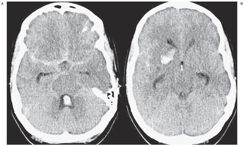
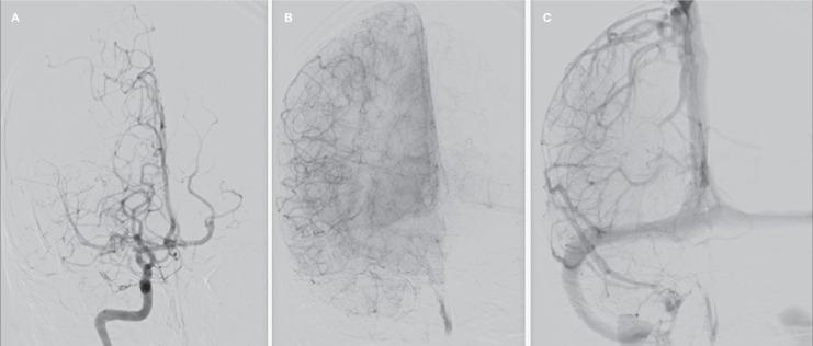
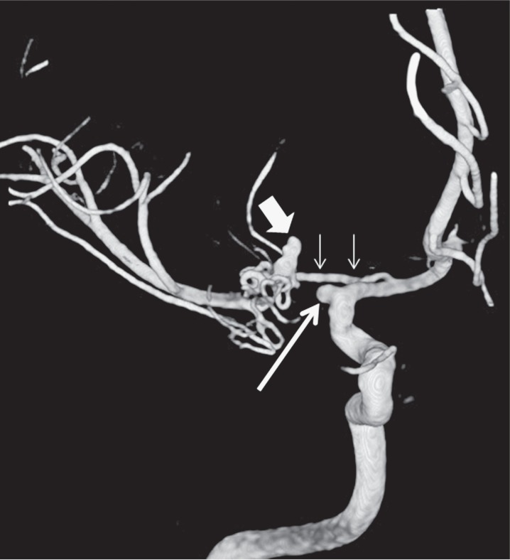
A 24-year old previously healthy Caucasian woman presented to the emergency department with sudden headache, nausea and vomiting. She was drowsy but had no neurologic deficits. Brain CT scan showed a subarachnoid and intraventricular haemorrhage with an intraparenchymal component in the basal ganglia (Figure 1). The same day 2D and 3D angiography showed a right M1 occlusion at its origin. The M2 branches were primarily supplied by a deep moyamoya collateral network via the Heubner artery originating from the right A1. In addition, cortical leptomeningeal collaterals were present originating in the contralateral hemisphere and ipsilateral anterior cerebral artery (Figure 2). A deeply located aneurysm distal on the artery of Heubner was responsible for the haemorrhage (Figures 2 and 3).
Figure 1.
CT scan with subarachnoid, intraventricular and intraparenchymal blood.
Figure 2.
Frontal right internal carotid angiogram in early (A), mid (B) and late (C) phase showing M1 occlusion and perfusion of right hemisphere by the basal collateral moyamoya network and leptomeningeal collateral vessels.
Figure 3.
3D angiogram of the right internal carotid artery shows the occluded middle cerebral artery at its origin (long arrow), hypertrophied Heubner artery as part of a collateral network (small arrows) and ruptured aneurysm on the distal Heubner artery (broad arrow).
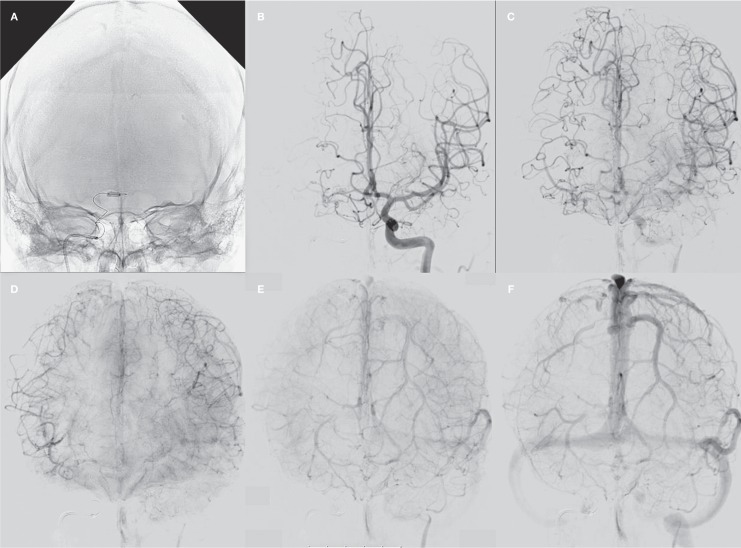
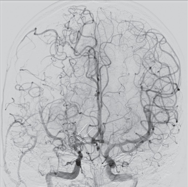
Because of the deep location of the aneurysm surgical treatment was not a first option. Direct endovascular treatment of the aneurysm was not possible. After ample discussion, we suggested another treatment. If the leptomeningeal collateral circulation over the hemispheres were sufficient, proximal Heubner artery occlusion would exclude the aneurysm from the circulation. Under general anaesthesia, the extent of the leptomeningeal collateral circulation was tested by placing a balloon in the right A1. The balloon occluded the origin of the Heubner artery (Figure 3A). Concomitant angiography of the left internal carotid artery demonstrated abundant leptomeningeal collateral supply with synchronous venous phase in both cerebral hemispheres (Figure 3B-F). Subsequently, via the left internal carotid artery and over the anterior communicating artery a microcatheter was placed in the hypertrophic right artery of Heubner. With several small coils this vessel could be proximally occluded, excluding the aneurysm from the circulation (Figure 4). The patient recovered without neurologic deficits after the procedure. A post-procedural MRI including diffusion-weighted imaging showed no ischaemic lesions. At six months, the patient was clinically fully recovered and MRI showed no (silent) infarctions.
Figure 4.
Test occlusion of the right A1. A) Balloon inflated in the right A1 occluding the origin of the Heubner artery. B-F) Arterial, parenchymal and venous phases of left internal carotid angiogram during test occlusion: abundant leptomeningeal collateral supply over the hemispheres with synchronous venous phase filling.
Figure 5.
Bilateral internal carotid angiogram after proximal coil occlusion of the Heubner artery (arrows).
Discussion
Spontaneous occlusion of the middle cerebral artery may be associated with moyamoya phenomenon around the occlusion site in the proximal segment of the artery. The lack of involvement of the internal carotid artery differentiates it from moyamoya disease and the moyamoya syndrome. The aetiology of this rare entity is unclear. Moyamoya can be distinguished from arteriosclerotic occlusion as there is no evidence of arteriosclerotic plaques or stenosis in other cerebral arteries 1. Some authors propose a congenital aetiology for this disease 2,3. In childhood, the condition is called transient cerebral arteriopathy and is considered a self-limiting inflammatory disease that in the early stage may not be distinguished from unilateral moyamoya or other progressive arteriopathies such as vasculitis 4. On pathological examination, collateral vessels in moyamoya have weakened media with segmentation of the internal elastic lamina and fibrosis. The attenuated arterial wall acquires a predisposition for micro-aneurysm formation under haemodynamic stress 5. Aneurysms formed on the collaterals may rupture and lead to intracerebral, intraventricular and subarachnoid haemorrhage. After bleeding, the chance of rebleeding is high and treatment is indicated 6.
Direct surgery carries a risk of additional brain damage, while the aneurysm is usually deeply located and the brain is in pre-existing ischaemic condition. Surgical injury of the collateral vessels can cause additional ischaemic brain damage 6. Surgical revascularization procedures have been proposed to reduce haemodynamic stress on the collateral vessels but efficiency remains in question 7,8. Endovascular treatment by occluding the aneurysm with its parent artery with glue has been successfully applied in several cases 9. In addition, endovascular treatment with coils or Onyx has been performed with good results 10.
In our patient the M1 was completely occluded at its origin. The hypertrophied Heubner artery harbouring the ruptured aneurysm appeared a major collateral pathway to the M2 and “blind” endovascular occlusion of this artery was considered dangerous in view of possible ischaemic damage. Therefore, we first performed a test occlusion by occluding the origin of the Heubner artery with a balloon in the A1. This demonstrated ample leptomeningeal collateral circulation over the hemispheres with synchronous venous phase filling indicative of equal circulation times and hence tolerance to permanent occlusion 11. We presumed that the Heubner artery was functional as a collateral only and not for supplying brain parenchyma. Subsequently, the Heubner artery could be safely occluded with coils. We opted for coils and not glue because the artery was large enough to accommodate 1.5 and 2 mm coils. With the use of glue, basal ganglia infarction would have been possible.
Few cases of ruptured aneurysms have been reported in patients with a middle cerebral artery occlusion and moyamoya phenomenon. Only one case has been described with an aneurysm of the Heubner artery. This earlier case presented with an infarction rather than a subarachnoid haemorrhage. Only three cases with a ruptured aneurysm on a collateral network have been described 3,7-10,12.
In moyamoya disease or syndrome associated with ruptured aneurysms endovascular treatment is the modality of choice. This is the first reported case of spontaneous occlusion of the middle cerebral artery associated with an aneurysm on the collateral network managed by endovascular parent vessel coil occlusion after angiographic testing of leptomeningeal collaterals. This case shows that the leptomeningeal circulation can be evaluated by selective balloon test occlusion in selected cases followed by simple coil occlusion of the vessel harbouring the aneurysm.
References
- 1.Fukawa O, Aihara H, Ishii K, et al. Middle cerebral artery occlusion with moyamoya phenomenon. First report: clinical course and angiographic findings.; Proceedings of the 10th Conference of Surgery for Cerebral Stroke; Tokyo. 1981. pp. 36–41. [Google Scholar]
- 2.Yasargil MG, Smith RD. Association of middle cerebral artery anomalies with saccular aneurysms and Moyamoya disease. Surg Neurol. 1976;6(1):39–43. [PubMed] [Google Scholar]
- 3.Seki Y, Fujita M, Mizutani N, et al. Spontaneous middle cerebral artery occlusion leading to moyamoya phenomenon and aneurysm formation on collateral arteries. Surg Neurol. 2001;55:58–62. doi: 10.1016/s0090-3019(00)00339-6. Discussion 62. [DOI] [PubMed] [Google Scholar]
- 4.Yeon JY, Shin HJ, Seol HJ, et al. Unilateral intracranial arteriopathy in pediatric stroke: Course, outcome, and prediction of reversible arteriopathy. Stroke. 2014;5(4):1173–6. doi: 10.1161/STROKEAHA.113.004125. doi: 10.1161/STROKEAHA.113.004125. [DOI] [PubMed] [Google Scholar]
- 5.Yamashita M, Oka K, Tanaka K. Histopathology of the brain vascular network in moyamoya disease. Stroke. 1983;14(1):50–58. doi: 10.1161/01.str.14.1.50. doi: 10.1161/01.STR.14.1.50. [DOI] [PubMed] [Google Scholar]
- 6.Yoshida Y, Yoshimoto T, Shirane R, et al. Clinical course, surgical management, and long-term outcome of moyamoya patients with rebleeding after an episode of intracerebral hemorrhage: An extensive follow-up study. Stroke. 1999;30(11):2272–2276. doi: 10.1161/01.str.30.11.2272. doi: 10.1161/01.STR.30.11.2272. [DOI] [PubMed] [Google Scholar]
- 7.Karasawa J, Touho H, Ohnishi H, et al. Long-term follow-up study after extracranial-intracranial bypass surgery for anterior circulation ischemia in childhood moyamoya disease. J Neurosurg. 1992;77(1):84–89. doi: 10.3171/jns.1992.77.1.0084. doi: 10.3171/jns.1992.77.1.0084. [DOI] [PubMed] [Google Scholar]
- 8.Takahashi M, Fujimoto T, Suzuki R, et al. A case of spontaneous middle cerebral artery occlusion associated with a cerebral aneurysm angiographically disappearing after STA-MCA anastomosis. No Shinkei Geka. 1997;25(8):727–732. [PubMed] [Google Scholar]
- 9.Kim SH, Kwon OK, Jung CK, et al. Endovascular treatment of ruptured aneurysms or pseudoaneurysms on the collateral vessels in patients with moyamoya disease. Neurosurgery. 2009;65(5):1000–1004. doi: 10.1227/01.NEU.0000345648.46096.CE. Discussion 1004. doi: 10.1227/01.NEU.0000345648.46096.CE. [DOI] [PubMed] [Google Scholar]
- 10.Yu JL, Wang HL, Xu K, et al. Endovascular treatment of intracranial aneurysms associated with moyamoya disease or moyamoya syndrome. Interv Neuroradiol. 2010;;16(3):240–248. doi: 10.1177/159101991001600302. [DOI] [PMC free article] [PubMed] [Google Scholar]
- 11.van Rooij WJ, Sluzewski M, Slob MJ, et al. Predictive value of angiographic testing for tolerance to therapeutic occlusion of the carotid artery. Am J Neuroradiol. 2005;26(1):175–178. [PMC free article] [PubMed] [Google Scholar]
- 12.Rivera R, Sordo J, Badilla L, et al. Middle cerebral artery occlusion with moyamoya-like vessels and aneurysms: A report of two cases. Interv Neuroradiol. 2014;20(1):96–99. doi: 10.15274/INR-2014-10014. doi: 10.15274/INR-2014-10014. [DOI] [PMC free article] [PubMed] [Google Scholar]


