Abstract
Introduction: To investigate the ultrastructural changes of dentin irradiated with 980-nm diode laser under different parameters and to observe the morphological alterations of odontoblasts and pulp tissue to determine the safety parameters of 980-nm diode laser in the treatment of dentin hypersensitivity (DH).
Methods: Twenty extracted human third molars were selected to prepare dentin discs. Each dentin disc was divided into four areas and was irradiated by 980-nm diode laser under different parameters: Group A: control group, 0 J/cm2; Group B: 2 W/CW (continuous mode), 166 J/cm2; Group C: 3W/CW, 250 J/cm2; and Group D: 4W/CW, 333 J/cm2. Ten additional extracted human third molars were selected to prepare dentin discs. Each dentin disc was divided into two areas and was irradiated by 980-nm diode laser: Group E: control group, 0 J/cm2; and Group F: 2.0 W/CW, 166 J/cm2. The morphological alterations of the dentin surfaces and odontoblasts were examined with scanning electron microscopy (SEM), and the morphological alterations of the dental pulp tissue irradiated by laser were observed with an upright microscope.
Results: The study demonstrated that dentinal tubules can be entirely blocked after irradiation by 980-nm diode laser, regardless of the parameter setting. Diode laser with settings of 2.0 W and 980-nm sealed exposed dentin tubules effectively, and no significant morphological alterations of the pulp and odontoblasts were observed after irradiation.
Conclusions: Irradiation with 980-nm diode laser could be effective for routine clinical treatment of DH, and 2.0W/CW (166 J/cm2) was a suitable energy parameter due to its rapid sealing of the exposed dentin tubules and its safety to the odontoblasts and pulp tissue.
Keywords: dentin sensitivity, lasers, scanning electron microscopy
Introduction
Dentin hypersensitivity (DH) is a type of common disease in cariology and endodontics, which is characterized by acute, non-spontaneous, short- or long-lasting pain originating from exposure of the dentin to thermal, chemical, mechanical, or osmotic stimuli and which cannot be attributed to any other dental pathology 1-7. Abrasion, wedge-shaped defects, odontagma and periodontal atrophy can all result in dentin hypersensitivity. Several theories have been proposed to explain the mechanism of dentin hypersensitivity, including the transducer theory, the gate-control and vibration theory, and the hydrodynamic theory. Currently, the hydrodynamic theory has been widely accepted, and it states that external stimuli cause movement of fluid inside dentinal tubules inwardly or outwardly, promoting mechanical deformation of nerve endings at the pulp/ dentin interface, which is transmitted as a painful sensation6 , 7. Under normal conditions, the dentin is covered by enamel or cementum and is not sensitive to direct stimulation. DH occurs only with the exposure of the peripheral terminations of the dentinal tubules3 , 6, 8. Therefore, blocking the exposed dentinal tubules is critical to the treatment of dentin hypersensitivity 3 , 9
. Laser systems have been used widely in the endodontic field, including Neodymium-Doped Yttrium Aluminium Garnet (Nd:YAG), Erbium- Doped Yttrium Aluminum Garnet (Er:YAG), Carbon Dioxide Laser (CO2) and diode lasers7 ,10 - 19,. Recently, an innovative 980-nm diode wavelength laser was introduced, which was first reported on 200420. It is a high-energy laser, with low purchase and maintenance costs, as well as greater versatility because of its compact size20 , 21. Currently, several studies have reported that the application of 980-nm diode laser could be used safely in endodontic treatment and in root canal disinfection22 , 23. However, few studies have reported on the interaction of 980-nm diode laser energy with the dentin surface and the ensuing structural alterations. Whether the 980-nm diode laser can treat dentine hypersensitivity effectively, like other types of lasers, requires more studies to determine.
However, laser energy should be carefully applied due to the adverse thermal effects on the pulp and adjacent structures. Pulpal space pressure and temperature increase with increases in the energy density of laser24- 27. However, most laser systems produce an increase in pulpal temperature that is dependent on the power setting26- 29. Odontoblasts, which are located at the dentin-pulp interface, like a densely packed layer sending long cytoplasmic processes into the dentinal tubules, have been recognized as playing important roles in the defense reaction of the dental pulp30 , 31. Thus, odontoblasts might the first cells to be damaged by contact with irradiation energy. Few studies have reported on the morphological alterations of dental pulp tissue, especially the odontoblasts, after irradiation with 980-nm diode laser. Furthermore, the safety parameters of 980-nm diode laser are unknown.
Considering the aforementioned issues, the aims of this in vitro study were to investigate, by scanning electron microscopy (SEM), the ultrastructural changes in dentin irradiated with 980-nm diode laser under different parameters and to observe the morphological alterations in odontoblasts and pulp tissue to determine the safety parameters of 980-nm diode laser in the treatment of DH.
Methods
Morphological alterations of dentin irradiated by different parameters of 980-nm diode laser
Twenty extracted human third molars were used for this study. Crowns with caries, restorations, or fractures were discarded. The enamel was removed with plaincut tungsten carbide fissure burs (Densply; Maillefer Instruments Holding Co., Ballaigues, Switzerland) at high speed under a continuous water spray, and crown dentin discs, with a thickness of 2 mm, were cut perpendicular to the long axis of tooth. The dentin discs were polished with a600-grit carborundum paper (600- grit carborundum paper; Weiyi Metallographic Test Instrument Co., Guangzhou, China) and were washed with distilled water and ultrasound. Each specimen was etched with 35% phosphoric acid (Gluma Etch 35Gel; Heraeus Kulzer, Hanau, Germany) for 1 min to expose the dentinal tubules and then was rinsed with copious distilled water, as well as ultrasound once more. All of the discs were stored in 4ºC distilled water containing 0.2% thymol to inhibit microbial growth until use.
Each dentin disc was divided into four areas (3 mm×4 mm), with one area chosen randomly for Group A and then Group B, Group C, and Group D assigned in a counterclockwise direction. According to the parameters of a 980-nm diode laser system (SIROlaser 2.2; Sirona Dental Systems GmbH,Bensheim, Germany), the group characteristics were as follows: Group A: control group, 0 J/cm2; Group B: 2 W/CW (continuous mode), 166 J/cm2; Group C: 3 W/CW, 250 J/cm2; and Group D: 4 W/CW, 333 J/cm2. Each area was irradiated twice for 5 seconds. The laser was delivered through a 320-μm optic fiber with a straight handpiece, and the laser tip was held perpendicular to the irradiated surface at a distance of 1 mm to prevent contamination from dentin. The discs were dehydrated at 70ºC for 24 h and then were fixed on stubs with double-faced carbon tape, covered with a 30-lm gold-platinum layer in a vacuum apparatus and examined with SEM (HITACHI S-3000N; Hitachi Ltd., Tokyo, Japan).
Morphological alterations of dentin and pulp irradiated by 2.0-W 980-nm diode laser
According to the results of the above study, we chose 2.0 W/CW (166 J/cm2) as the energy parameter of 980-nm diode laser to be tested.
Ten extracted human third molars were selected. Crowns with caries, restorations, or fractures were discarded. Immediately after extraction, 2 mm of enamel and dentin were removed perpendicular to the long axis of the tooth with plain-cut tungsten carbide fissure burs at high speed under a continuous water spray. The dentin did not receive etching to avoid stimulation of the dental pulp tissue and was washed with distilled water.
Each dentin surface was divided into two areas (3 mm×6 mm), with one area chosen randomly as Group E: control group, 0 J/cm2; with the other becoming part of Group F: 2.0W/CW, 166 J/cm2. Each area was irradiated twice for 5 seconds. A plain-cut tungsten carbide fissure bur, at high speed under a continuous water spray, was used to create a sulcus along the cementoenamel junction of the tooth. The teeth were fractured with a bitapered chisel and a surgical hammer, exposing the entire pulp tissue. The pulp tissues from under the dentin, with or without 980-nm diode laser treatment, were removed from the tooth. The pulp tissues were fixed in 4% formaldehyde (Formaldehyde; Shanghai Hanguang Chemical Reagent Factory, Shanghai, China) for 2 h, and the crowns were fixed in 2.5% dilute glutaral (Glutaral; Shanghai Hanguang Chemical Reagent Factory, Shanghai, China) solution for 2 h. Otherwise, a plain-cut tungsten carbide fissure bur, at high speed under a continuous water spray, was used to create a longitudinal sulcus along the buccal and palatal surfaces of the crown. The teeth were fractured longitudinally with a bitapered chisel and a surgical hammer, exposing the entire dentinal tubule to observe the morphological alterations in the tubules. Fixed crowns were dehydrated by a series of graded ethanol solutions and then were coated with a gold layer after drying. The morphological alterations of the odontoblasts and dentin surface were examined with an SEM.
Dental pulp tissue from Group E and Group F was fixed in 4% formaldehyde and then was dehydrated in a series of graded ethanol solutions; it was then paraffin embedded and cut sagittally at a thickness of 4 μm and stained with hematoxylin (Hematoxylin H9627; Sigma-Aldrich Corporation, St. Louis, USA) and eosin (EosinY E4009; Sigma-Aldrich Corporation, St. Louis, USA). The morphological alterations in the dental pulp tissue were observed by light microscopy (Olympus BX51; Olympus Optical Co., Ltd., Tokyo, Japan).
Results
Morphological alterations of dentin irradiated by different parameters of 980-nm diode laser
SEM analysis showed that the morphological aspects of the specimens treated with 980-nm diode laser were sealed by melting dentin, regardless of the parameter settings.
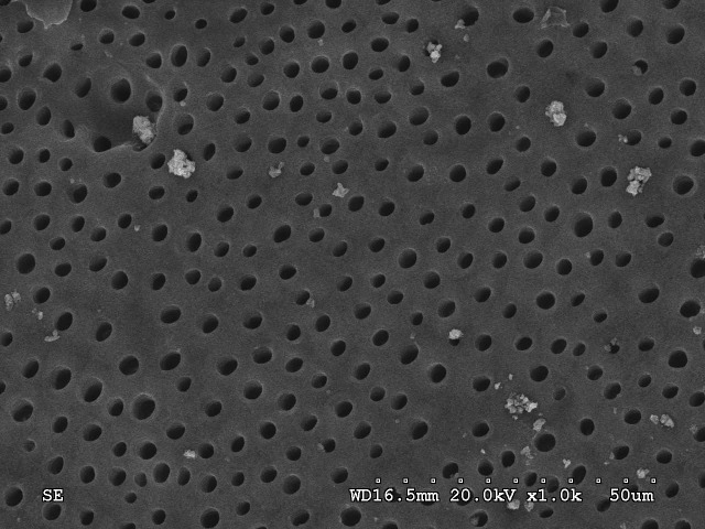
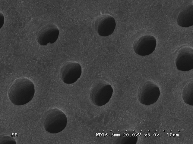
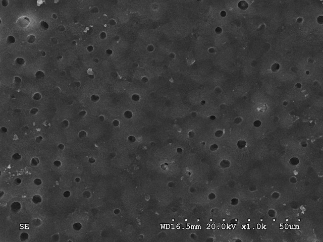
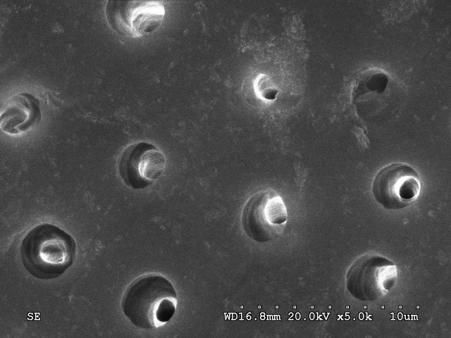
In Group A (control group), photomicrographs of the specimens showed that the dentin surfaces were smooth, and dentinal tubules were visible and well distributed. The dentin tubules were round or oval, and their diameters were 2 μm to 5 μm, with intervals of 3 μm to 8 μm (Figure 1).
Figure 1.
SEM micrograph of dentin surface without laser treatment. (a) A homogeneous surface with visible dentine tubules (×1000). (b) The diameters of the dentin tubules were 2 μm to 5 μm, and the intervals were 3 μm to 8 μm (×5000).
a.
b.
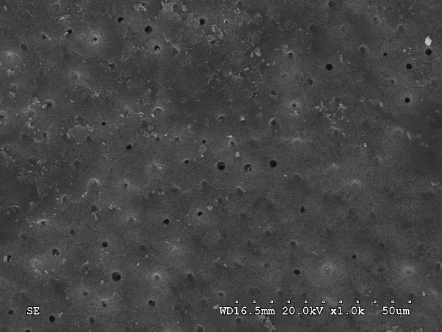
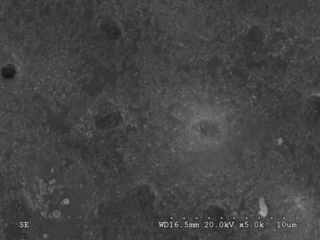
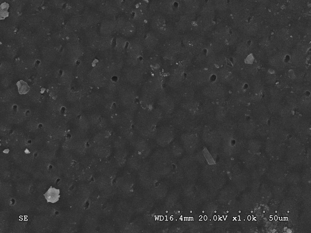
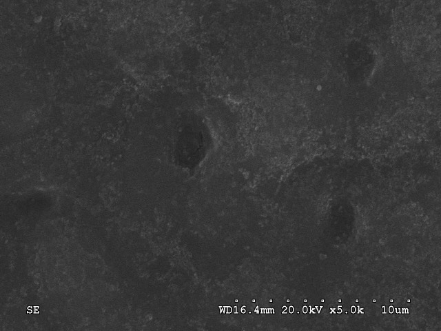
In Group B, the specimens were treated with 2 W/CW (166 J/cm2) 980-nm diode laser. Little or no open tubules were observed, the dentinal tubules were sealed or partially sealed, and the diameters ranged from 0 μm to 2 μm. Melting areas were present on the specimens’ surfaces, without fissures and craters (Figure 2).
Figure 2.
SEM micrograph of dentin surface after 2 W/CW (166 J/cm2) of 980-nm diode laser treatment. (a) Little or no open tubules were observed (×1000). (b) Melting areas were present on the specimens’ surfaces, without fissures or craters (×5000).
a.
b.
In Group C, the specimens were treated with 3 W/CW (250 J/cm2) 980-nm diode laser. There were many more open tubules in this group than in Group B, and the melting degree of the dentin surface was also strengthened. Double-layer structures of tubules were observed (Figure 3).
Figure 3.
SEM micrograph of dentin surface after 3 W/CW (250 J/cm2) of 980-nm diode laser treatment. (a) The melting degree of the dentin surface was strengthened (×1000). (b) Double layer structures of tubules were observed (×5000).
a.
b.
In Group D, the specimens were treated with 4 W/ CW (333 J/cm2) 980-nm diode laser. The overwhelming majority of the dentinal tubules were sealed by melting dentin. The peritubular dentin was deeply colored like scales because of the increased degree of dentin melting (Figure 4).
Figure 4.
SEM micrograph of dentin surface after 4 W/CW (333 J/cm2) of 980-nm diode laser treatment. (a) The overwhelming majority of dentin tubules were sealed by melting dentin (×1000). (b) The dentin surfaces were covered by homogeneous and melted dentin, without exposure of dentinal tubules (×5000).
a.
b.
Morphological alterations of dentin and pulp irradiated by 2.0-W 980-nm diode laser
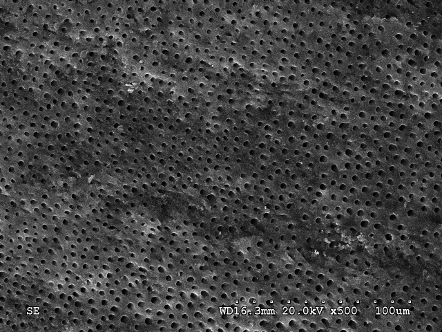
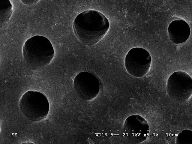
In Group E (control group), open dentinal tubules were found. The longitudinal section revealed that the dentinal tubule orifices were not occluded, and the diameters measured from 2 μm to 4 μm (Figure 5).
Figure 5.
SEM micrograph of dentin surface without laser treatment. (a) Homogeneous surface with visible dentin tubules (×500). (b) Dentin tubules were round or oval, and the diameters were 2 μm to 5 μm, with intervals of 3 μm to 8 μm (×5000).
a.
b.
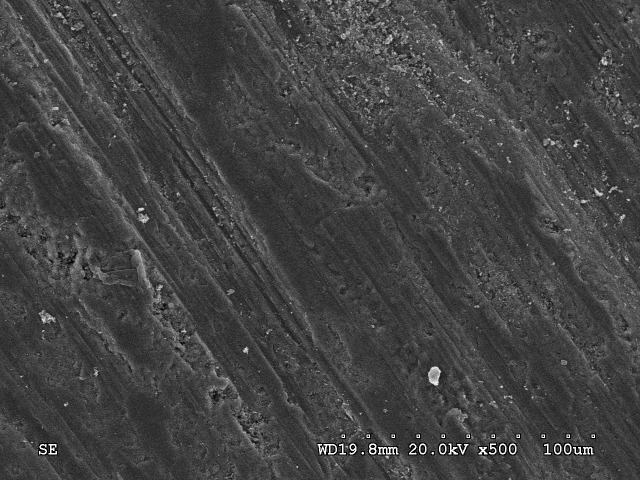
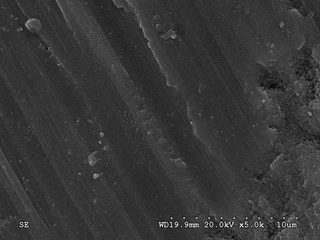
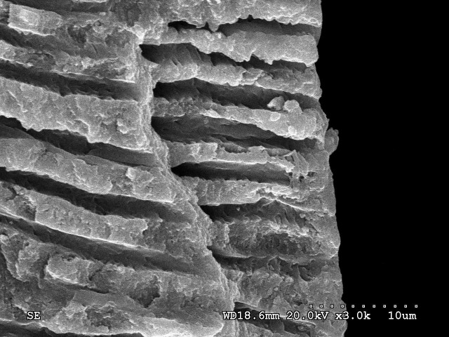
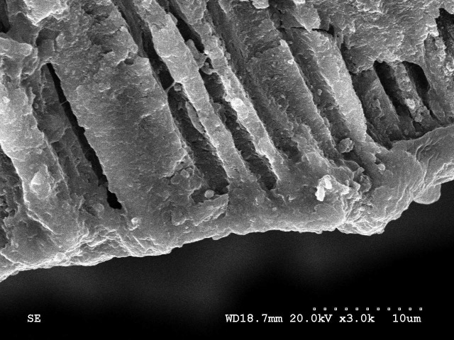
Conversely, Group F (treated with 2.0W/CW, 166 J/ cm2 of 980-nm diode laser) exhibited homogeneous aspects of the surfaces, totally covered by the melting dentin and without exposure of dentinal tubules (Figure 6). From the longitudinal section parallel to the direction of the dentinal tubules’ alignment, the sealing depth of the melted dentin was approximately 8 mm into dentinal tubules. No gap between the melted dentin and tubular walls was noted (Figure 7).
Figure 6.
SEM micrograph of dentin surface after 2.0 W/CW (166 J/ cm2) of 980-nm diode laser treatment. (a) A homogeneous surface covered by melting dentin, without exposure of dentinal tubules (×500). (b) No opened dentin tubules were found. (×5000).
a.
b.
Figure 7.
SEM micrograph of longitudinal section parallel to the direction of the dentinal tubules’ alignment. (a) SEM micrograph of control group; the dentinal tubules were open (×3000). (b) SEM micrograph of dentin after 2.0 W/CW (166 J/cm2) of 980-nm diode laser treatment. The sealing depth of the melted dentin reached 8 mm (×3000).
a.
b.
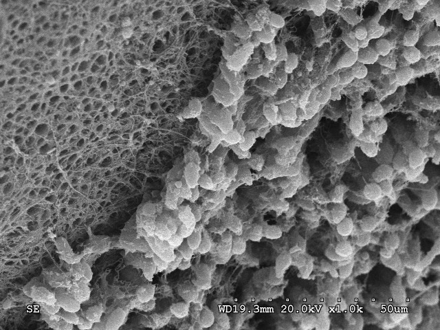
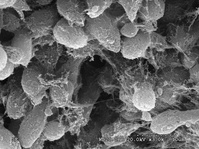
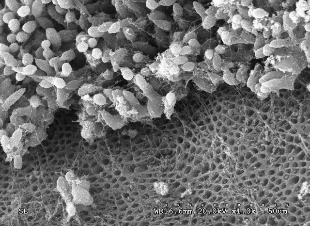
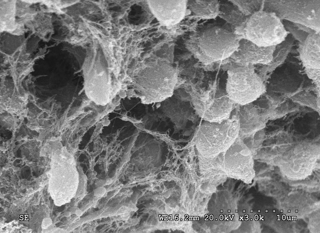
Odontoblasts on the inner surface of the pulpal chamber were observed under SEM, after the removal of the pulp. In Group E, SEM micrography revealed the integrity of the odontoblast cell layer and the well-preserved morphology of individual odontoblast cells. Odontoblasts located at the dentin surface appeared like a densely packed layer. Potato-like cell bodies were observed, approximately 4 μm to 7 μm long, with collagenic and neural fibers. The cytoplasmic processes of the odontoblasts were connected to each other, and they extended into the dentinal tubules. The opened dentinal tubules were visible, and their diameter ranged from 4 μm to 8 μm (Figure 8). Similar results were observed in Group F. There was no significant difference between the two groups regarding the morphology of the odontoblasts (Figure 9).
Figure 8.
SEM micrograph of odontoblasts in the control group. (a) SEM micrography revealed the integrity of the odontoblast cell-layer and opened dentinal tubules (×1000). (b) The well-preserved morphology of the odontoblast cells (×3000).
a.
b.
Figure 9.
SEM micrograph of odontoblasts after 2.0 W/CW (166 J/cm2) of 980-nm diode laser treatment.
a.
b.
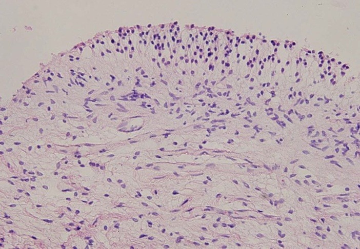
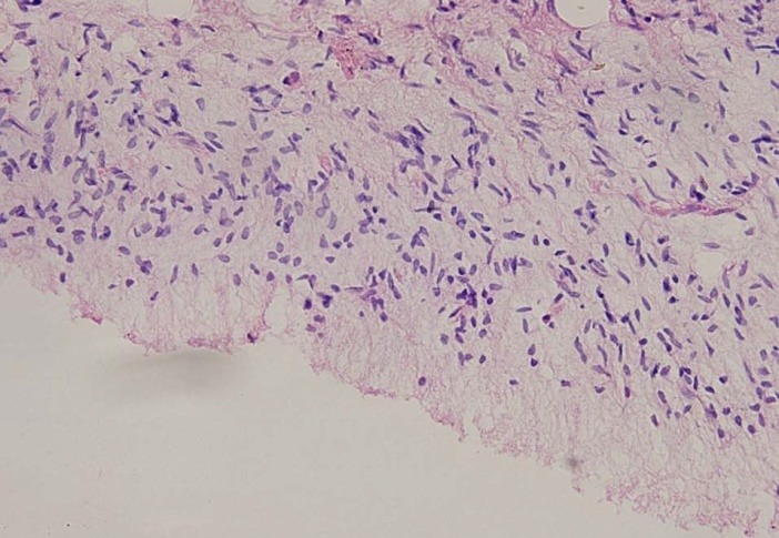
The morphology of the pulp was observed by light microscopy after HE staining. No significant differences were observed between the two groups. The morphology of the odontoblast cells and vessels, as well as of the collagenic and neural fibers, was clear and healthy (Figure 10).
Figure 10.
Micrograph of pulp tissue after HE staining. (a) Micrograph of pulp tissue in the control group (×200). (b) Micrograph of pulp tissue after 2.0 W/CW (166 J/cm2) of 980-nm diode laser treatment (×200).
a.
b.
Discussion
Hard-tissue modification by means of laser irradiation is becoming popular in dentistry. The role of Nd:YAG laser, Er:YAG laser and 830-nm wavelength diode laser in DH has been proved. However, the effect of 980-nm wavelength diode laser on DH is not clear. The results of this study show that different parameters of 980-nm wavelength diode laser can seal dentinal tubules. The dentin structure was changed because of the thermal effects caused by laser energy23 , 32 - 35. A crystalline arrangement and melting can be attributed to some of the energy being absorbed by the dentin’s mineral content, which includes carbonate and phosphate32. The mechanism of action of 980-nm diode laser is likely to be similar to that of 1064-nm Nd:YAG laser because both are located in the nearinfrared region of the electromagnetic spectrum36.
Morphological alterations in dentin irradiated with 980-nm diode laser could be observed by SEM, and they depended mainly on the parameters of the laser, including the output power, frequency, and application mode, because these parameters are directly related to the energy transmitted to the dentin. According to the manufacturer (Sirona Dental Systems), the power of this device ranges from 0.5 to 7.0 W and from 1 to 10 kHz, with a continuous wave operating mode, which consists of an uninterrupted laser beam. In this study,2 W/CW, 3 W/CW, and 4 W/CW 980-nm diode laser was used, and the energy density was 166 J/cm2, 250 J/cm2, and 333 J/cm2, respectively. The laser’s effect on the melting degree of the dentin surface was intensified when the parameters were increased from 2 to 4 W in the continuous mode. The reason for this result was that the energy of 980-nm diode laser absorbed by the dentin’s mineral content was increased, resulting in crystalline arrangement and melting32, 37, 38.
We found that the number of sealed dentin tubules in Group C was less than that in Group B. The differences could be attributed to the double-layer structure of the tubules. We speculate that, at first, the superficial layer melted and the crystalline arrangement occurred because low energy was absorbed by the superficial layer of the dentin’s mineral content. With increasing energy, the superficial layer of dentin melted again and collapsed into the dentinal tubules, so the double-layer structure formed. In Group D, the surface and deep layer of dentin were both melted by the high energy of laser to form a homogeneous and regular layer that covered the surface of the dentinal tubules.
This study demonstrated that 2 W/CW (166 J/cm2) was suitable parameter for a 980-nm diode laser to seal dentinal tubules without excess melting of the dentin.
The energy of laser can cause adverse thermal effects to the pulp, so lasers should be carefully operated. Studies have shown that pulp responds to externally applied heat 25 , 39, with 15% of teeth failing to recover from an intrapulpal temperature increase of 5.5 ºC. If the temperature increase was 11 ºC, 60% of teeth could not recover, so energy has a close relationship with the health of pulp tissue. Based on the above studies, 2.0 W/CW (166 J/cm2) was chosen to evaluate the role of 980-nm diode laser in the sealing of dentin tubules, as well as its safety in dental pulp tissue. Sealed dentin tubules were observed after irradiation by 980-nm diode laser (2.0 W/CW, 166 J/cm2). Compared with the control group, the dentinal surfaces in the laser groups were melted, and the overwhelming majority of dentinal tubules were sealed. From the longitudinal sections parallel to the direction of dentinal tubules’ alignment, melted dentin entered the dentinal tubules to as deep as 8 μm. No gap between the melted dentin and tubular walls was noted, most likely due to the same components in the dentin and pipe plug. Sealing exposed dentinal tubules, with melted and recrystallized dentin, were caused by the thermal and occlusive effects of laser, resulting in a prolonged role.
From SEM micrographs, no significant morphological alterations of the odontoblasts were found after treatment with 2.0 W/CW (166 J/cm2) 980-nm diode laser and a similar result was observed in the scan of pulp tissue stained by HE. Parameters of 2.0 W/CW (166 J/cm2) could be safe for 980-nm diode laser treatment for DH.
Irradiation with 980-nm diode laser seems to be effective for routine clinical treatment of DH, and 2.0 W/CW (166 J/cm2) is suitable energy parameter due to the rapid sealing of exposed dentinal tubules and its safety to odontoblasts and pulp tissue. Further studies are necessary to evaluate the rapid and prospective clinical effectiveness, as well as the adverse reactions, after 980-nm diode laser application for DH treatment.
Acknowledgments
This study was supported by the Science and Technology Planning Project of Guangdong Province, China (no. 2010B060900053).
Please cite this article as follows:
Liu Y, Gao J, Gao Y, XU Sh, Zhan X, Wu B. In Vitro Study of Dentin Hypersensitivity Treated by 980-nm Diode Laser. J Lasers Med Sci 2013; 4(3):111-9
References
- 1.Holland GR, Narhi MN, Addy M, Gangarosa L, Orchardson R. Guidelines for the design and conduct of clinical trials ondentine hypersensitivity. J Clin Periodontol. 1997;24:808–13. doi: 10.1111/j.1600-051x.1997.tb01194.x. [DOI] [PubMed] [Google Scholar]
- 2.Dowell P, Addy M. Dentine hypersensitivity – a review Aetiology, symptoms and theories of pain production. J Clin Periodontol. 1983;10:341–50. doi: 10.1111/j.1600-051x.1983.tb01283.x. [DOI] [PubMed] [Google Scholar]
- 3.Hasan Guney Yilmaz a, Sevcan Kurtulmus-Yilmaz, Esra Cengiz, Hakan Bayindir, Yasar Aykac. Clinical evaluation of Er,Cr:YSGG and GaAlAs laser therap for treating dentine hypersensitivity: A randomized controlled clinical trial. J Dent. 2011;39:249–54. doi: 10.1016/j.jdent.2011.01.003. [DOI] [PubMed] [Google Scholar]
- 4.Hmud Hmud, Kahler Kahler, George George, Walsh Walsh. Cavitational effects in aqueous endodontic irrigants generated by near-infrared lasers. J Endod. 2010;36:275–8. doi: 10.1016/j.joen.2009.08.012. [DOI] [PubMed] [Google Scholar]
- 5.Bergmans Bergmans, Moisiadis Moisiadis, Teughels Teughels, Van Meerbeek, Quirynen Quirynen, Lambrechts Lambrechts. Bactericidal effect of Nd:YAG laser irradiation on some endodontic pathogens exvivo. Int Endod J. 2006;39:547–57. doi: 10.1111/j.1365-2591.2006.01115.x. [DOI] [PubMed] [Google Scholar]
- 6.Augusto R. Elias Boneta, Karol Ramirez, Joselyn Naboa, Luis RMateo, Bernal Stewart, Foti Panagokos, William De Vizio. Efficacy in reducing dentine hypersensitivity of a regimen using a toothpaste containing 8% arginine and calcium carbonate, a mouthwash containing 08% arginine, pyrophosphate and PVM/MA copolymer and a toothbrush compared to potassium and negative control regimens: an eight-week randomized clinical trial. J Dent. 2013;41:42–9. doi: 10.1016/j.jdent.2012.11.011. [DOI] [PubMed] [Google Scholar]
- 7. Romeo Umberto, Russo Claudia, Palaia Gaspare, Tenore Gianluca, and Del Vecchio Alessandro.Treatment of Dentine Hypersensitivity by Diode Laser: A Clinical Study.Int J Dent 2012 Jun 25. [Epub ahead of print]. [DOI] [PMC free article] [PubMed]
- 8.Consensus-based recommendations for the diagnosis andmanagement of dentin hypersensitivity. J Can Dent Assoc. 2003;69:221–6. [PubMed] [Google Scholar]
- 9.Bor-Shiunn Leea, Chun-Wei Changb, Weng-Pin Chenc, Wan-Hong Lana, Chun-Pin Lina. In vitro study of dentin hypersensitivity treated by Nd:YAP laser and bioglass. Dent Mater. 2005;21:511–9. doi: 10.1016/j.dental.2004.08.002. [DOI] [PubMed] [Google Scholar]
- 10.Aranha AC, Eduardo Cde P. In vitro effects of Er,Cr:YSGG laser on dentine hypersensitivityDentine permeability and scanning electron microscopy analysis. Lasers Med Sci. 2012;27:827–34. doi: 10.1007/s10103-011-0986-y. [DOI] [PubMed] [Google Scholar]
- 11.Yilmaz HG, Kurtulmus-Yilmaz S, Cengiz E. Long-term effect of diode laser irradiation compared to sodium fluoride varnish in the treatment of dentine hypersensitivity in periodontal maintenance patients: a randomized controlled clinical study. Photomed Laser Surg. 2011;29:721–5. doi: 10.1089/pho.2010.2974. [DOI] [PubMed] [Google Scholar]
- 12.Maden M, Görgül G, Sultan MN, Akça G, Er O. Determination of the effect of Nd:YAG laser irradiation through dentinal tubules on several oral pathogens. Lasers Med Sci. 2013;28:281–6. doi: 10.1007/s10103-012-1150-z. [DOI] [PubMed] [Google Scholar]
- 13.Tavares JG, Eduardo CD, Burnett LH Jr, Boff TR, Freitas PM. Argon and Nd:YAG Lasers for Caries Prevention in Enamel. Photomed Laser Surg. 2012;30:433–7. doi: 10.1089/pho.2011.3104. [DOI] [PubMed] [Google Scholar]
- 14.Azevedo DT, Faraoni-Romano JJ, Derceli Jdos R, Palma-Dibb RG. Effect of Nd:YAG laser combined with fluoride on the prevention of primary tooth enamel demineralization. Braz Dent J. 2012;23:104–9. doi: 10.1590/s0103-64402012000200003. [DOI] [PubMed] [Google Scholar]
- 15.Maamary S, De Moor R, Nammour S. Treatment of dentin hypersensitivity by means of the Nd:YAG laserPreliminary clinical study. Rev Belge Med Dent (1984) 2009;64:140–6. [PubMed] [Google Scholar]
- 16.Moshonov J, Orstavik D, Yamauchi S, Pettiette M, Trope M. Nd:YAG laser irradiation in root canal disinfection. Endod Dent Traumatol. 1995;11:220–4. doi: 10.1111/j.1600-9657.1995.tb00492.x. [DOI] [PubMed] [Google Scholar]
- 17.Perin FM, França SC, Silva-Sousa YT, Alfredo E, Saquy PC, Estrela C, Sousa-Neto MD. Evaluation of the antimicrobial effect of Er:YAG laser irradiation versus 1% sodium hypochlorite irrigation for root canal disinfection. Aust Endod J. 2004;30:20–2. doi: 10.1111/j.1747-4477.2004.tb00162.x. [DOI] [PubMed] [Google Scholar]
- 18.Raucci-Neto W, Pécora JD, Palma-Dibb RG. Thermal effects and morphological aspects of human dentin surface irradiated with different frequencies of Er:YAG laser. Microsc Res Tech. 2012;75:1370–5. doi: 10.1002/jemt.22076. [DOI] [PubMed] [Google Scholar]
- 19.Birang R, Poursamimi J, Gutknecht N, Lampert F, Mir M. Comparative evaluation of the effects of Nd:YAG and Er:YAG laser in dentin hypersensitivity treatment. Lasers Med Sci. 2007;22:21–4. doi: 10.1007/s10103-006-0412-z. [DOI] [PubMed] [Google Scholar]
- 20.Gutknecht N, Franzen R, Schippers M, Lampert F. Bactericidal effect of a 980-nm diode laser in the root canal wall dentin of bovine teeth. J Clin Laser Med Surg. 2004;22:9–13. doi: 10.1089/104454704773660912. [DOI] [PubMed] [Google Scholar]
- 21.Raqueli Viapiana, Manoel D. Sousa-Neto, Aline Evangelista Souza-Gabriel, Edson Alfredo, Yara TCSilva-Sousa, Microhardness of Radicular Dentin Treated with 980-nm Diode Laser and Different Irrigant Solutions. Photomed Laser Surg. 2012;30:102–6. doi: 10.1089/pho.2011.3109. [DOI] [PubMed] [Google Scholar]
- 22.Faria MI, Sousa-Neto MD, Souza-Gabriel AE, Alfredo E, Romeo U, Silva-Sousa YT. Effects of 980-nm diode laser on the ultrastructure and fracture resistance of dentine. Lasers Med Sci. 2013;28:275–80. doi: 10.1007/s10103-012-1147-7. [DOI] [PubMed] [Google Scholar]
- 23.Marchesan MA, Brugnera-Junior A, Souza-Gabriel AE, Correa-Silva SR, Sousa-Neto MD. Ultrastructural analysis of root canal dentine irradiated with 980-nm diode laser energy at different parameters. Photomed Laser Surg. 2008;26:235–40. doi: 10.1089/pho.2007.2136. [DOI] [PubMed] [Google Scholar]
- 24.Srimaneepong V, Palamara JE, Wilson PR. Pulpal space pressure and temperature changes from Nd:YAG laser irradiation of dentin. 2002;30:291–6. doi: 10.1016/s0300-5712(02)00023-4. [DOI] [PubMed] [Google Scholar]
- 25. E. Alfredo, Marchesan, Sousa-Neto, A. Brugnera-Ju ´niora, Silva-Sousa.Temperature variation at the external root surface during 980-nm diode laser irradiation in the root canal. J Dent. 2008; 36:529–34. [DOI] [PubMed]
- 26. Hubbezoglu I, Unal M, Zan R, Hurmuzlu F. Temperature Rises During Application of Er:YAG Laser Under Different Primary Dentin Thicknesses. Photomed Laser Surg 2013 Mar 12. [Epub ahead of print]. [DOI] [PubMed]
- 27.Secilmis A, Bulbul M, Sari T, Usumez A. Effects of different dentin thicknesses and air cooling on pulpal temperature rise during laser welding. Lasers Med Sci. 2013;28:167–70. doi: 10.1007/s10103-012-1108-1. [DOI] [PubMed] [Google Scholar]
- 28.White JM, Fagan MC, Goodis HE. Intrapulpal temperatures during pulsed Nd:YAG laser treatment of dentin, in vitro. J Periodontol. 1994;65:255–9. doi: 10.1902/jop.1994.65.3.255. [DOI] [PubMed] [Google Scholar]
- 29.Mehl A, Kremers L, Salzmann K, Hickel T. 3D volumeablation rate and thermal side effects with the Er:YAG and Nd:YAG laser. Dent Mater. 1997;13:246–51. doi: 10.1016/S0109-5641(97)80036-X. [DOI] [PubMed] [Google Scholar]
- 30.Durand SH, Flacher V, Roméas A, Carrouel F, Colomb E, Vincent C, Magloire H, Couble ML, Bleicher F, Staquet MJ, Lebecque S, Farges JC. Lipoteichoic acid increases TLR and functional chemokine expression while reducing dentin formation in in vitro differentiated human odontoblasts. J Immunol. 2006;176:2880–7. doi: 10.4049/jimmunol.176.5.2880. [DOI] [PubMed] [Google Scholar]
- 31.Farges JC, Keller JF, Carrouel F, Durand SH, Romeas A, Bleicher F, Lebecque S, Staquet MJ. Odontoblasts in the dental pulp immune response. J Exp Zool B Mol Dev Evol. 2009;312B:425–36. doi: 10.1002/jez.b.21259. [DOI] [PubMed] [Google Scholar]
- 32.Alfredo Alfredo, Souza–Gabriel Souza–Gabriel, Silva Silva, Sousa–Neto Sousa–Neto, Brugnera– Junior, Silva–Sousa Silva–Sousa. Morphological alterations of radicular dentine pretreated with different irrigations solutions and irradiated with 980 nm diode laser. Microsc Res Tech. 2009;72:22–7. doi: 10.1002/jemt.20638. [DOI] [PubMed] [Google Scholar]
- 33.Faria Faria, Souza–Gabriel Souza–Gabriel, Marchesan Marchesan, Sousa–Neto Sousa–Neto, and Silva–Sousa. Ultrastructuralevaluation of radicular dentin after Nd:YAG laser irradiation combined with different chemical substances. Gen Dent. 2008;56:641–6. [PubMed] [Google Scholar]
- 34.Stabholz A, Neev J, Liaw HL, Khayat A, Torabinejad M. Sealing of human dentinal tubules by XeCl 308-nm excimer laser. J Endod. 1993;19:267–71. doi: 10.1016/s0099-2399(06)80454-1. [DOI] [PubMed] [Google Scholar]
- 35.Faria MI, Souza-Gabriel AE, Alfredo E, Messias DC, Silva-Sousa YT. Apical microleakage and SEM analysis of dentin surface after 980 nm diode laser irradiation. Braz Dent J. 2011;22:382–7. doi: 10.1590/s0103-64402011000500006. [DOI] [PubMed] [Google Scholar]
- 36.Coluzzi Coluzzi. An overview of laser wavelengths used in dentistry. Dent Clin North Am. 2000;44:753–65. [PubMed] [Google Scholar]
- 37.Faria MIA, Souza-Gabriel AE, Marchesan MA, Sousa- Neto MD, Silva-Sousa YTC. Ultrastructural evaluation of radicular dentin after Nd:YAG laser irradiation combined with different chemical substances. Gen Dent. 2008;56:641–646. [PubMed] [Google Scholar]
- 38.Garcia LF, Naves LZ, Farina AP, Walker CM, Consani S, Pires-de-Souza FC. The effect of a 980 nm diode laser with different parameters of irradiation on the bond strength of fiberglass posts. Gen Dent. 2011;59:31–37. [PubMed] [Google Scholar]
- 39.Zach L, Cohen G. Pulp response to externally applied heat. Oral Surg Oral Med Oral Pathol. 1965;19:515–30. doi: 10.1016/0030-4220(65)90015-0. [DOI] [PubMed] [Google Scholar]


