

Sir,
A 52-year-old Chinese female weighing 60 kg, previously community ambulant, presented with concurrent cervical stenosis and thoracic myelopathy. She was administered general anaesthesia, and posterior spine decompression, instrumentation and fusion from T6 to T11 was carried out in the prone position.
Nasal intubation was performed in the supine position using the awake fibreoptic intubation technique to avoid aggravation of the cervical cord pathology. After confirmation of endotracheal tube placement, general anaesthesia was induced using standard anaesthetic agents with standard monitoring. Patient was then turned to the prone position. Confirmation of proper positioning of head and face was done through frequent visual inspections by the anaesthesiologist.
Surgery proceeded uneventfully with the periodic use of intraoperative high-voltage motor evoked potentials (MEP) spinal cord monitoring. Duration of surgery was 5½ h, and patient was repositioned in supine position. At this time, we observed that the anterior half of the patient's tongue was dusky and congested. Further, there was a 1 cm laceration on the dorsum of the tongue and a 3 cm laceration on the undersurface. Teeth marks were also noted on the tongue [Figure 1]. An otolaryngologist's opinion was sought, who performed toilet and suture of the tongue lacerations before anaesthesia reversal.
Figure 1.
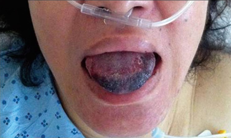
Tongue showing laceration
Postoperatively, the patient complained of dysarthria and tongue pain affecting feeding, necessitating liquid diet. Over the next few days, the swelling and discolouration of the tongue improved markedly. Subsequently, the sutures were removed 2 weeks later. Patient's tongue was found to be healing well, and her speech and feeding were back to normal. Two months later, the tongue lacerations had healed, and no long-term complications were noted.
Tongue lacerations associated with anaesthesia is an uncommon occurrence. Advanced age and higher Mallampati class, representing the difficult palatal anatomy, are factors that contribute to difficulty in intubation and increase the risk of upper airway injuries such as tongue and lip lacerations.[1] When a patient is placed in the prone position under general anaesthesia, as in our case, the risk of injury is aggravated. This is due to the gravity induced protrusion and compromise of venous return and subsequent oedema.[2] In our case, the most likely mechanism is a combination of the prone position causing tongue oedema, with subsequent bite injury to the oedematous tongue.
In MEP, the most likely mechanism is direct electrical stimulation of the masseter and temporalis muscles leading to contraction, especially with C3/4 stimulation.[3] Hence, whenever MEP monitoring is used, tongue lacerations can occur even if the surgery is carried out in the supine position.[4] In a review of 17,273 surgical procedures requiring MEP monitoring, Tamkus and Rice reported a slightly higher percentage of bite injuries in anterior versus posterior surgical approaches (54.1% vs. 35.8%, although this did not reach significance [P = 0.078]).[5]
Tongue bite injuries occurring after MEP monitoring are distressing to both the patient and the medical team. Our patient suffered from pain, dysphagia and dysarthria. Previous studies report even more serious complications arising from bite injuries, including endotracheal tube rupture, mandibular fracture, and delayed extubation due to severe tongue swelling.[5] Hence, whenever patients undergo spinal surgery with the use of MEP monitoring, measures to prevent such injuries need to be considered. We propose the routine placement of soft bite blocks into the oral cavity whenever MEP monitoring is used, irrespective of patient position. A bite block can be easily created from rolled gauze pads secured with tape, which resembles a “cigar.” One should be placed between the molars on each side, and each should be large enough to prevent contact between the teeth and the tongue [Figure 2]. They should be properly secured and should also be regularly inspected to ensure they have not been displaced. Hard plastic bite blocks are not recommended as they increase the risk of tooth injury.[5]
Figure 2.

Bite blocks made from rolled gauze
In conclusion, tongue lacerations occurring after general anaesthesia are a distressing complication for both the patient and the medical team. This complication is uncommon, but the risk is increased when the patient is placed in a prone position and use of intraoperative MEP monitoring. A simple rolled gauze “cigar” placed between the molars on each side may reduce the risk of this complication.
REFERENCES
- 1.Hua M, Brady J, Li G. The epidemiology of upper airway injury in patients undergoing major surgical procedures. Anesth Analg. 2012;114:148–51. doi: 10.1213/ANE.0b013e318239c2f8. [DOI] [PMC free article] [PubMed] [Google Scholar]
- 2.Chae YJ, Kim JY, Yoo JY, Choi YH, Park KS. Tongue bite in a patient with tracheostomy after prone position-A case report. Korean J Anesthesiol. 2011;60:365–8. doi: 10.4097/kjae.2011.60.5.365. [DOI] [PMC free article] [PubMed] [Google Scholar]
- 3.MacDonald DB. Safety of intraoperative transcranial electrical stimulation motor evoked potential monitoring. J Clin Neurophysiol. 2002;19:416–29. doi: 10.1097/00004691-200210000-00005. [DOI] [PubMed] [Google Scholar]
- 4.Williams A, Singh G. Tongue bite injury after use of transcranial electric stimulation motor-evoked potential monitoring. J Anaesthesiol Clin Pharmacol. 2014;30:439–40. doi: 10.4103/0970-9185.137297. [DOI] [PMC free article] [PubMed] [Google Scholar]
- 5.Tamkus A, Rice K. The incidence of bite injuries associated with transcranial motor-evoked potential monitoring. Anesth Analg. 2012;115:663–7. doi: 10.1213/ANE.0b013e3182542331. [DOI] [PubMed] [Google Scholar]