Abstract
Objective:
We investigated the feasibility of delayed computed tomography (CT) arthrography for evaluation of human knee cartilage in vivo. Especially, the diffusion of contrast agent out of the joint space and the optimal time points for imaging were determined.
Design:
Two patients were imaged using delayed CT arthrography and delayed gadolinium-enhanced magnetic resonance imaging of cartilage (dGEMRIC) techniques.
Results:
Two hours after injection, the concentration of contrast agent in the joint space was still high enough (20% to 24.5% of the initial concentration at 0 minutes) to allow delayed CT arthrography. The half-life of the contrast agent in the joint space varied from 30 to 60 minutes. The contrast agent concentration in patellar and femoral cartilage reached the maximum after 30 and 60 minutes, respectively. According to dGEMRIC, there were no differences between patients. However, in delayed CT arthrography, the penetration of the contrast agent was higher in the osteoarthritic knee cartilage.
Conclusions:
Contrast agent remained in the joint space long enough to enable delayed CT arthrography of cartilage. After 30 minutes, the normalized contrast agent concentration was higher in the cartilage of the osteoarthritic knee in comparison with the healthy knee. To conclude, delayed CT arthrography exhibited potential for use in the clinical evaluation of cartilage integrity.
Keywords: contrast-enhanced computed tomography (CT), delayed CT arthrography, cartilage, knee, osteoarthritis, in vivo
Introduction
Delayed gadolinium-enhanced magnetic resonance imaging of cartilage (dGEMRIC) and contrast-enhanced computed tomography (CECT) have been proposed for diagnostics of proteoglycan loss in osteoarthritis (OA).1-4 In these methods, anionic contrast agents are hypothesized to distribute into the cartilage in inverse proportion to the spatial distribution of fixed charge density in the cartilage matrix.1,5 In addition, it has been claimed that the steric hindrance induced by the cartilage matrix can significantly affect the contrast agent diffusion. This is supported by our recent study, which described a significant increase in contrast agent uptake after an impact injury.6
It has been reported in vitro that clinical contrast agents may reach diffusion equilibrium only after 8 to 9 hours of immersion.5 However, it is not feasible to use such long delays in vivo, and the contrast agent washout from the joint also prevents the reaching of the Donnan equilibrium. Importantly, in our earlier in vitro study, fresh cartilage injuries could be detected in vitro already after 30 to 60 minutes of immersion in a contrast agent bath.6 Earlier, CECT of cartilage has been applied in vivo only with rats.7,8 However, because of the faster metabolism and smaller size of the joint, the contrast agent dynamics is probably faster in a rat model than in a human joint. In animal models, the contrast agent leakage from the joint capsule is often prevented by mixing a vasoconstrictive agent, epinephrine, with the contrast agent.7,8 When using epinephrine, the x-ray attenuation decreases only 15% in about 50 minutes in the knee joint space of a rat.8
CT arthrography is a method that has been traditionally used for the evaluation of cartilage morphology.9 In arthrography, the contrast agent is injected into the joint cavity and the CT image is acquired immediately. However, there are studies in which the effect of a time delay between the injection and imaging has been investigated.10,11 The visual quality of the image has been reported to decrease in the first 20 minutes because of the diffusion of the contrast agent into the cartilage and out of the joint cavity.10,11 Furthermore, delayed CT arthrography has been used to detect articular ganglion cysts.12,13 Ionic contrast agents were applied in these studies, and CT images were acquired immediately after the injection and then again 1 to 2 hours after the injection.12,13 Although the articular cartilage was not investigated in the above studies, the contrast agent remained in the joint cavity for this period and the articular cartilage could also be visualized from the obtained images.
The first steps toward clinical application of CECT were taken recently, as the delayed CECT (10-minute delay) was used to assess the glycosaminoglycan concentration of cadaver knee cartilage.14 However, the clinical feasibility of the CECT method has not been evaluated. For example, it is not known whether the contrast agent would remain in the joint space long enough to enable CECT. In addition, the optimal imaging time point after injection, the optimal contrast agent volume and concentration are not known.
The aim of this study was to evaluate the potential of the delayed CT arthrography (referred to as CECT when discussing in vitro studies) method for in vivo imaging of articular cartilage of clinical patients. Therefore, we investigated the contrast agent leakage out of the joint space and determined the optimal time points for imaging. In the present study, 2 clinical patients underwent delayed CT arthrography and as a reference dGEMRIC was conducted for both patients.
Methods
Participants
Two patients from the orthopaedic outpatient clinic at Kuopio University Hospital were recruited for this study. Both patients provided an informed consent before the study. Based on the clinical symptoms (both patients) and native x-ray examination (patient 2), an orthopedic surgeon (HK) referred the patients for CT arthrography. The left knee of patient 1 (male, age 31 years) was imaged as an intact reference to his right knee with degeneration related to an earlier anterior cruciate ligament tear. Patient 2 (male, age 50 years) had suffered from pain and swelling of his right knee for 2 years and the x-ray showed signs of early osteoarthritis (slightly narrowed joint space, Kellgren–Lawrence grade 2). The left knee of patient 1 and the right knee of patient 2 were imaged with a clinical CT (Siemens Somatom AS, Siemens, Germany) and 3T MRI scanners (Siemens, Germany, patient 1, and Philips Achieva. 3.0 Tx, Philips, Netherlands, patient 2).
Delayed Computed Tomography Arthrography
First, a series of test tubes, filled with contrast agent at different concentrations, was imaged using the CT scanner. The acquired attenuation values were plotted against the contrast agent concentration values to confirm linearity and to determine the saturation concentration, that is, the concentration value after which the Hounsfield unit values did not increase. The relationship between x-ray attenuation and contrast agent concentration was linear at concentrations below 37% and above that, the CT scanner readings were seen to saturate. However, it was assumed that the synovial fluid in the knee joint would dilute the injected contrast agent and it was decided to use 50% solution.
In delayed CT arthrography, the knees were first imaged without contrast agent, after which a dose of clinical anionic contrast agent (q = −1, M = 1269 g/mol, Hexabrix) was injected into the knee joint through the superolateral portal (12 mL and 24 mL for patients 1 and 2, respectively). In this study, epinephrine was not used because of the risk of inflammation when mixed with the charged iodine based contrast agents.15 In the intact knee of patient 1, a 12 mL injection of contrast agent was sufficient. Patient 2 had a history of osteoarthritic symptoms and occasional swelling of the knee and thus had an enlargened knee joint capsule. For this reason, we injected a larger amount of contrast agent into this OA knee. To enhance the contrast agent distribution in the knee joint, the patient exercised the knee lightly for 1 minute without bearing weight on it. The knee was then imaged at 0, 30, 60, and 120 minutes after the exercise. CT measurements were conducted using the following settings: total scanning time 10 seconds, tube voltage 120 kV, tube current 180 mA, slice collimation 0.3 mm, pitch 0.85, and voxel size of 0.21 × 0.21 × 0.40 mm3. The upper limit for the total effective radiation dose was estimated to be 1.5 mSv by using dose estimation software (CT-Expo).16
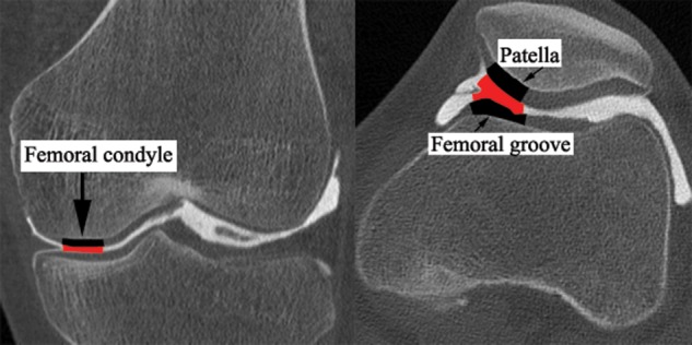
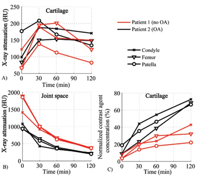
Three anatomical areas were analyzed from the delayed CT arthrography images: patellar cartilage, cartilage of femoral groove, and lateral femoral condyle (Fig. 1). First, all the image stacks were registered in 3 dimensions (3D), separately for femur and patella. As the cartilage boundaries are most clearly visible immediately after administration of the contrast agent, the segmentation was applied to that image stack. A nonlinear anisotropic diffusion gradient filtering was applied, this being followed by an edge enhancement procedure. Manual boundaries were drawn to restrict the area of segmentation inside the joint. A seed point was chosen within the volume of interest (VOI) and the optimal grayscale values were chosen for thresholding. Finally, an automatic region growing procedure was applied and the desired VOI was acquired as a 3D object map. All the analyses were conducted with Analyze (Analyze 10.0., Analyze Direct, Inc., Overland Park, KS). The volumes of the segmented VOIs are shown in Table 1. The mean x-ray attenuation is calculated for each VOI in cartilage and in the intra-articular space at all time points. Then, the average values of attenuation obtained from the images before administration of contrast agent are subtracted from the average attenuation values obtained from images acquired at all time points after the administration of the contrast agent. This data are presented in Figure 2A (cartilage) and Figure 2B (intra-articular space). Finally, the x-ray attenuation in cartilage (Fig. 2A) is normalized with the x-ray attenuation in the intra-articular space (Fig. 2B) at each time point (Fig. 2C).
Figure 1.

The analyzed anatomical locations (patient 1) in the lateral femoral condyle, femoral groove, and patella are indicated with black markings. The analyzed contrast agent locations in the intra-articular space are indicated with gray markings (red in electric material). Similar anatomical locations were analyzed in patient 2.
Table 1.
The Segmented VOIs in Cartilage and in the Joint Space
| VOI (mm3) |
||
|---|---|---|
| Location | Patient 1 (no OA) | Patient 2 (OA) |
| Patella (cartilage) | 708 | 385 |
| Condyle (cartilage) | 493 | 264 |
| Groove (cartilage) | 441 | 104 |
| Condyle (contrast) | 364 | 661 |
| Patella/groove (contrast) | 920 | 401 |
Note: VOI = volume of interest; OA = osteoarthritis.
Figure 2.

(A) X-ray attenuation in cartilage at different time points. (B) X-ray attenuation in synovial fluid as a function of time. The half-life of the contrast agent varied between 30 and 60 minutes, depending on the anatomical location. (C) Normalized contrast agent concentration in cartilage (Ccartilage/Csynovial × 100%) as a function of time after the injection. The difference in the contrast agent partition between osteoarthritis (OA) and intact cartilage increased with time, and already at 30 to 60 minutes after the injection, the difference was detectable.
dGEMRIC
In 3T dGEMRIC, the patients were first imaged using a spin echo sequence with variable inversion times (T1 = 100, 200, 400, 800, 1600, and 3200 ms). Then a double dose of anionic gadolinium-based contrast agent (0.2 mmol/kg, q = −2, M = 548 g/mol, Magnevist, Bayer Schering Pharma, Berlin, Germany) was administered intravenously. In our hospital, no specific exercise is included in the dGEMRIC protocol. After the injection and a few minutes of walking, the patients continued with routine activities and came back after 90 minutes for the postcontrast imaging. The acquisition of the T1 map took 20 minutes. After 90 minutes, the MR imaging was repeated (T1 = 50, 100, 200, 400, 800, 1600, and 3200 ms) and the T1 maps were calculated. For all MR images, the TR was 3280 ms and the TE was 12 ms. The imaging matrix size was 256 × 256 and the field of view was 120 × 120 mm2, yielding an in-plane pixel size of 0.47 × 0.47 mm2 and a slice thickness of 3 mm. The mean T1 times for cartilage of femoral medial condyle and medial tibial plateau (from the areas shown in the last figure of this article) were calculated for both patients before and after the administration of the contrast agent. Unfortunately, the x-ray contrast agent did not distribute properly into medial condyle. For this reason, dGEMRIC and CT analyses were not conducted on the same area. For the postcontrast T1 times, correction was done for the body mass index.17 Furthermore, the difference in the relaxation rate (ΔR) was calculated using the following equation:
| (1) |
Cartilage thickness was determined for the corresponding sites in the CT and MR images. Cartilage thickness was measured at 9 points in the center of each VOI and averaged to estimate the mean thickness.
As the acquisition of T1 map is a relatively long process, only one slice was imaged. For this reason, only femoral condyle and tibial plateau were included in the image. In addition to quantitative analysis, as part of the routine clinical procedure, an experienced musculoskeletal radiologist (JSS) analyzed the precontrast CT and MR images. The joint space narrowing, osteophyte growth, periarticular cysts, periarticular sclerosis, and bone deformation were estimated from the images. In addition, radiography-based Kellgren–Lawrence grade was assigned to patient 2.
Results
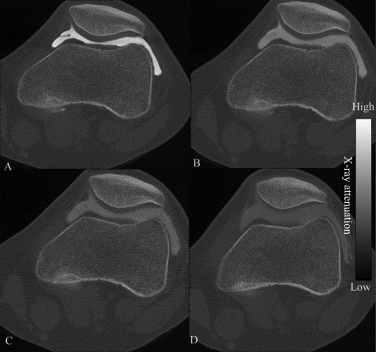
Two hours after the injection, there was still a significant amount of contrast agent left in the joint spaces of both patients (Figs. 2B and 3). The half-life of the contrast agent in the joint space varied from 30 to 60 minutes, depending on the patient and the anatomical location (Figure 2A). The contrast agent concentration in the cartilage (patella and femoral condyle) reached its maximum value after 30 minutes and gradually decreased after that time (Figure 2A). In femoral groove cartilage, the maximum concentration was achieved after 60 minutes. In the OA knee (patient 2), the cartilage contrast agent concentration (23.1% to 44.0% at 30 minutes) was systematically higher than in cartilage of the intact knee (patient 1, 13.9% to 20.7%, at 30 minutes; Fig. 2A and C). The difference in the contrast agent partition between OA and intact cartilage increased with time, and already 30 to 60 minutes after the injection, a clear difference was present.
Figure 3.
Contrast agent diffusion into cartilage in a nonarthritic joint (patient 1) is demonstrated in the delayed computed tomography (CT) arthrography images acquired at the time points of (A) 0 minutes, (B) 30 minutes, (C) 60 minutes, and (D) 120 minutes after the contrast agent injection. Although the contrast agent concentration in the joint space declines, the contrast agent is still detectable after 2 hours.
Cartilage thickness was evaluated from the CT and MRI images at patella, femoral groove, and femoral condyle (Table 2). The thickness of OA cartilage (patient 2) was lower than that of intact cartilage (patient 1). Cartilage thickness values obtained from CT and MRI were in agreement.
Table 2.
Cartilage Thickness (in Millimeters) at Patella, Femoral Groove, and Medial Femoral Condyle, Measured From the CT and MR Images
| Patient 1 (intact) |
Patient 2 (OA) |
|||||
|---|---|---|---|---|---|---|
| Patella | Groove | Condyle | Patella | Groove | Condyle | |
| CT | 3.42 | 2.28 | 2.35 | 2.03 | 1.57 | 2.16 |
| MRI | 2.99 | 2.28 | 2.47 | 1.88 | 1.68 | 2.10 |
Note: The cartilage of OA patient is thinner than that of the healthy patient. The values of cartilage thickness determined with the CT and MRI are in agreement. CT = computed tomography; MRI = magnetic resonance imaging; OA, osteoarthritis.
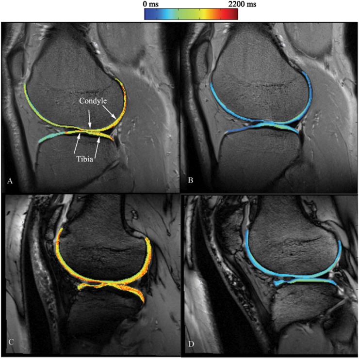
In intact cartilage, the contrast agent decreased the average T1 times from 1332 to 597 ms in femoral condyle and from 1255 to 687 ms in tibial plateau (Table 3, Fig. 4). In OA cartilage, contrast agent decreased the average T1 times from 1347 to 638 ms in femoral condyle and from 1487 to 664 ms in tibial plateau. The corresponding differences in relaxation rates were for intact cartilage ΔRFemur = 0.92 and ΔRTibia = 0.66, and for OA cartilage ΔRFemur = 0.83 and ΔRTibia = 0.83 (Table 3).
Table 3.
The Mean T1 Relaxation Times (in Milliseconds) Before and After the Injection of the Contrast Agent
| Femoral cartilage |
Tibial cartilage |
|||||
|---|---|---|---|---|---|---|
| Before | After | ΔR | Before | After | ΔR | |
| Patient 1 (intact) | 1332 | 597 | 0.92 | 1255 | 687 | 0.66 |
| Patient 2 (OA) | 1347 | 638 | 0.83 | 1483 | 664 | 0.83 |
Note: There are no systematic differences between the patients in the postcontrast T1 relaxation times or relaxation rates. OA = osteoarthritis.
Figure 4.
dGEMRIC images of the investigated knee joints. The T1 map of the cartilage is shown in color on top of the anatomical grayscale images. (A and C) Magnetic resonance (MR) images before the contrast agent injection for patient 1 (intact) and patient 2 (osteoarthritis, OA), respectively. (B and D) MR images at 90 minutes after the injection of contrast agent in patient 1 (intact) and patient 2 (OA), respectively. The analyzed regions for mean T1 time in medial femoral condyle and medial tibial plateau are shown in subfigure A as the area between arrows. No visible cartilage lesions or degradation can be detected from the images.
The x-ray attenuation was found to exhibit linear relationship with the contrast agent concentration and the saturation concentration for the contrast agent was found to be 37% with the present scanner. Furthermore, the 12 mL injection of 50% contrast agent became diluted in the intact knee into a concentration of 16.7% to 22.5% and the 24 mL injection in the OA knee into a concentration of 10.9% to 12.6%.
Discussion
In this study, clinical feasibility of the delayed CT arthrography was evaluated for the first time. Three different locations in the knees of 2 patients were analyzed at 5 time points after administration of the contrast agent. The contrast agent concentration in cartilage of patella and lateral femoral condyle reached its maximum at 30 minutes after injection. In femoral cartilage opposing the patella, the maximum was reached at 60 minutes after injection. In OA cartilage (patient 2), the normalized contrast agent concentration in cartilage was higher (at all time points) than in the intact cartilage (patient 1).
Experiments in animals have raised the issue that the contrast agent seems to leave the joint cavity rapidly and vasoconstrictive agents are commonly used to diminish this effect.8 Rapid contrast agent extraction from knee joint could prevent the imaging of the diffusion process into the cartilage. Based on the present results, this does not seem to be a major concern in human cartilage, as at 30 minutes after the injection, half of the initial concentration was present in the joint space. Even 2 hours after injection, 20% to 24.5% of the initial contrast agent concentration remained in the joint space. This is important as an adequate amount of contrast agent has to be present in the joint space during imaging for good image quality.
In dGEMRIC, the T1 maps were calculated for the precontrast and postcontrast images. In addition, the average T1 times for cartilage in medial femoral condyle and medial tibial plateau were determined. In intact cartilage, contrast agent decreased the average T1 times from 1332 to 597 ms in femoral condyle and from 1255 to 687 ms in tibial plateau. In OA cartilage, the contrast agent decreased the average T1 times from 1347 to 638 ms in femoral condyle and from 1487 to 664 ms in tibial plateau. These values are in line with the earlier in vivo studies.18,19 It is common to use the postcontrast T1 time (i.e., dGEMRIC index) as an indicator of cartilage quality. The more badly degraded the tissue, the more contrast agent will diffuse into the cartilage and the shorter will be the relaxation time. However, as the patients were imaged using different MRI scanners, it was considered better to use the difference in the relaxation rate (ΔR) when comparing the patients. This method minimizes the differences between the protocols and MRI scanners. According to the postcontrast T1 times, there was no difference between the patients. However, the ΔR values do suggest that the intact femur (patient 1) is the most degraded site with the intact tibia (patient 1) being the most intact site. In the intact cartilage, the radiologist did not find any signs of OA. According to the delayed CT arthrography, after 30 minutes the normalized contrast agent concentration in the OA cartilage was already higher than in the intact cartilage. Thus, delayed CT arthrography is a promising method for diagnosing cartilage degeneration. However, to evaluate the true diagnostic potential of the technique, more research with a larger number of patients will be required.
Although dGEMRIC has been used in cartilage diagnostics it does suffer from certain limitations. The in-plane pixel size and especially the slice thickness are compromised because of the lengthy imaging times. In an attempt to minimize the imaging time only one slice is often acquired, which obviously reduces the potential of this technique to detect small injuries and lesions. Although delayed CT arthrography enables the acquisition of isotropic voxels and high resolution with fast scan times, it also has some limitations. Delayed CT arthrography induces a radiation dose and requires intra-articular contrast agent injection. Fortunately, the knee joint is only minimally sensitive to radiation and the applied contrast agent is well tolerated. However, there is a minor risk of infection when injecting contrast agent into the joint. In MRI, the contrast agent is traditionally administered intravenously. Because of lower sensitivity of CT imaging, excessive amounts of contrast agent need to be injected intravenously to ensure an adequate concentration in cartilage. Thus, use of intra-articular injection may be the only feasible option for delayed CT arthrography. In addition, as the volume of the joint capsule varies from patient to patient, the amount of injected contrast agent needs to be tailored individually for each patient. For this reason, the contrast agent concentration in cartilage must be normalized with the concentration in the joint capsule.
Before the clinical experiment, a series of different concentrations of contrast agents were imaged to confirm the linearity of the relationship between x-ray attenuation and contrast agent. The attenuation was found to exhibit a linear relationship with the contrast agent concentration and the saturation concentration for the contrast agent to be 37%. This value indicates the maximum concentration that can be used to obtain reliable numerical information. However, the dilution of contrast agent into the synovial fluid has to be taken into account when determining the optimal concentration. In this study, the 12 mL injection of 50% contrast agent became diluted in the intact knee and resulted in a concentration of 16.7% to 22.5% and the 24 mL injection in the OA knee led to concentration of 10.9% to 12.6%. Thus, significantly higher contrast agent concentrations could be used. However, high contrast agent concentrations are hyperosmolaric. In our recent study, we found out that this hyperosmolarity will cause a temporary softening of articular cartilage, which might jeopardize the well-being of the tissue if the patient undertakes intense exercise immediately after imaging.20
In normal CT or x-ray images, cartilage is not visible and the cartilage thickness can only be estimated based on the joint space width. In the clinic, cartilage thickness is often evaluated from MR images.21-24 Using contrast-enhanced methods, namely CT arthrography, the cartilage is more easily detectable and acquisition of isotropic voxels allows the measurement of the cartilage thickness of 3D cartilage segments. In this study, the cartilage thickness was estimated from the CT and anatomical MRI images of patella, femoral groove, and lateral femoral condyle. Even though the values of cartilage thickness acquired were similar with both methods, there are a few shortcomings related to the thickness measurements from the MR images. MRI slice was 3 mm thick, which could affect the reliability of the results at locations where there are rapid changes in cartilage thickness. Furthermore, the used knee coil was centered on the condyle area, which caused the signal from patellar region to be of lower quality, complicating the cartilage thickness estimation. In delayed CT arthrography, the diffusion of the contrast agent into the cartilage makes segmentation of cartilage challenging. For this reason, the cartilage has to be segmented from the image acquired immediately after the administration of contrast agent. In this study, the thickness of intact cartilage (patient 1) was greater than that of OA cartilage (patient 2). As the delayed CT arthrography method could allow at least semiautomatic segmentation, the cartilage thickness could be easily mapped throughout the entire joint. The development of this technique is one of our future aims, as it could enable the use of delayed CT arthrography data in creation of 3D models of joint function.25 The volumes of analyzed regions were not the same in the 2 patients undergoing delayed CT arthrography. However, a larger volume should only diminish noise and should not significantly affect the average attenuation in the analyzed volume.
In dGEMRIC, at the tibial plateau, the OA cartilage showed a higher change in the relaxation rate than intact cartilage. In addition, in delayed CT arthrography the contrast agent partition (Fig. 2C) at 60 minutes after the injection was higher in OA cartilage at lateral femoral condyle than in intact cartilage. However, the change in the relaxation rate at the medial femoral condyle was higher in intact cartilage than in OA cartilage. This is surprising and further studies will be needed to evaluate the correlation between dGEMRIC and delayed CT arthrography.
This study was designed to gather information about the practical issues regarding application of the delayed CT arthrography technique in vivo. The first issue was the optimization of the amount of contrast agent to be injected into the joint space. Patient 1 had an intact knee joint and the volume of 12 mL of contrast agent that was used was nearly enough. Patient 2 had OA in the knee with occasional swelling, the joint capsule was much larger in size, and it also included a higher amount of synovial fluid. For these reasons, 24 mL of contrast agent was used for this patient. To ensure the availability of contrast agent in the joint space we propose that the amount of contrast agent injected into the joint should be increased and a cuff could be placed just above the knee to restrict the size of the joint. In addition, should the joint be full of fluid before imaging, then draining of the joint should be considered.
In the present study, after the injection the patients exercised the knee gently without bearing any weight on it. However, the contrast agent had not optimally distributed in the first images but at later time points the distribution was seen to be uniform. For this reason, we propose that the knee should be gently exercised (e.g., cycling in the air when lying on the back) for 2 to 3 minutes before acquisition of the contrast enhanced images.
For future studies, we would recommend using three acquisitions. One acquisition before contrast agent injection, one immediately after the injection, and one at 30 to 60 minutes after the injection. During this time, the contrast agent in the joint space will not be diluted too much and the sensitivity of the technique will be maximal.
To conclude, delayed CT arthrography is a promising new method for use in the diagnostics of cartilage degeneration and it has the potential to detect differences between the intact and OA cartilage.
Footnotes
Acknowledgments and Funding: This work was supported by Sigrid Juselius Foundation, Strategic Funding of the University of Eastern Finland and Kuopio University Hospital (EVO project 5041715).
Declaration of Conflicting Interests: The authors declared no potential conflicts of interest with respect to the research, authorship, and/or publication of this article.
References
- 1. Bashir A, Gray ML, Boutin RD, Burstein D. Glycosaminoglycan in articular cartilage: in vivo assessment with delayed Gd(DTPA)(2-)-enhanced MR imaging. Radiology. 1997;205:551-8. [DOI] [PubMed] [Google Scholar]
- 2. Kallioniemi AS, Jurvelin JS, Nieminen MT, Lammi MJ, Töyräs J. Contrast agent enhanced pQCT of articular cartilage. Phys Med Biol. 2007;52:1209-19. [DOI] [PubMed] [Google Scholar]
- 3. Nieminen MT, Rieppo J, Silvennoinen J, Töyräs J, Hakumäki JM, Hyttinen MM, et al. Spatial assessment of articular cartilage proteoglycans with Gd-DTPA-enhanced T(1) imaging. Magn Reson Med. 2002;48:640-8. [DOI] [PubMed] [Google Scholar]
- 4. Palmer AW, Robertson GC, Guldberg RE, Levenstone ME. Contrast-enhanced microcomputed tomography detects cartilage degradation. In: Proceedings of the 51st Annual Meeting of the Orthopaedic Research Society; 2005 Feb 20-23 Washington, DC: Orthopaedic Research Society; 2005. [Google Scholar]
- 5. Silvast TS, Kokkonen HT, Jurvelin JS, Quinn TM, Nieminen MT, Töyräs J. Diffusion and near-equilibrium distribution of MRI and CT contrast agents in articular cartilage. Phys Med Biol. 2009;54:6823-36. [DOI] [PubMed] [Google Scholar]
- 6. Kokkonen HT, Jurvelin JS, Tiitu V, Töyräs J. Detection of mechanical injury of articular cartilage using contrast enhanced computed tomography. Osteoarthritis Cartilage. 2011;19:295-301. [DOI] [PubMed] [Google Scholar]
- 7. Piscaer TM, van Osch GJ, Verhaar JA, Weinans H. Imaging of experimental osteoarthritis in small animal models. Biorheology. 2008;45:355-64. [PubMed] [Google Scholar]
- 8. Piscaer TM, Waarsing JH, Kops N, Pavljasevic P, Verhaar JA, van Osch GJ, et al. In vivo imaging of cartilage degeneration using microCT-arthrography. Osteoarthritis Cartilage. 2008;16:1011-7. [DOI] [PubMed] [Google Scholar]
- 9. Winalski CS, Rajiah P. The evolution of articular cartilage imaging and its impact on clinical practice. Skeletal Radiol. 2011;40:1197-222. [DOI] [PubMed] [Google Scholar]
- 10. Carlier RY, Akeson P, Morvan G, Kristoffersen DT, Amar C, Vallee C. Time dependence of iopamidol and iodixanol in arthrography of the knee. Acta Radiol. 1999;40:107-10. [DOI] [PubMed] [Google Scholar]
- 11. Railhac JJ, Brekke O. Iopentol (Imagopaque 300) compared with ioxaglate (Hexabrix 320) in knee arthrography. A clinical trial assessing immediate and late adverse events and diagnostic information. Eur Radiol. 1997;7(Suppl 4):S135-9. [DOI] [PubMed] [Google Scholar]
- 12. Malghem J, Vande berg BC, Lebon C, Lecouvet FE, Maldague BE. Ganglion cysts of the knee: articular communication revealed by delayed radiography and CT after arthrography. AJR Am J Roentgenol. 1998;170:1579-83. [DOI] [PubMed] [Google Scholar]
- 13. Omoumi P, de Gheldere A, Leemrijse T, Galant C, Van den Bergh P, Malghem J, et al. Value of computed tomography arthrography with delayed acquisitions in the work-up of ganglion cysts of the tarsal tunnel: report of three cases. Skeletal Radiol. 2010;39:381-6. [DOI] [PubMed] [Google Scholar]
- 14. Siebelt M, van Tiel J, Waarsing JH, Piscaer TM, van Straten M, Booij R, et al. Clinically applied CT arthrography to measure the sulphated glycosaminoglycan content of cartilage. Osteoarthritis Cartilage. 2011;19:1183-9. [DOI] [PubMed] [Google Scholar]
- 15. Corbetti F, Malatesta V, Camposampiero A, Mazzi A, Punzi L, Angelini F, et al. Knee arthrography: effects of various contrast media and epinephrine on synovial fluid. Radiology. 1986;161:195-8. [DOI] [PubMed] [Google Scholar]
- 16. Stamm G, Nagel HD. [CT-expo—a novel program for dose evaluation in CT]. Rofo. 2002;174:1570-6. [DOI] [PubMed] [Google Scholar]
- 17. Tiderius C, Hori M, Williams A, Sharma L, Prasad PV, Finnell M, et al. dGEMRIC as a function of BMI. Osteoarthritis Cartilage. 2006;14:1091-7. [DOI] [PubMed] [Google Scholar]
- 18. Watanabe A, Wada Y, Obata T, Ueda T, Tamura M, Ikehira H, et al. Delayed gadolinium-enhanced MR to determine glycosaminoglycan concentration in reparative cartilage after autologous chondrocyte implantation: preliminary results. Radiology. 2006;239:201-8. [DOI] [PubMed] [Google Scholar]
- 19. Williams A, Mikulis B, Krishnan N, Gray M, McKenzie C, Burstein D. Suitability of T(1Gd) as the dGEMRIC index at 1.5T and 3.0T. Magn Reson Med. 2007;58:830-4. [DOI] [PubMed] [Google Scholar]
- 20. Turunen MJ, Töyräs J, Lammi MJ, Jurvelin JS, Korhonen RK. Hyperosmolaric contrast agent in cartilage tomography may expose cartilage to overload-induced cell death. J Biomech. 2012;45:497-503. [DOI] [PubMed] [Google Scholar]
- 21. Draper CE, Besier TF, Gold GE, Fredericson M, Fiene A, Beaupre GS, et al. Is cartilage thickness different in young subjects with and without patellofemoral pain? Osteoarthritis Cartilage. 2006;14:931-7. [DOI] [PubMed] [Google Scholar]
- 22. Kladny B, Bail H, Swoboda B, Schiwy-Bochat H, Beyer WF, Weseloh G. Cartilage thickness measurement in magnetic resonance imaging. Osteoarthritis Cartilage. 1996;4:181-6. [DOI] [PubMed] [Google Scholar]
- 23. Kladny B, Martus P, Schiwy-Bochat KH, Weseloh G, Swoboda B. Measurement of cartilage thickness in the human knee-joint by magnetic resonance imaging using a three-dimensional gradient-echo sequence. Int Orthop. 1999;23:264-7. [DOI] [PMC free article] [PubMed] [Google Scholar]
- 24. Koo S, Gold GE, Andriacchi TP. Considerations in measuring cartilage thickness using MRI: factors influencing reproducibility and accuracy. Osteoarthritis Cartilage. 2005;13: 782-9. [DOI] [PubMed] [Google Scholar]
- 25. Mononen ME, Julkunen P, Töyräs J, Jurvelin JS, Kiviranta I, Korhonen RK. Alterations in structure and properties of collagen network of osteoarthritic and repaired cartilage modify knee joint stresses. Biomech Model Mechanobiol. 2011;10:357-69. [DOI] [PubMed] [Google Scholar]


