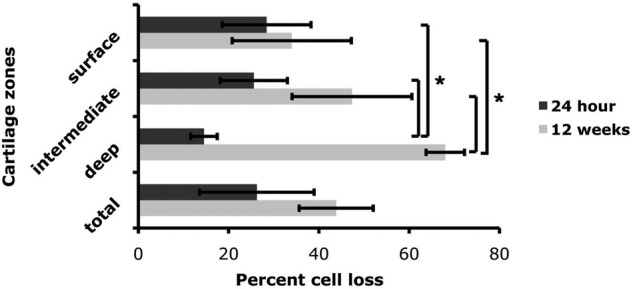
Figure 6.

Quantification of cell death in impacted cartilage in vivo. Three sections from impacted femoral condyles at 24 hours and 12 weeks (n = 3 for each group) were stained with Hoechst 33342 nuclear staining. Cell loss in each zone (superficial, intermediate, and deep) of impacted cartilage is expressed as a percentage of total cell number in each zone as compared with neighboring unimpacted cartilage. Values are the mean (SD of 3 samples from one experiment (*P < 0.01).
