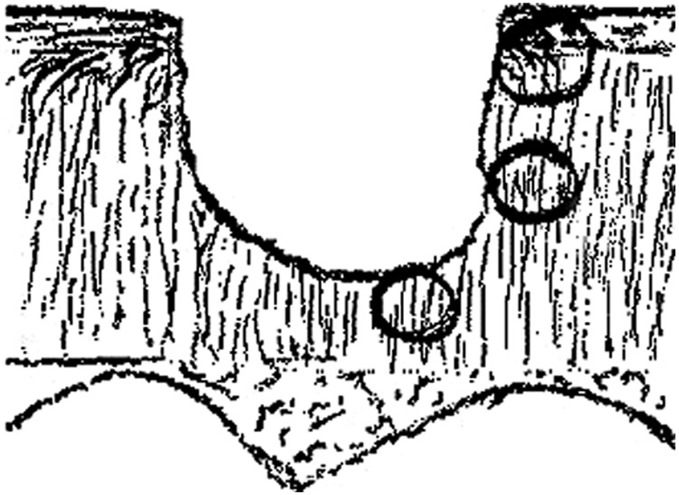
Figure 1.

Drawing depicting a partial-thickness cartilage injury and relative predicted average collagen orientation in 3 regions adjacent to the defect. In the superficial zone (top circle) and deep zone (bottom circle), the majority of collagen fibrils are perpendicular to the defect edge. In the middle zone, most fibrils are predicted to run parallel to the defect edge.
