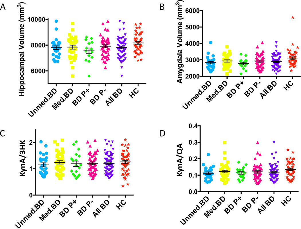
Figure 1.
Scatterplots of group differences in (A) hippocampal volume, (B) amygdalar volume, (C) KynA/3HK, and (D) KynA/QA across the BD and healthy control groups. The unmedicated BD group is shown in blue circles, the medicated BD group is displayed with yellow squares, the BD subjects with a history of psychosis (BD P+) are shown in green diamonds, the BD subjects without a history of psychosis (BD P−) are shown in pink triangles, the combined unmedicated and medicated BD group is shown in inverted purples triangles, and the healthy controls are illustrated with orange stars. The mean and standard error of the mean is displayed for each group in black. The healthy control group had significantly larger hippocampal and amygdalar volumes than all five of the BD groups, but the results were no longer significant after regressing out the effects of age, sex, and intracranial volume. The unmedicated BD group had reduced levels of KynA/3HK compared with the healthy controls but the results were no longer significant after regressing out the effects of age and sex. The unmedicated BD (F=4.4, p=0.039) and the combined BD (F=4.2, p=0.043) group had reduced levels of KynA/QA compared with the healthy control group after controlling for age and sex.

