Abstract
Wnt proteins are thought to bind to their receptors on the cell surfaces of neighboring cells. Wnt8 likely substitutes for the dorsal determinants in Xenopus embryos to dorsalize early embryos via the Wnt/β-catenin pathway. Here, we show that Wnt8 can dorsalize Xenopus embryos working cell autonomously. Wnt8 mRNA was injected into a cleavage-stage blastomere, and the subcellular distribution of Wnt8 protein was analyzed. Wnt8 protein was predominantly found in the endoplasmic reticulum (ER) and resided at the periphery of the cells; however, this protein was restricted to the mRNA-injected cellular region as shown by lineage tracing. A mutant Wnt8 that contained an ER retention signal (Wnt8-KDEL) could dorsalize Xenopus embryos. Finally, Wnt8-induced dorsalization occurred only in cells injected with Wnt8mRNA. These experiments suggest that the Wnt8 protein acts within the cell, likely in the ER or on the cell surface in an autocrine manner for dorsalization.
Keywords: endoplasmic reticulum, immunohistochemistry, KDEL, Wnt, Xenopus
Introduction
Early development of Xenopus embryos is established through the interaction of cytoplasmic determinants. Cytoplasmic transplantation studies revealed the presence of a “dorsal determinant” in future dorsal cells of the 16-cell stage embryo (Yuge et al. 1990) as well as in the vegetal pole of the fertilized egg (Fujisue et al. 1993; Holowacz & Elinson 1993). The dorsal determinant in the vegetal pole is necessary for Xenopus dorsal axial development (Kikkawa et al. 1996; Sakai 1996). However, the dorsal determinant (DD) alone is not sufficient because vegetal cortex transplantation resulted in dorsal structure formation only when it is transplanted into the subequatorial region of the embryo (Kageura 1997). These data suggest that the Spemann organizer forms as a result of mixing the dorsal and subequatorial determinants (Sakai 2008). Dorsal endoderm is thought to induce the Spemann organizer during the cleavage stage; however, this induction seems to occur after the end of the cleavage stages (Gurdon et al. 1985; Wylie et al. 1996; Nagano et al. 2000). The “mesoderm inducer” is proposed to be the organizer factor chordin (Vonica & Gumbiner 2007) which is expressed after the cleavage stages. Therefore, we suggest that formation of the organizer is the result of cell autonomous functions of the determinants but not the result of so-called mesoderm induction (Sakai 2008).
Wnt/β-catenin signaling is likely involved in this cell-autonomous process (Moon 2005). Wnt8 is not present in cleavage stage embryos, and its function in normal development is the posteriorization, not the dorsalization, of the embryo (Christian & Moon 1993; Fujii et al. 2008). Interestingly, however, Wnt8 mRNA induces a complete secondary body when injected into a ventral blastomere of early Xenopus embryos (Christian et al. 1991; Smith & Harland 1991). Additionally, this molecule acts upstream of β-catenin (Heasman et al. 1994), supporting the idea that Wnt8 substitutes for the dorsal determinant (DD). Heasman and colleagues postulated that Wnt5a and Wnt11 work synergistically as the DD (Cha et al. 2008). On the other hand, zebrafish Wnt8a has been recently proposed to be the intrinsic dorsal determinant in this species (Lu et al. 2011).
Permanent blastula-type embryos (PBEs) are obtained when >60% of the vegetal region (including both the surface and cytoplasm) is ablated from fertilized Xenopus embryos. Because PBEs neither have dorsal nor subequatorial determinants, the molecular nature of the latter is VegT mRNA (Zhang et al. 1998), they do not form dorsal structures or express dorsal marker genes (Fujii et al. 2002, 2008). When Wnt8 and VegT mRNAs were co-injected into a PBE blastomere, it forms dorsal structures and expresses the organizer marker gene chordin. However, when the two mRNAs were injected separately, the resulting embryo showed no sign of dorsalization (Katsumoto et al. 2004). This finding led us to hypothesize that Wnt8 acts with VegT in a cell-autonomous manner.
Although the dorsal determinant has been proposed to act in a cell-autonomous manner to form the Spemann organizer (Sakai 1996; Nagano et al. 2000; Katsumoto et al. 2004; Vonica & Gumbiner 2007), Wnt proteins are believed to act in a non-cell autonomous manner on the cell surface (Clevers 2006). It has been shown that Wnt8 proteins are localized at the plasma membrane (Yang-Snyder et al. 1996; Takada et al. 2006), which supports the idea that secreted Wnt proteins act on the cell surface; however, if Wnt8 is capable of substituting for the dorsal determinant, it is reasonable to hypothesize that the Wnt8 protein acts in a cell-autonomous manner within the cell. Recent studies revealed that FGF8 and Protogenin, thought to act on the cell surface, can be internalized to the nucleus and can act in a cell-autonomous manner (Suzuki et al. 2012; Watanabe & Nakamura 2012). To examine the potential cell-autonomous role of Wnt8 in the dorsalization process of Xenopus embryos, we focused on subcellular distribution of Wnt8 using immunofluorescence and confocal microscopy.
We hypothesized that we could precisely determine the axis-forming activity of injected Wnt8 mRNA using PBEs as a test system because they do not form the dorsal structure (proboscis) or express dorsal marker genes if they receive neither Wnt8 nor VegT mRNAs. However, they can be dorsalized when these mRNAs are co-injected (Katsumoto et al. 2004). Furthermore, because PBEs do not have large vegetal yolk granules, they should facilitate visibility in our immunohistochemical observations.
Materials and methods
Experimental animals
We have maintained all animals in accordance with the ARRIVE guidelines on the care and use of experimental animals of Kagoshima University.
Permanent blastula-type embryos (PBEs)
Fertilization and embryo culture were performed as described (Katsumoto et al. 2004). PBEs were obtained by ablating >60% of the vegetal region, including the cytoplasm, yolk, and cell membrane (Fujii et al. 2002). The denuded egg was inclined 90° off its vertical axis. A glass rod (diameter 300 μm, length 2 cm) was placed on the egg to divide it into animal and vegetal fragments (Fig.1). The PBE derives from the animal part of the embryo, and therefore lacks most of the large yolk platelets. This characteristic facilitated the observation of HA staining.
Figure 1.

Permanent blastula-type embryo (PBE) as a test system for Wnt8 distribution. (A) Schematic drawing of the cytoplasmic contents of the PBE (upper area). PBEs do not contain both the dorsal determinant and the VegT (endomesodermal determinant). (B–D) Deletion of the vegetal cytoplasm (>60% of the egg surface) with a glass rod results in the formation of the PBE. (B) Just after placing the glass rod. (C) Just after the separation into an animal egg fragment (PBE) and a vegetal egg fragment. (D) A PBE at the neurula stage. The PBE does not form dorsal or endomesodermal structures. (E) A normal embryo at the same stage.
Plasmid construction and mRNA synthesis
Xwnt8-HA was generated using polymerase chain reaction (PCR). The cDNA was inserted with the HA epitope tag sequence (5′-tatccatacgatgtaccagattacgca-3′; YPYDVPDYA) at the same position that harbored the Myc epitope tag described by Christian & Moon (1993). The cDNA was subcloned into pCS2+. Xwnt8-HA-KDEL was generated using PCR. The cDNA was added at the C-terminus with spacer sequences (5′-agatcgtacaag-3′; RSYK) and KDEL sequences (5′-aaggacgagctg-3′) and was subcloned into pCS2+. ΔSP-Xwnt8-HA, which lacks the predicted signal peptides (2nd-QNTTLFILATLLIFCPFFTASA-23th a.a.), was generated using an inverse PCR technique. Synthetic capped mRNAs were produced using mMessage mMachine SP6 kit (Ambion, AM1340).
Microinjection
For microinjection, mRNAs were diluted in sterile nuclease-free water containing fluorescent lineage tracers as described below. In the experiments shown in Figures2 and 5, mRNAs were injected with Alexa Fluor 647 dextran into a single blastomere of 4 to 8-cell stage PBEs with a glass capillary with a diameter of 5–7 μm, using a Narishige micromanipulator. For the experiment shown in Figure 7, Wnt8 mRNA and Oregon Green dextran were injected into one C4 blastomere of normal embryos with regular cleavage pattern (Kageura 1990). The amounts of injected RNA and lineage tracers are indicated in the respective figure legends.
Figure 2.
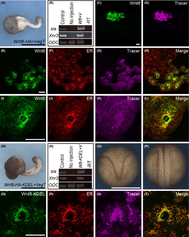
Wnt8 protein is localized in the endoplasmic reticulum (ER) of mRNA-injected cells. (A) A stage-17 permanent blastula-type embryo (PBE) injected with VegT (15 pg) and Wnt8-HA (3 pg) into a blastomere at the 4-cell stage. Co-injection of VegTmRNA is necessary for proboscis formation (Katsumoto et al. 2004), which is a marker of dorsalization. Formation of the proboscis (34 out of 34) indicates dorsalization. (B) Reverse transcription–polymerase chain reaction (RT–PCR) analyses for dorsalization markers siamois and Xnr3 expression in control PBEs and PBEs injected with the two mRNAs. ODC serves as loading control. Control, normal embryos; No injection, PBEs with no mRNA injection; W8+V, PBEs injected with Wnt8-HA (3 pg) and VegT (15 pg) mRNAs; -RT, PCR with cDNA synthesized without reverse transcriptase. (C,D) A transverse section of a PBE injected with 250 pg Wnt8-HA and 15 pg VegTmRNAs containing 0.1% Alexa Fluor 647 dextran into an 8-cell stage blastomere. The sample was fixed and processed for immunostaining 2 h before stage 10. (E–L) Low-magnification (E–H) and high magnification (I–L) images showing the same region of a section. (E,I) Subcellular distribution of Wnt8-HA protein (green). (F,J) PDI immunostaining (red) showing subcellular distribution of ER. (G,K) Alexa Fluor 647 staining (magenta) showing the lineage of the mRNA-injected cells. (H,L) Merged images of Wnt (E, I) and PDI staining (F, J). Yellow regions indicate the overlap of these two molecules. Note the strong fluorescence of Alexa Fluor 647 in the nucleus (G,K), which is connected to the cytosol via the nucleopore, whereas HA and PDI staining was absent in the nucleus (I,J and L). (M) A PBE injected with VegT (15 pg) and Wnt8-HA-KDEL (10 pg). Proboscises (12 out of 12) formed as shown in (A). (N) RT-PCR for siamois and Xnr3. (O) An embryo with duplicated axis: This embryo was injected with 15 pg Wnt8-HA-KDEL mRNA into a ventral vegetal blastomere of 8-cell stage embryo. (P) A control embryo at the same stage (stage 17). (Q–T) Wnt8-HA-KDEL protein also overlapped with ER staining. Staining was the same as for the above-mentioned panels for Wnt8-HA (I–L). Bar in A and M, 1 mm. Bar in C for C and D, 100 μm. Bar in E for E–H, 100 μm. Bar in I for I–L, 50 μm. Bar in O for O-R, 50 μm. Bar in O for O and P, 1 mm.
Figure 5.
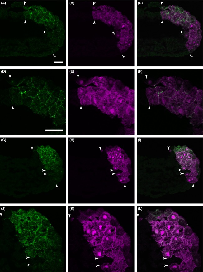
Wnt8 is localized at the cell boundaries in the absence of methanol treatment, but is restricted within the tracer injected domain. Wnt8-HA or Wnt8-HA-KDEL proteins were detected by HA-immunostaining (green), and the lineage of mRNA-injected cells was shown by Alexa Fluor 647 dextran (magenta) as in Figure2. Samples were not treated with methanol in these experiments. (A–F) A Wnt8-HA injected sample. (A) Low magnification image of Wnt8-HA staining. Note that weak but substantial staining within the cell, likely in the endoplasmic reticulum (ER), was also detected. (B) Alexa Fluor 647 staining (magenta) showing the lineage of the mRNA-injected cells. (C) Composite image of A and B. Alexa Fluor 647 (lineage tracer) is indicated in the red and blue channel (thus shown in magenta) whereas Wnt8-HA is represented in the green channel. (D–F) High magnification view of A–C. (G–L) Wnt8-HA-KDEL injected sample. Staining within the cell is stronger as compared to Wnt8-HA (A–F). (G) A Wnt8-HA-KDEL injected sample. (H) Alexa Fluor 647 staining (magenta) (I) Composite image of G and H. (J–L) High magnification view of G–I. White arrowheads indicate the border of the lineage-labeled/negative cells. Bar in A for A–C and G–I, and bar in D for D–F and J–L: 100 μm.
Figure 7.
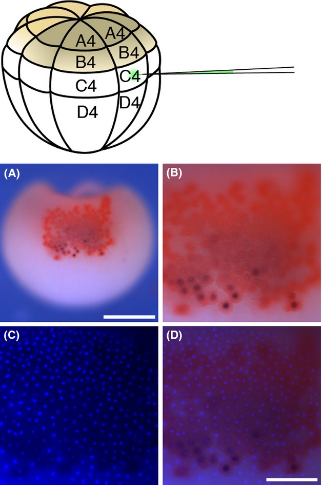
Chordin is expressed only in Wnt8mRNA-injected cells. (Top) Schematic drawing of the injection of Wnt8-HAmRNA and Oregon Green dextran. Normal embryos with a typical cleavage pattern were selected for injection. Wnt8-HAmRNA and Oregon Green dextran were injected into a C4 blastomere. The embryo was fixed 30–60 min before the onset of gastrulation and processed for in situ hybridization combined with histochemistry for lineage tracing (see Materials and methods). (A) Lateral view of an embryo showing the injected area. Expression of chordin is shown as dark blue dots, whereas lineage tracing by immunostaining of Oregon Green dextran is shown in red. (B) Enlarged image of A. (C) Counterstaining with Hoechst 33258. Note that the distribution pattern of nuclei precisely matches that of chordin expression. (D) The nuclei pattern can be superimposed on the image and therefore can be used as a single-cell level map. Bar in A, 500 μm. Bar in D for B–D, 200 μm.
Immunohistochemistry
Permanent blastula-type embryos (PBEs) injected with mRNAs and Alexa Fluor 647 dextran (lineage tracer) were fixed in MEMPFA [4% paraformaldehyde, 100 mmol/L MOPS (pH 7.4), 2 mmol/L EGTA, 1 mmol/L MgSO4] for 1 h at 20°C at the 4000-cell stage, just before the gene expression commences (Newport & Kirschner 1982) to avoid possible effects of the newly synthesized transcripts. For the correct timing of the fixation, embryos were continuously scored for each cleavage. Typically (Figs2, 6), the embryos were dehydrated and stored in methanol at −20°C for at least 3 days. In some cases, the methanol storage step was skipped. Embryos were embedded in OTC compound (Tissue-Tek) and cut into 8-μm sections using a cryostat. After treatment with 1% skim milk in phosphate-buffered saline (PBS) for 1 h, the sections were incubated overnight at 4°C in a mixture of the rat anti-HA monoclonal antibody (mAb; 3F10, Roche, 1:200), mouse anti-PDI mAb (ab12225, Abcam, 1:4000), and goat anti-Frzb (sFRP3) Ab (AF192, R&D, 1:2000). Anti-rat IgG-Alexa488 (Invitrogen, 1:250), anti-mouse IgG-Alexa594 (Invitrogen, 1:250), and anti-goat IgG-Alexa594 (Invitrogen, 1:250) were used as secondary antibodies, and samples were incubated for 2 h at room temperature with these antibodies. Observations were performed using a Leica TCS SP2 confocal laser microscope.
Preparation of cell lysates and concentration of culture media
We seeded HEK293 cells at a density of 3.8 × 10⁵ cells in 60-mm dishes containing modified Eagle's media supplemented with 10% horse serum. Cells were cultured for 24 h. Aspirated media were replaced with 4 mL Opti-MEM (Invitrogen). HEK293 cells were transfected with expression plasmids using FuGENE6 (Roche). Culture media were collected after 24 h (1 day) or 72 h (3 days), and cell lysates were prepared. Protein concentrations were determined as described previously (Lintern et al. 2009). Culture media were clarified to remove debris. Supernatants were concentrated 35- to 50-fold and stored in aliquots at −80°C.
Western blotting
Total cell lysate protein (5 μg) and concentrated culture media (an equivalent volume of 500 μL of non-concentrated culture media) were resolved by electrophoresis on 15% sodium dodecyl sulfate–polyacrylamide gels (SDS–PAGE) and probed with the primary anti-HA antibody (Roche, 3F10, 1:1000) and the secondary anti-rat IgG-(Fab)'2(HRP) antibody (Abcam, ab6517, 1:40 000). Proteins were detected using a 1:10 dilution of the reagent mixture provided with the ECL Prime System (GE Healthcare).
In situ hybridization combined with histochemistry for lineage tracing
Wnt8 mRNA (10–20 pg/nL) and Oregon Green dextran (1%) in 1 nL of distilled water (DW) were injected into a C4 blastomere of the 32-cell embryo (Nakamura & Kishiyama 1971). The embryos were fixed 30–60 min before stage 10 in MEMFA overnight at room temperature, washed thoroughly, and stored in 100% methanol (−20°C) for at least 3 days. Whole-mount in situ hybridization was carried out following the method of Sive et al. (Sive et al. 2000), without proteinase K treatment. After in situ hybridization, embryos were cut vertically to facilitate the immunostaining. The cell lineage tracer Oregon Green dextran was detected by immunostaining using anti-fluorescein-AP Fab fragments (Roche) and Fast Red (Roche) following the method of Koga et al. (2007).
RT–PCR
Total RNA was extracted from a single embryo or three PBEs using the SV Total RNA Isolation System (Promega). cDNA was synthesized from total RNA (3.1 μg) using Ready-To-Go You-Prime First-Strand Beads (GE Healthcare). Each PCR reaction was performed using 1 μL of the reverse transcription reaction product as template, 0.25 U rTaq DNA polymerase (Takara Bio, Shiga, Japan), 10 μmol/L each primer, and 2.5 mmol/L dNTPs in volume of 10 μL of 1× PCR buffer. The denaturation step proceeded at 95°C for 30 s. The annealing temperatures were 65°C for ODC, 58°C for siamois, and 54°C for Xnr3. Annealing proceeded for 30 s before final extension at 72°C for 30 s. There were 23 cycles for ODC, 25 cycles for siamois, and 30 cycles for Xnr3. PCR products were analyzed on a 6% acrylamide gel. The primers were as follows: ODC, 5′-GCAAAGCCATTGTGAAGACTCTCTCCATTC-3′ and 5′-AAGCTTTGCATTCGGGTGATTCCTTGCCAC-3′; siamois, 5′-AGACATGACCTATGAGGCTG-3′ and 5′-AGTCAGTTTGGGTAGGGTAGG-3′; and Xnr3, 5′-TAATCTGTTGTGCCGATCCA-3′ and 5′-ATCAATGTTGCCCTTTTTCA-3′.
Results
The Wnt8-HA protein is localized in the endoplasmic reticulum (ER) of mRNA-injected cells
We injected HA-tagged Wnt8 mRNA (Wnt8-HA) together with VegT mRNA into a PBE blastomere (Fig.1) (Sakai 1996; Fujii et al. 2002). PBEs do not form dorsal structures (Fig.1D) or express dorsal genes, but these embryos do form dorsal structures and express dorsal genes following injection of Wnt8 and VegT (Katsumoto et al. 2004). Because HA-tagged Wnt8 dorsalized PBE (Fig2A,B), HA appears to have no inhibitory effects on Wnt activity.
Immunohistochemistry with anti HA tag showed that HA positive cells are restricted within the Wnt8 mRNA-injected cells that are assessed by co-injected Alexa Fluor 647 fluorescence (compare 2C and D, 2E and G). It was also shown that Wnt8 protein was mainly co-localized with the ER (endoplasmic reticulum) marker PDI (compare Fig.2E and F, I and J). Merged images clearly show that Wnt8 proteins are mainly localized in the ER (Fig.2H,L). These results support our hypothesis that Wnt8 protein could act within the cell, presumably in the ER.
ER-retention signal does not inhibit dorsalization by Wnt8
To further confirm our hypothesis, we constructed a Wnt8 construct tagged with the ER-retention signal KDEL (Munro & Pelham 1987; Pelham 1988) (Wnt8-HA-KDEL). As we expected, PBEs injected with Wnt8-HA-KDEL and VegT mRNA expressed siamois and Xnr3 (Fig.2N), and formed a proboscis (Fig.2M). Further, Wnt8-HA-KDEL generated duplicated axis in normal embryos (Fig.2O) when injected into a ventral vegetal blastomere of 8- to 16-cell stage embryos (23 out of 23 embryos formed Y-shaped secondary axis). These results indicate that Wnt8-HA-KDEL has dorsalizing activity. The dose needed for an identical result was approximately three times higher than Wnt8-HA, which might be due to the general inhibitory effect of the additional tag sequence KDEL or Wnt8-HA-KDEL might not have dorsalizing activity acting on the cell surface which might be present in Wnt8-HA. Upon immunofluorescence examination of the injected embryos, Wnt8-HA-KDEL protein was also localized in the ER (Fig.2Q–T).
Wnt8-HA-KDEL is retained within the cell in cell culture
To examine ER retention of Wnt8-HA-KDEL, we analyzed the Wnt8-HA-KDEL protein in culture media from HEK293 cells transfected with Wnt8-HA-KDEL expression plasmids (Fig.3). Wnt8-HA was observed in both the cell lysate and the culture media 3 days after transfection. In contrast, Wnt8-HA-KDEL was observed in the cell lysate, but not in the culture media 3 days after transfection. These data suggest that the Wnt8-HA-KDEL protein is retained in the ER.
Figure 3.
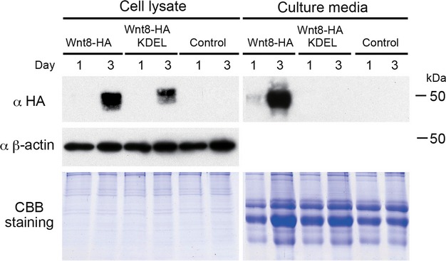
Wnt8-HA-KDEL protein is retained in the cell in cell culture. Plasmid constructs of Wnt8-HA or Wnt8-HA-KDEL were transfected into HEK293 cells. Proteins (40 μg) from total cell lysates were resolved by sodium dodecyl sulfate–polyacrylamide gel electrophoresis (SDS–PAGE) and examined by Western blotting using the primary anti-HA antibody. β-actin was used as a loading control (cell lysates). Coomassie brilliant blue (CBB) staining of acrylamide gels was used as a loading control (cell lysates and culture media).
The signal peptide is required for dorsalizing activity
Moreover, to test whether entry of the Wnt8 protein into the ER is required for dorsalizing activity, we constructed a mutant Wnt8 that lacks a signal peptide for secretion (ΔSP-Wnt8-HA). ΔSP-Wnt8-HA was present in the cytoplasm and in the nucleus (Fig.4C–F) of mRNA-injected cells. Some part of ΔSP-Wnt8-HA seems to overlap with ER marker PDI (Fig.4F). But the merged image show that green area, staining for ΔSP-Wnt8-HA, is present, which means that some part of ΔSP-Wnt8-HA is localized apart from ER (Fig.4F). ΔSP-Wnt8-HA was also localized in the nucleus (Fig.4F). We speculated that large part of ΔSP-Wnt8-HA left ER after translation is completed. ΔSP-Wnt8-HA did not dorsalize PBEs (Fig.4A,B), suggesting that ER entry of the Wnt8 protein is required for its activity.
Figure 4.
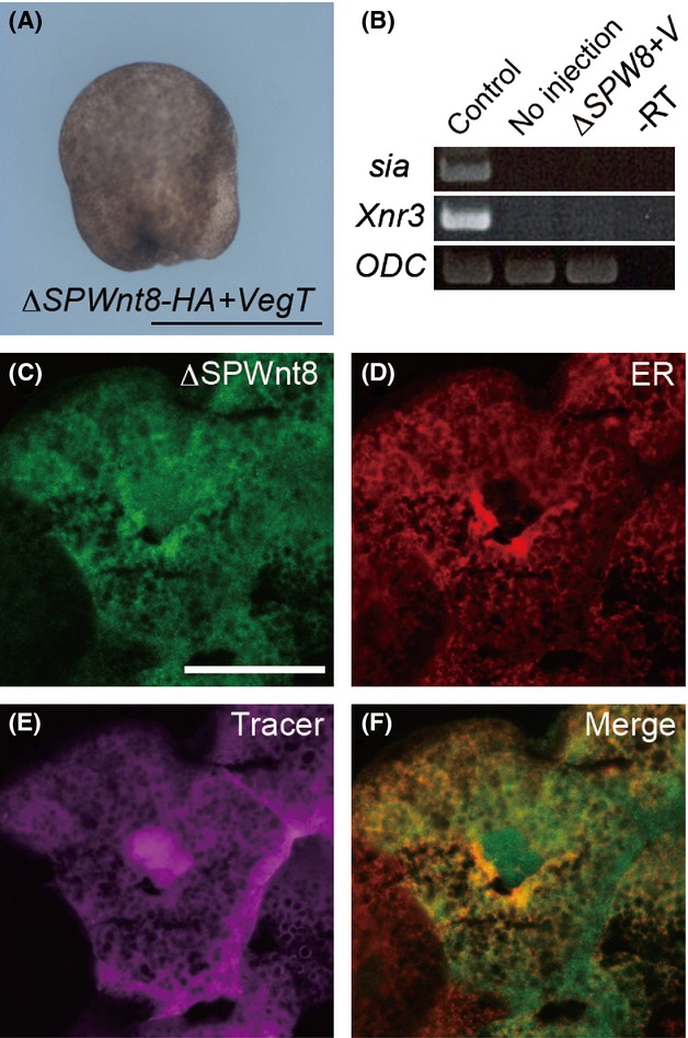
Wnt8 lacking a signal peptide (ΔSP-Wnt8-HA) did not cause dorsalization. (A) ΔSP-Wnt8-HA-injected PBEs do not form dorsal structures. Proboscises were not observed (0 out of 10). (B) Reverse transcription–polymerase chain reaction (RT–PCR) analyses for dorsalization markers siamois and Xnr3 expression in control PBEs (no injection) and PBEs injected with ΔSP-Wnt8-HA and VegT. (C–F) ΔSP-Wnt8-HA (green) was broadly expressed in the cytosol of the injected cells. Note that strong HA staining (C) was observed in the nucleus, whereas ER staining was not found in the nucleus (D), suggesting that ΔSP-Wnt8-HA is in the cytosol and not in the ER.
Localization of Wnt8 at the cell boundaries is revealed in the absence of methanol treatment, although its distribution is restricted to the area of mRNA injection
Although our experiments revealed that Wnt8 was localized in the ER, it has been accepted that Wnt proteins are localized mainly at the cell boundaries (Yang-Snyder et al. 1996; Takada et al. 2006). The main difference in the protocols between the previous studies and the present study was methanol treatment after fixation. In our protocol, embryos were stored in methanol for longer than 3 days for permeabilization (see Materials and methods), whereas methanol was not used or only briefly used in the previous experiments. Wnt proteins in the ER may not have been detected effectively due to insufficient permeabilization. According to our protocol, most parts of Wnt8 protein on the cell surface may have been lost by the long-term storage in methanol. Therefore, we analyzed the distribution of Wnt8 without using methanol (Fig.5). Wnt8 protein was detected mainly at the cell boundaries in the absence of methanol treatment (Fig.5A–F). As shown in previous studies (Yang-Snyder et al. 1996; Takada et al. 2006), weak but substantial staining within the cell, likely in the ER, was also detected (Fig.5A,D). Most importantly, Wnt8 staining was restricted to the region of mRNA injection as shown by the lineage tracer (compare Fig.5A,B). High-resolution images also show the absence of HA staining in tracer-negative cells (compare Fig.5D,E).
Furthermore, we examined the distribution of the Wnt8-KDEL protein in the absence of methanol treatment (Fig.5G–L). As compared to Wnt8, the Wnt8-KDEL protein tends to be localized inside of the cell (compare Fig.5A and G, D and J) although the staining was stronger at the cell boundary (Fig.5G,J). Wnt8-HA-KDEL staining also did not exceed outside of the lineage positive cells (Fig.5G,H,J,K).
Co-injection of Frzb mRNA and Wnt8 or Wnt8-KDEL mRNA displayed distinct distributions
We co-injected Frzb mRNA, which is an inhibitor of Wnt8 (Leyns et al. 1997; Wang et al. 1997) and examined whether Frzb changes the distribution of Wnt8-HA, using our methanol method (Fig.6). Interestingly, Frzb (Fig.6B) and Wnt8-HA (Fig.6C) were both observed predominantly at the cell boundaries and were even observed in the lineage-negative cell domain (Fig.6D,E). Since Wnt8-HA is localized in the ER when the samples were treated with methanol (Fig.2E,H,I,L), the distribution at the cell boundary should be the effect of Frzb, which moves out of the cell (Mii & Taira 2009). Frzb was found on the cell surface of the whole PBE while Wnt8 was noted two to three cells apart from the lineage-labeled cells (compare Fig.6B and C, D and E). These results indicate that these proteins are secreted outside of the mRNA-injected cells, and that using the methanol method of immunohistochemistry we can visualize the secreted proteins.
Figure 6.

Co-injection of Frzb (500 pg) and Wnt8-HA (250 pg) or Wnt8-HA-KDEL (250 pg) mRNAs. Frzb and Wnt8-HA stains the cell boundaries, outside of the mRNA-injected domain while Wnt8-HA-KDEL is restricted in the lineage positive domain. (A–F) Co-injection of Frzb with Wnt8-HA. Methanol was used for immunohistochemistry (see Materials and methods). (A) Alexa Fluor 647 staining (magenta) showing the lineage of the mRNA-injected cells. (B) Frzb staining is seen predominantly at the cell boundaries, even outside of the mRNA-injected domain. (C) Staining for Wnt8-HA. Wnt8-HA staining was also at the cell boundaries but it diffused less extensively as compared to Frzb. (D) Composite image of A and B. Alexa647 is in the red and blue channel (thus shown in magenta) and originally red Frzb staining is represented in the green channel. Note that green Frzb staining is observed at the cell boundaries of both the lineage-injected domain and the lineage-negative domain, but not overlapped with the lineage tracer which represents inside of the cell. (E) Composite image of A and C. Note some cell boundaries outside of the tracer-positive region are stained in green. (F) Composite image of B and C. These images were from methanol-treated samples. These results indicate that methanol treatment does not eliminate secreted proteins. Wnt8 can be detected on the cell surface when it is secreted. (G–L) Co-injection of Frzb with Wnt8-HA-KDEL. (G) Alexa Fluor 647 staining (magenta). (H) Frzb is seen both within the lineage positive cells and cell boundaries throughout the PBE. (I) Wnt8-HA-KDEL staining (green) is restricted in the lineage positive cells. (J) Composite image of G and H. (K) Composite image of G and I. Note that the outer region of the lineage positive cells is colored magenta. (L) Composite image of H and I. Note that the outer region of the lineage positive cells is colored red, suggesting that Frzb exists on the surface while Wnt8-HA-KDEL is absent on the cell surface. Bar in A for A–L, 500 μm.
On the contrary, when Wnt8-HA-KDEL was co-injected with Frzb, the resulting PBE showed distinct staining pattern (Fig.6G–L). Although Frzb was found at the cell boundaries throughout the PBE, some staining still existed within the cell of the lineage positive region (Fig.6H). Wnt8-HA-KDEL was seen only inside of the lineage positive cells (Fig.6I,K). The Frzb staining at the cell boundaries does not overlapped with Wnt-HA-KDEL (Fig.6L, note red Frzb staining outside of the green Wnt-HA-KDEL staining) suggesting that the Frzb staining resides outside of the cell while Wnt-HA-KDEL resides inside of the cell and Frzb does not alter the distribution pattern of Wnt-HA-KDEL. These data suggest that the Wnt8-HA-KDEL protein behaves differently from the Wnt8-HA protein: the former tends to stay within the cell in which the mRNA was injected, presumably in the ER while the latter going out of the cell with Frzb.
Dorsalization occurs in cells following Wnt8 mRNA injection
Immunohistochemical analyses of the Wnt8 protein revealed that Wnt8 resides in the ER and on the cell surface. Because Wnt8-KDEL can dorsalize Xenopus embryos as described above and resides predominantly in the ER, we hypothesized that Wnt8 protein in the ER is functioning as the dorsalizing factor.
Therefore, we examined whether dorsalization occurs in cells adjacent to cells that were injected with Wnt8 mRNA. To follow the lineage of the injected blastomere, we used Oregon Green dextran (Koga et al. 2007). A mixture of Wnt8 mRNA and Oregon Green dextran was injected into a C4 blastomere of a normal embryo (Nakamura & Kishiyama 1971) at the 32-cell stage, reared until 30–60 min before the onset of gastrulation (30–60 min before stage 10), and the embryo was fixed overnight in MEMFA. We examined chordin expression as a marker of the organizer (Sasai et al. 1994) using in situ hybridization. Chordin is not the first gene to be expressed in the organizer domain; however, expression of this factor is readily observed just before gastrulation (Vonica & Gumbiner 2007). After in situ hybridization, samples were processed for Oregon Green immunostaining using Fast Red and then bleached. In general, the expression pattern of chordin overlapped with the Fast Red staining (Fig.7). This overlapping pattern should be reliable because the majority of embryos showed a very precise overlapping pattern. Punctate chordin perinuclear staining was observed exclusively in cells injected with Wnt mRNA based on Fast Red immunostaining against Oregon Green (Fig.7A,B). Distribution pattern of the nuclei, which was stained with Hoechst 33248 (Fig.7C), facilitated observation at the single cell level. The nuclei adjacent to the chordin- and lineage tracer-positive cells did not exhibit a chordin signal in the absence of Fast Red staining (Fig.7B–D).
We repeated these experiments using 21 batches of embryos (n = 463). Although some embryos (n = 160) were not stained clearly, 243 embryos exhibited an overlapping pattern whereas in 60 embryos, chordin was expressed outside of the Wnt-8-injected cells. If the latter is due to Wnt signaling at the cell surface, chordin-positive cells should be adjacent to the injected cells because Wnt protein distribution was restricted to the Wnt mRNA-injected cells and their descendants (Figs2C–F, 5A,B). Chordin expression outside of the lineage usually showed a cluster-like pattern or a broad spreading pattern, suggesting that expression was due to leakage of the injected Wnt mRNA through a cytoplasmic bridge or that chordin-induced chordin expression (Nagano et al. 2000) occurred after the mid-blastula transition (Newport & Kirschner 1982).
Even with these data, it is still possible that Wnt8 acts in an autocrine manner: Wnt8 may be secreted and act on the “source” cells in an autocrine manner.
Discussion
The first step of signal transduction generally involves the interaction of a ligand with its receptor on the surface of neighboring cells. Here, we show that the signaling protein Wnt8 could act in a cell-autonomous manner, in the dorsalizing process of early Xenopus embryos. This finding initially appeared curious. However, if we consider that signaling molecules or ligands are synthesized in the ER and are thereafter transported to the cell surface, and that the receptor is also synthesized in the ER and incorporated into the ER membrane before being translocated to the plasma membrane, it is reasonable that these two molecules interact within the ER. The outer space of the ER is cytoplasm, and the inner space of the plasma membrane is also cytoplasm. The cytoplasmic domain of the Wnt8 receptor on the cell membrane or on the ER membrane might transduce the signal to the cytoplasm.
We first analyzed the distribution of Wnt8 protein in Xenopus PBEs which had been injected with Wnt8 and VegT mRNA. Wnt8 was distributed within the domain of the descendants of Wnt8 mRNA-injected cells. Furthermore, Wnt8 was mainly distributed in the ER. It has been accepted that Wnt proteins are distributed at the cell boundaries, presumably on the cell surface (Yang-Snyder et al. 1996; Takada et al. 2006). We used methanol to permeabilize the embryo, which was not used in the previous studies. Owing to permeabilization, we could see inside the cell more clearly, and we could show that Wnt8 is distributed within the cell in the early stage of Xenopus embryos. We also confirmed that Wnt8 was localized at the cell boundaries in the absence of methanol treatment. These findings indicate that Wnt8 is distributed in the ER and on the cell boundaries.
It should be noted that the subcellular localization of the Wnt proteins was not analyzed with lineage tracers in previous studies. In the present study, however, we used a lineage tracer to demonstrate that the Wnt8 protein is distributed within the domain where Wnt8 mRNA was injected. We used an artificial embryoid PBE to facilitate observation of dorsalization, since we can assay dorsal gene expression using this system easily: PBEs never express dorsal gene while co-injection of Wnt8/VegT and Wnt8-KDEL/VegT resulted in the expression of siamois and Xnr3. It should be emphasized that when Wnt8 and VegT are injected separately into the PBE, it neither formed dorsal structure nor expressed an organizer marker chordin (Katsumoto et al. 2004).
Co-injection of Frzb and Wnt8-HA or Wnt8-HA followed by the immunostaining with the present methanol method revealed interesting results. In both cases, Frzb was found on the cell surface throughout the embryo. Further, Wnt8-HA and Wnt8-HA-KDEL showed distinct pattern of localization. Wnt8-HA was localized overlapping with Frzb at the cell boundaries and in the lineage tracer-negative cell domain. Wnt8-HA may have been transported by Frzb to be localized predominantly to the cell surface, and secreted. It has been reported that sFRPs transports and enhance the diffusion of Wnts (Mii & Taira 2009). On the contrary, Wnt8-KDEL was retained within the cell. In the latter case, Frzb was also retained within the mRNA-injected cells.
It has been reported that signal peptide is required for the activity of cWnt3a (Narita et al. 2007). Our study revealed that the absence of the signal peptide inhibits dorsalization by Wnt8 protein. Wnt8 protein lacking signal peptide accumulated in the cytoplasm rather than the ER, These results suggest that Wnt8 must enter into ER to act in the dorsalization process.
The presence of Wnt8 in the ER may be more important than Wnt8 on the cell boundaries for dorsalizing activity. First, Wnt8 that is deleted of signal peptide did not have dorsalizing activity. Second, even when ER retention signal KDEL was attached to Wnt8, Wnt8 maintained its activity to dorsalize Xenopus embryos. Further, the distribution of Wnt8-KDEL did not extend to cells neighboring the descendants of the injected cells. Therefore, Wnt8-KDEL in the ER might be sufficient for dorsalization. Upon co-injection with Frzb, the KDEL-tagged Wnt8 was not found on the cell surface (Fig.6I) while Wnt8-HA was found predominantly on the cell surface (Fig.6C).
Because Wnt8 protein is observed on the cell surface, we could not rule out the possibility that Wnt8 acts on the cell surface of neighboring cells to induce dorsal gene expression. Therefore, we examined chordin expression combined with lineage tracing to investigate this possibility. Dorsalization occurred only in cells that were injected with Wnt8 mRNA. Therefore, the present study, together with previous studies describing the cell-autonomous functions of Wnt/β-catenin pathway molecules (Logan et al. 1999; Weitzel et al. 2004; Itoh et al. 2005; Vonica & Gumbiner 2007), reveals that the initial step of the Wnt/β-catenin pathway in early development occurs in a cell autonomous manner.
In the canonical Wnt pathway, important downstream events are known to occur in the nucleus. β-catenin and Xdsh are intracellular components of the Wnt/β-catenin pathway. The first visible sign of this pathway in Xenopus development is enrichment of β-catenin in the nucleus on the dorsal side of the embryo. Xdsh is thought to act upstream of β-catenin (Clevers 2006) and in order to promote dorsalization, Xdsh should reside in the nucleus (Itoh et al. 2005). Given that these molecules function in the nucleus, the first signal should therefore initiate from near the nucleus. Because extensive ER membrane exists around the nucleus, signaling through the ER membrane is likely very efficient. In the present study, the Wnt8 protein was found predominantly in the ER near the nucleus, suggesting that the canonical Wnt pathway in this system signals via the ER membrane. Recently, Wnt signaling was reported to require endocytosis (Bellen & Seto 2006; Blitzer & Nusse 2006; Yamamoto et al. 2006), and Wnt proteins outside the cell were proposed to be internalized via endocytosis and function in the endosome (Niehrs & Boutros 2010).
Multicellular organisms evolved from unicellular organisms. Signaling in general may have originally evolved in a unicellular organism where “neighboring cells” are not present. Intracellular signaling is noticed in unicellular eukaryote yeast cells (Pahl 1999). Therefore, signaling within the cell, as presented here, may also occur elsewhere, including unicellular organism. Signal transduction pathways other than Wnt/β-catenin pathway may also initiate within the cell. Additional studies in the future should further explore this notion.
In the general canonical Wnt pathway model, Wnt8 protein is secreted from the source cell and the Wnt co-receptors (He et al. 1997) that reside on the cell surface of the surrounding cells receive this ligand (Fig.8A). In the present study, Wnt8 protein was found in the mRNA-injected cells and chordin, a sign of dorsalization, was found only in these cells. We conclude that Wnt8 protein, presumably in the ER, has dorsalizing activity in Xenopus embryos.
Figure 8.
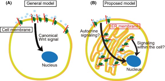
Two models for Wnt signaling. (A) General model. Wnt (light blue) is secreted on the plasma membrane where it interacts with the Wnt receptors (red and green). (B) Our new model. Wnt signaling occurs cell autonomously. It may acts within the cell and/or on the cell surface in an autocrine manner.
Acknowledgments
We thank Hidefumi Fujii, Jun-ya Doi, and Masaaki Koga for advice on in situ hybridization and immunohistochemistry. We would also like to thank Shinji Takada for critical evaluation of the manuscript. This study was supported by a grant from the Japan Society of Promotion of Science (22570206).
Author's contribution
EM performed the embryological and molecular biological studies and drafted the manuscript. TN performed the molecular biological and immunohistochemical studies and drafted the manuscript. YN carried out the embryological and molecular biological studies. HK participated in the in situ hybridization experiments with lineage tracing. AS participated in the immunological studies. SN participated in the design of the study. MS performed the embryological study, conceived the study, and participated in its design and coordination, as well as helped draft the manuscript. All authors read and approved the final manuscript.
References
- Bellen HJ. Seto ES. Internalization is required for proper Wingless signaling in Drosophila melanogaster. J. Cell Biol. 2006;173:95–106. doi: 10.1083/jcb.200510123. [DOI] [PMC free article] [PubMed] [Google Scholar]
- Cha SW, Tadjuidje E, Tao Q, Wylie C. Heasman J. Wnt5a and Wnt11 interact in a maternal Dkk1-regulated fashion to activate both canonical and non-canonical signaling in Xenopus axis formation. Development. 2008;135:3719–3729. doi: 10.1242/dev.029025. [DOI] [PubMed] [Google Scholar]
- Christian JL, Mcmahon JA, Mcmahon AP. Moon RT. Xwnt-8, a Xenopus Wnt-1/int-1-related gene responsive to mesoderm-inducing growth factors, may play a role in ventral mesodermal patterning during embryogenesis. Development. 1991;111:1045–1055. doi: 10.1242/dev.111.4.1045. [DOI] [PubMed] [Google Scholar]
- Christian JL. Moon RT. Interactions between Xwnt-8 and Spemann organizer signaling pathways generate dorsoventral pattern in the embryonic mesoderm of Xenopus. Genes Dev. 1993;7:13–28. doi: 10.1101/gad.7.1.13. [DOI] [PubMed] [Google Scholar]
- Clevers H. Wnt/beta-catenin signaling in development and disease. Cell. 2006;127:469–480. doi: 10.1016/j.cell.2006.10.018. [DOI] [PubMed] [Google Scholar]
- Fujii H, Nagai T, Shirasawa H, Doi JY, Yasui K, Nishimatsu S, Takeda H. Sakai M. Anteroposterior patterning in Xenopus embryos: egg fragment assay system reveals a synergy of dorsalizing and posteriorizing embryonic domains. Dev. Biol. 2002;252:15–30. doi: 10.1006/dbio.2002.0843. [DOI] [PubMed] [Google Scholar]
- Fujii H, Sakai M, Nishimatsu S, Nohno T, Mochii M, Orii H. Watanabe K. VegT, eFGF and Xbra cause overall posteriorization while Xwnt8 causes eye-level restricted posteriorization in synergy with chordin in early Xenopus development. Dev. Growth Differ. 2008;50:169–180. doi: 10.1111/j.1440-169X.2008.01014.x. [DOI] [PubMed] [Google Scholar]
- Fujisue M, Kobayakawa Y. Yamana K. Occurrence of dorsal axis-inducing activity around the vegetal pole of an uncleaved Xenopus egg and displacement to the equatorial region by cortical rotation. Development. 1993;118:163–170. doi: 10.1242/dev.118.1.163. [DOI] [PubMed] [Google Scholar]
- Gurdon JB, Fairman S, Mohun TJ. Brennan S. Activation of muscle-specific actin genes in Xenopus development by an induction between animal and vegetal cells of a blastula. Cell. 1985;41:913–922. doi: 10.1016/s0092-8674(85)80072-6. [DOI] [PubMed] [Google Scholar]
- He X, Saint-Jeannet JP, Wang Y, Nathans J, Dawid I. Varmus H. A member of the Frizzled protein family mediating axis induction by Wnt-5A. Science. 1997;275:1652–1654. doi: 10.1126/science.275.5306.1652. [DOI] [PubMed] [Google Scholar]
- Heasman J, Crawford A, Goldstone K, Garner-Hamrick P, Gumbiner B, McCrea P, Kintner C, Noro CY. Wylie C. Overexpression of cadherins and underexpression of beta-catenin inhibit dorsal mesoderm induction in early Xenopus embryos. Cell. 1994;79:791–803. doi: 10.1016/0092-8674(94)90069-8. [DOI] [PubMed] [Google Scholar]
- Holowacz T. Elinson RP. Cortical cytoplasm, which induces dorsal axis formation in Xenopus, is inactivated by UV irradiation of the oocyte. Development. 1993;119:277–285. doi: 10.1242/dev.119.1.277. [DOI] [PubMed] [Google Scholar]
- Itoh K, Brott BK, Bae GU, Ratcliffe MJ. Sokol SY. Nuclear localization is required for Dishevelled function in Wnt/beta-catenin signaling. J. Biol. 2005;4:3. doi: 10.1186/jbiol20. [DOI] [PMC free article] [PubMed] [Google Scholar]
- Kageura H. Spatial distribution of the capacity to initiate a secondary embryo in the 32-cell embryo of Xenopus laevis. Dev. Biol. 1990;142:432–438. doi: 10.1016/0012-1606(90)90365-p. [DOI] [PubMed] [Google Scholar]
- Kageura H. Activation of dorsal development by contact between the cortical dorsal determinant and the equatorial core cytoplasm in eggs of Xenopus laevis. Development. 1997;124:1543–1551. doi: 10.1242/dev.124.8.1543. [DOI] [PubMed] [Google Scholar]
- Katsumoto K, Arikawa T, Doi J-Y, Fujii H, Nishimatsu S-I. Sakai M. Cytoplasmic and molecular reconstruction of Xenopus embryos: synergy of dorsalizing and endo-mesodermalizing determinants drives early axial patterning. Development. 2004;131:1135–1144. doi: 10.1242/dev.01015. [DOI] [PubMed] [Google Scholar]
- Kikkawa M, Takano K. Shinagawa A. Location and behavior of dorsal determinants during first cell cycle in Xenopus eggs. Development. 1996;122:3687–3696. doi: 10.1242/dev.122.12.3687. [DOI] [PubMed] [Google Scholar]
- Koga M, Kudoh T, Hamada Y, Watanabe M. Kageura H. A new triple staining method for double in situ hybridization in combination with cell lineage tracing in whole-mount Xenopus embryos. Dev. Growth Differ. 2007;49:635–645. doi: 10.1111/j.1440-169X.2007.00958.x. [DOI] [PubMed] [Google Scholar]
- Leyns L, Bouwmeester T, Kim SH, Piccolo S. De RE. Frzb-1 is a secreted antagonist of Wnt signaling expressed in the Spemann organizer. Cell. 1997;88:747–756. doi: 10.1016/s0092-8674(00)81921-2. [DOI] [PMC free article] [PubMed] [Google Scholar]
- Lintern KB, Guidato S, Rowe A, Saldanha JW. Itasaki N. Characterization of wise protein and its molecular mechanism to interact with both Wnt and BMP signals. J. Biol. Chem. 2009;284:23159–23168. doi: 10.1074/jbc.M109.025478. [DOI] [PMC free article] [PubMed] [Google Scholar]
- Logan CY, Miller JR, Ferkowicz MJ. Mcclay DR. Nuclear beta-catenin is required to specify vegetal cell fates in the sea urchin embryo. Development. 1999;126:345–357. doi: 10.1242/dev.126.2.345. [DOI] [PubMed] [Google Scholar]
- Lu FI, Thisse C. Thisse B. Identification and mechanism of regulation of the zebrafish dorsal determinant. Proc. Natl Acad. Sci. USA. 2011;108:15876–15880. doi: 10.1073/pnas.1106801108. [DOI] [PMC free article] [PubMed] [Google Scholar]
- Mii Y. Taira M. Secreted Frizzled-related proteins enhance the diffusion of Wnt ligands and expand their signalling range. Development. 2009;136:4083–4088. doi: 10.1242/dev.032524. [DOI] [PubMed] [Google Scholar]
- Moon RT. Xenopus egg Wnt/beta-catenin pathway. Sci. STKE. 2005;2005 , cm2. [Google Scholar]
- Munro S. Pelham HR. A C-terminal signal prevents secretion of luminal ER proteins. Cell. 1987;48:899–907. doi: 10.1016/0092-8674(87)90086-9. [DOI] [PubMed] [Google Scholar]
- Nagano T, Ito Y, Tashiro K, Kobayakawa Y. Sakai M. Dorsal induction from dorsal vegetal cells in Xenopus occurs after mid-blastula transition. Mech. Dev. 2000;93:3–14. doi: 10.1016/s0925-4773(00)00251-3. [DOI] [PubMed] [Google Scholar]
- Nakamura O. Kishiyama K. Prospective fates of blastomeres at the 32-cell stage of Xenopus laevis embryos. Proc. Jpn. Acad. 1971;47:407–412. [Google Scholar]
- Narita T, Nishimatsu S, Wada N. Nohno T. A Wnt3a variant participates in chick apical ectodermal ridge formation: distinct biological activities of Wnt3a splice variants in chick limb development. Dev. Growth Differ. 2007;49:493–501. doi: 10.1111/j.1440-169X.2007.00938.x. [DOI] [PubMed] [Google Scholar]
- Newport J. Kirschner M. A major developmental transition in early Xenopus embryos 2. Control of the onset of transcription. Cell. 1982;30:687–696. doi: 10.1016/0092-8674(82)90273-2. [DOI] [PubMed] [Google Scholar]
- Niehrs C. Boutros M. Trafficking, acidification, and growth factor signaling. Sci. Signal. 2010;3 doi: 10.1126/scisignal.3134pe26. pe126. [DOI] [PubMed] [Google Scholar]
- Blitzer JT. Nusse R. A critical role for endocytosis in Wnt signaling. BMC Cell Biol. 2006;7:28. doi: 10.1186/1471-2121-7-28. [DOI] [PMC free article] [PubMed] [Google Scholar]
- Pahl HL. Signal transduction from the endoplasmic reticulum to the cell nucleus. Physiol. Rev. 1999;79:683–701. doi: 10.1152/physrev.1999.79.3.683. [DOI] [PubMed] [Google Scholar]
- Pelham HR. Evidence that luminal ER proteins are sorted from secreted proteins in a post-ER compartment. EMBO J. 1988;7:913–918. doi: 10.1002/j.1460-2075.1988.tb02896.x. [DOI] [PMC free article] [PubMed] [Google Scholar]
- Sakai M. The vegetal determinants required for the Spemann organizer move equatorially during the first cell cycle. Development. 1996;122:2207–2214. doi: 10.1242/dev.122.7.2207. [DOI] [PubMed] [Google Scholar]
- Sakai M. Cell-autonomous and inductive processes among three embryonic domains control dorsoventral and anterior-posterior development of Xenopus laevis. Dev. Growth Differ. 2008;50:49–62. doi: 10.1111/j.1440-169X.2007.00975.x. [DOI] [PubMed] [Google Scholar]
- Sasai Y, Lu B, Steinbeisser H, Geissert D, Gont LK. Robertis EMD. Xenopus chordin: a nobel dorsalizing factor activated by organizer-specific homeobox genes. Cell. 1994;79:779–790. doi: 10.1016/0092-8674(94)90068-x. [DOI] [PMC free article] [PubMed] [Google Scholar]
- Sive H, Grainger R. Harland R. Early Development of Xenopus laevis: A Laboratory Manual. Cold Spring Harbor, NY: Cold Spring Harbor Laboratory; 2000. [Google Scholar]
- Smith WC. Harland RM. Injected Xwnt-8 RNA acts early in Xenopus embryos to promote formation of a vegetal dorsalizing center. Cell. 1991;67:753–765. doi: 10.1016/0092-8674(91)90070-f. [DOI] [PubMed] [Google Scholar]
- Suzuki A, Harada H. Nakamura H. Nuclear translocation of FGF8 and its implication to induce Sprouty2. Dev. Growth Differ. 2012;54:463–473. doi: 10.1111/j.1440-169X.2012.01332.x. [DOI] [PubMed] [Google Scholar]
- Takada R, Satomi Y, Kurata T, Ueno N, Norioka S, Kondoh H, Takao T. Takada S. Monounsaturated fatty acid modification of Wnt protein: its role in Wnt secretion. Dev. Cell. 2006;11:791–801. doi: 10.1016/j.devcel.2006.10.003. [DOI] [PubMed] [Google Scholar]
- Vonica A. Gumbiner BM. The Xenopus Nieuwkoop center and Spemann-Mangold organizer share molecular components and a requirement for maternal Wnt activity. Dev. Biol. 2007;312:90–102. doi: 10.1016/j.ydbio.2007.09.039. [DOI] [PMC free article] [PubMed] [Google Scholar]
- Wang S, Krinks M, Lin K, Luyten FP. Moos MJ. Frzb, a secreted protein expressed in the Spemann organizer, binds and inhibits Wnt-8. Cell. 1997;88:757–766. doi: 10.1016/s0092-8674(00)81922-4. [DOI] [PubMed] [Google Scholar]
- Watanabe Y. Nakamura H. Nuclear translocation of intracellular domain of Protogenin by proteolytic cleavage. Dev. Growth Differ. 2012;54:167–176. doi: 10.1111/j.1440-169X.2011.01315.x. [DOI] [PubMed] [Google Scholar]
- Weitzel HE, Illies MR, Byrum CA, Xu R, Wikramanayake AH. Ettensohn CA. Differential stability of beta-catenin along the animal-vegetal axis of the sea urchin embryo mediated by dishevelled. Development. 2004;131:2947–2956. doi: 10.1242/dev.01152. [DOI] [PubMed] [Google Scholar]
- Wylie C, Kofron M, Payne C, Anderson R, Hosobuchi M, Joseph E. Heasman J. Maternal β-catenin establishes a ‘dorsal signal’ in early Xenopus embryos. Development. 1996;122:2987–2996. doi: 10.1242/dev.122.10.2987. [DOI] [PubMed] [Google Scholar]
- Yamamoto H, Komekado H. Kikuchi A. Caveolin is necessary for Wnt-3a-dependent internalization of LRP6 and accumulation of beta-catenin. Dev. Cell. 2006;11:213–223. doi: 10.1016/j.devcel.2006.07.003. [DOI] [PubMed] [Google Scholar]
- Yang-Snyder J, Miller JR, Brown JD, Lai CJ. Moon RT. A frizzled homolog functions in a vertebrate Wnt signaling pathway. Curr. Biol. 1996;6:1302–1306. doi: 10.1016/s0960-9822(02)70716-1. [DOI] [PubMed] [Google Scholar]
- Yuge M, Kobayakawa Y, Fujisue M. Yamana K. A Cytoplasmic determinant for dorsal axis formation in an early embryo of Xenopus laevis. Development. 1990;110:1051–1056. doi: 10.1242/dev.110.4.1051. [DOI] [PubMed] [Google Scholar]
- Zhang J, Houston DW, King ML, Payne C, Wylie C. Heasman J. The role of maternal VegT in establishing the primary germ layers in Xenopus embryos. Cell. 1998;94:515–524. doi: 10.1016/s0092-8674(00)81592-5. [DOI] [PubMed] [Google Scholar]


