



Abstract
The TSH receptor (TSHR) has the propensity to form dimers and oligomers. Our data using ectodomain-truncated TSHRs indicated that the predominant interfaces for oligomerization reside in the transmembrane (TM) domain. To map the potentially interacting residues, we first performed in silico studies of the TSHR transmembrane domain using a homology model and using Brownian dynamics (BD). The cluster of dimer conformations obtained from BD analysis indicated that TM1 made contact with TM4 and two residues in TM2 made contact with TM5. To confirm the proximity of these contact residues, we then generated cysteine mutants at all six contact residues predicted by the BD analysis and performed cysteine cross-linking studies. These results showed that the predicted helices in the protomer were indeed involved in proximity interactions. Furthermore, an alternative experimental approach, receptor truncation experiments and LH receptor sequence substitution experiments, identified TM1 harboring a major region involved in TSHR oligomerization, in agreement with the conclusion from the cross-linking studies. Point mutations of the predicted interacting residues did not yield a substantial decrease in oligomerization, unlike the truncation of the TM1, so we concluded that constitutive oligomerization must involve interfaces forming domains of attraction in a cooperative manner that is not dominated by interactions between specific residues.
G protein-coupled receptors (GPCRs) form constitutive homo- and heterodimers and oligomers (1). There is increasing evidence that dimeric and oligomeric receptor forms may be the minimal functional unit of these heptahelical receptors (2, 3). Furthermore, despite studies trying to dismiss oligomers as functional units (4), there remains a lack of strong evidence to implicate monomeric receptors as the only receptor forms that are functional units (5, 6). Furthermore, the demonstration that two inactive LH receptor mutants can complement each other functionally in vivo establishes beyond reasonable doubt that glycoprotein hormone receptors are able to dimerize/oligomerize in vivo when expressed at their normal physiological concentrations (7, 8).
The TSH receptor (TSHR) is a member of the class A GPCR super family and is the primary regulator of thyroid epithelial cell function (9). The TSHR is also a major antigen in Graves' disease and the target of stimulating and blocking TSHR antibodies (10–12). The TSHR is a 764-amino acid protein comprising a large, heavily glycosylated ectodomain connected to a seven-helix transmembrane domain (TMD) (13). We have previously shown, by biochemical and biophysical methods, that TSHRs in native, as well as transfected cells, exist as dimeric and oligomeric units and that oligomerization may be regulated by exposure to the TSH ligand (14–16). We have also shown previously that dimerization involves contact between TSHR extracellular domains (17) but experimental data with truncated TSHRs presented here have indicated that the transmembrane (TM) region continued to dimerize and must also have a major role in TSHR dimerization and oligomerization.
In identifying the protein-protein interaction surfaces that result in receptor dimers/oligomers, the use of Brownian dynamics (BD) has been a well-established computational technique (18–20). To be able to use BD, we have developed a model of the TSHR transmembrane helical structure by homology modeling based on rhodopsin as a template (21–23) using the Modeler program (24). This allowed us to study the dimer interfacial regions of the TMD within a membrane environment using BD (19, 25, 26). Putative interacting residues that reside in the oligomerization interfaces of the TSHR-TMD were identified from the BD calculations. The experimental proof for this computational identification of the oligomerization interface in the TSHR-TMD was obtained by truncation and cysteine cross-linking experiments. The persistence of oligomerization after cysteine substitutions at the predicted interacting residues in the absence of cross-linking again demonstrated that TSHR constitutive oligomerzation is not restricted to any one set of defined residues in the transmembrane helices but rather cooperatively form domains of attraction, not dependent on specific pairs of residues.
Materials and Methods
Truncated receptor constructs
We had earlier constructed ectodomain-truncated TSHR β-subunits (27). Similarly, to examine the role of dimerization at the transmembrane domain, we truncated the various TM helices. The truncations were performed by the removal of 20–30 transmembrane amino acids corresponding to each helix fusing the extracellular loops to the corresponding helix.
Generation of transient and stable lines of mutant or truncated receptors
TSHR cDNA from wild-type (WT) and truncated receptors (TM1+TM2, TM3+TM4, TM5+TM6, or individual TM truncations) were used to generate stable or transiently transfected cells to study monomeric vs dimeric/oligomeric receptors. For transient transfections, the DNA was transfected into human embryonic kidney (HEK)-293 cells using Xfect reagent (CLONTECH Laboratories Inc) at different concentrations (10–20 μg of DNA for a 100 mm dish). To obtain stable lines, cDNA from various mutants or WT (2 μg) was electroporated into Chinese hamster ovary (CHO) cells and subsequently selected with either hygromycin (600 μg/mL) or G418 (800 μg/mL) for 2 weeks, and the best clone was selected after checking for the expression by flow cytometry using a TSHR-specific antibody.
Preparation of receptor protein
Transiently transfected HEK293 cells were grown in complete DMEM. Stable CHO cells expressing TSHR mutants and WT cells were maintained in complete F12 medium. To ascertain the expression of the full-length and truncated tagged receptors, we took 0.5 × 10⁶ unfixed cells and washed them twice with PBS before analyzing them by flow cytometry. Untagged TSHR CHO cells (JP09) were used as a control. The total membranes were then prepared from transfected cells by using a lysis buffer containing 1% Triton X-100 and 10% glycerol with protease and phosphatase inhibitors as described previously (1).
Modeling of extracellular and intracellular loops of the TSHR-TMD
The ab initio method implemented in the program Modeler (24) was used to model the three extra and intracellular loops of the TSHR-TMD (23). The initial atomic coordinates for the TSH-TMD and residue sequences without the intra- and extracellular loops were obtained from the glycoprotein-hormone receptors information system database (http://gris.ulb.ac.be). Residues 31–36, 113–122, and 190–204 (intracellular loops) and residues 57–79, 146–164, and 218–225 (extracellular loops) had to be generated by homology modeling. Applying rhodopsin as the template structure (28), the loops anchored by the TM helices were constructed using Modeler 9v7 (29). This involved the following steps: 1) alignment of the TSHR-TMD sequence to that of rhodopsin; 2) mutating/inserting the residues in which the two sequences differ and generating the coordinates accordingly; and 3) minimization of the structure to remove steric clashes introduced by the mutations or insertions. The quality and stability of the model was validated by PROCHEK (30). In general, modeling protein loops is the most difficult part of homology modeling, and the loops generated by such processes are likely to be only approximate. However, in our case the loops were relatively distant from the interfaces. This means that it is only the charged residues that will influence the interactions, but, due to the dielectric of 80 in the loop environment, their effect will be small so the influence of errors in the loop conformation will also be relatively low.
Brownian dynamics of the TSHR-TMD
The BD simulation was carried out with this new TMD structure of the TSHR. Recently the BD program called Macrodox (25, 26) was adapted to two-dimensional sampling to decrease the dimensionality of the search space (19) and was shown to be capable of reproducing the dimer conformation of the membrane-bound complex of outer membrane phospholipase A and glycophorin A. In the adapted version, the electrostatic and excluded volume terms were supplemented with Van der Waals repulsive and attractive terms. Because dimerization essentially occurs in the membrane bilayer, we restricted our transmembrane monomers' movements to the membrane plane; thus, the sampling space was reduced from three to two dimensions. The same software was used in the current work with the parametrization described previously (19) to predict the mode of the transmembrane dimerization.
Because we are interested only in the relative position and orientation of the monomers, the simulation protocol fixed monomer I centered at the origin and moved only monomer II, restricted to the membrane plane. The translational Brownian motion of two interacting proteins was simulated as the displacements Δr of the relative separation vector r between the centroids of the two proteins in a time step δt according to the relation:
| (1) |
where D is the translational diffusion coefficient for the relative motion and is assumed to be isotropic; k is the Boltzmann constant; T is the absolute temperature; and F is the interparticle force resulting from electrostatic and Van der Waals interactions, calculated at each conformation during the BD. The presence of solvents was accounted for in two different ways: the term S, introducing the frictional force due to solvent viscosity, and the dielectric constant of the surrounding medium to modulate the electrostatic force. Because the residues of the TMD are partially in water and the rest in membrane, hybrid dielectric constants of 2 and 78 were assigned to the membrane and water environment, respectively. For the protein medium, the dielectric constant of 4 was assigned. The stochastic component S arises from collisions of proteins with the solvent molecules. A similar equation governs the independent rotational Brownian motion Δθ of each particle, in which the force is replaced by a torque-τ and D is replaced by an isotropic rotational diffusion coefficient Dr is as follows:
| (2) |
The BD of TSHR-TMD dimers was performed to identify the most favorable conformation(s) of dimer complexes (low energy). Trajectories were started with monomer II at a random position, and its position and orientation evolved according to the equations (1 and 2). The positions, orientations, and interaction energies were recorded when the distances between the monomers were smaller than 35 Å.
Two-dimensional cluster analysis of BD trajectory
Of the 105 steps in BD, 486 low-energy dimer conformations (defined as interaction energy below −14.3 kJ/mol) were recorded as favorable because they also met the center-center cutoff criterion of 35 Å. The lowest energy observed was −76.2 kJ/mol. The root-mean-square deviation matrix was used to partition these dimer structures into two clusters, using the K-means clustering method implemented in the SIMULAID program (31–33). This method produces K clusters by iteratively optimizing the cluster centroids (one member of the cluster) starting from an initial set of K structures as centroids; in every iteration, each dimer structure was assigned to the cluster whose putative center is the nearest to it. The central structure of each cluster (defined as the structure whose largest root-mean-square deviation (with the rest of the cluster members is minimal) was further analyzed for putative residue contacts.
Modeling TMD cysteine interactions
To test the proposed interacting residue pairs contributing to the modeled dimer interface, we performed in silico and in vitro studies on cysteine-substituted mutants (34, 35). We mutated in silico each pair of interacting residues to cysteine while keeping the other two pairs unaltered. Table 1 shows that the proposed mutations did not alter significantly the interresidue distances, indicating that the cystein mutations do not affect the dimerization propensities of the TSHR-TMD monomer. BD simulations were also performed with the cysteine mutants under similar conditions as described above. The results were essentially the same as obtained with the WT protomers.
Table 1.
Details of Antibodies Used in the Study
| Peptide/Protein Target | Antigen Sequence (if Known) | Name of Antibody | Manufacturer, Catalog Number, and/or Name of Individual Providing the Antibody | Species Raised (Monoclonal or Polyclonal) | Dilution Used, μg/mL |
|---|---|---|---|---|---|
| TSHR | Amino acids 381–385 | RSR1 | RSR Ltd Cardiff | Mouse; monoclonal | 2 |
| TSHR | Amino acids 322–341 | RSR4 | RSR Ltd Cardiff | Mouse; monoclonal | 2 |
| TSHR | Conformational | MS1 | Generated in-house | Hamster; monoclonal | 5 |
Cross-linking and immunoprecipitation
In brief, receptors were first cross-linked and the cross-linked protein then resuspended with immunoprecipitation buffer (radioimmunoprecipitation assay). In more detail on the cross-linking, samples were normalized by taking 100 μg of total receptor protein obtained from mutant or WT stable cells. These were treated with 2.5 μM of CuSO4 and 7.5 μM of 1,10 phenanthroline (1:3 M ratio) for 30 minutes at room temperature and followed with immunoprecipitation. First, we used 5 μg/mL of TSHR-specific conformational antibody (hamster MS-1) (1) to the extracellular domain to pull down the complexes by incubating it overnight in radioimmunoprecipitation assay buffer at 4°C with gentle rotation. The following day 30 μL of Protein-G agarose beads were added to each tube and incubated further for 3 hours at 4°C. The immunoprecipitates were resolved on 12% SDS-PAGE and then probed with 2 μg/mL M4 antibody (M4-RSR4) that recognizes epitopes in the unique 50-amino acid region of the TSHR. Both antibodies are species specific and do not cross-react. Additional straight Western blots were performed by loading equal concentrations of total receptor protein per lane and resolved on 4%-15% gradient SDS-PAGE gels. After transfer the blots were probed with anti-TSHR (M1-RSR1; RSR Ltd), which recognizes amino acids 381–385 in the extracellular domain. Refer to Table 1 for details regarding the antibodies used in this study
Förster resonance energy transfer (FRET)
FRET measurements were carried out in live cells by the method of sensitized emission on a wide-field Nikon SE 2000 microscope using a ×60 Plan Apo VC oil objective with a of 1.4 numerical aperture using a motorized stage and filter wheels controlled by In Vivo software (Media Cybernetics Inc). In sensitized emission, the donor fluorophores, in this case cyan fluorescent protein (CFP)/yellow fluorescent protein (YFP), were excited at their specific wavelength (450 nm) and the signal gathered using two emission filter sets tuned for either the donor or acceptor fluorescence. Prior to the subtraction of spectral bleed-through, a background subtraction was performed from raw images using the built in plug-in background correction in the pFRET software. After this, regions of interest were choosen for the entire cell in the image again using the plug-in, automatically choose regions of interest, prior to FRET calculations. This was followed by subtracting out donor spectral bleed-through and acceptor spectral bleed-through from the images prior to calculating FRET efficiency. The data shown here are an average of at least 10 images collected for each test condition. This is a standard norm for doing a sensitized emission FRET. Images acquired for each of the transfected cells are as follows: 1) Dex-Dem, the donor excitation (Dex) wavelength and donor emission (Dem) filters; 2) Aex-Aem, the acceptor excitation (Aex) wavelength and acceptor emission (Aem) filters; 3) Dex-Aem, the donor excitation wavelength and acceptor emission filters were used (this was the FRET image); and 4) Aex-Dem, the acceptor excitation wavelength and acceptor emission filters were used.
All images were collected as 16-bit, gray-scale TIFF images. The energy transfer efficiency percentage (E%) was calculated by the pFRET software as follows:
E% = 100 × [1 − qdonor/udonor], where qdonor = quenched donor and udonor = unquenched donor, and r = Ro × [1 × E% − 1]^(1/6), where Ro = Förster distance between the fluorophores.
To demonstrate dimerization of the protomers of the WT receptor and the truncated receptors, HEK293 cells were double transfected as described as follows: 1) TSHR-CFP + TSHR-YFP tagged full-length receptors; and 2) δTM1/TM2TSHR-CFP + δTM1/TM2TSHR-YFP.
Single transfected cells were the internal controls in the FRET experiments as follows: 1) TSHR-CFP/δTM1/TM2TSHR-CFP only; and 2) TSHR-YFP/δTM1/TM2TSHR-YFP only CTV and C5V constructs tagged with CFP and venus reporter genes were used as positive controls for the sensitized FRET studies.
Results
TSHR β-subunits oligomerize and implicate the TMD
We first observed that a partial ectodomain-truncated TSHR (residues 366–764), encompassing the TSHR β-subunit (approximately TMD), exhibited oligomerization when expressed in CHO cells (Figure 1A), which is in agreement with earlier work (15, 36). This observation emphasized the role of the TMD in the formation of dimer/oligomer interfaces and reinforced the importance of TMD structures in this posttranslational process. This led us to generate a complete homology model of the TSHR-TMD with the intracellular and extracellular loops for BD studies as described in Materials and Methods.
Figure 1.
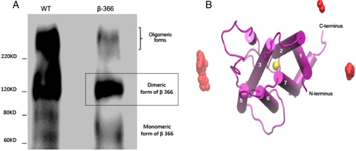
A, TSHR β-subunit dimerization. Western blotting of a truncated TSHR (β-366), partially devoid of the ectodomain, continued to demonstrate oligomerization with dimeric forms and oligomeric forms outnumbering the monomers (∼70 kDa). WT-type monomeric form 100- to 120-kDa and oligomeric forms (>220 kDa) are shown in the left lane. TMD appeared to be the primary site of the oligomer interface. B, Dimer poses of TSHR-TMD protomers. The individual centers of the second protomer (yellow ball) of the TSHR in the membrane plane are shown for the configurations in which the BD was successfully able to form a dimer. The likely contacts are restricted to essentially opposite sides of the central seven-helix bundle (shown as orange clusters).
Modeling TMD protomers
Using our model structure of the TMD, we saw that of 105 BD steps, only 486 dimeric poses met the energy cutoff condition of two protomers within the 35-Å interaction range. A K-mean cluster analysis was carried out on all the putative dimer conformations, and we obtained the best representative central structure in the correct orientation for residue proximity (Figure 1B). The chosen representative structure showed a well-defined membrane plane bound orientation. In fact, the clustering analysis showed that the center of the second copy of the receptor protomer in the membrane plane is capable of forming dimers, and likely contacts were restricted to the two opposite sides of the helix bundle (Figure 1B). The fact that the two contact surfaces were in essentially diagonal positions also explained the fact that the oligomerization of the TSHR is not limited to dimers because, after having formed a dimer, the complex still has two similar different contact surfaces free for interaction, which can result in oligomer formation.
This BD-defined dimeric structure was then analyzed for interfacial contact residues. The dimer pose showed three pairs of potential residue interfaces (Figure 2A and Table 2), two pairs of residues interacting between TM2 and TM5, and one pair of residues interacting between TM1 and TM4. The predicted interfacial associations were strongest between TM1 and TM4. In addition, the model also predicted an association of TM2 with TM5.
Figure 2.

A, Structure of a TSHR dimer. This is a representation of the dimer structure at the center of the largest cluster viewed from the side. The residues that make contact in this helical association are represented by black and steel gray ball structures. The residues involved were as follows: TM1 (V433) made contact with TM4 (L551); TM2 (L467) made contact with TM5 (T588); and TM2 (A470) made contact with TM5 (N590). Inset, A close-up top view of the interacting residues in the corresponding helices. TM5 and TM4 are in blue and TM1 and TM2 are in orange. B, Cross-linking of cysteine-mutated TSHRs. To confirm the proximity of the BD predicted residues, we generated a full-length TSHR construct having all six predicted residues (three pairs) mutated to cysteine and then transfected this construct into CHO cells to obtain a stable line (Mut6). The percent expression of surface receptors in WT TSHR- and Mut6-transfected cells was 83.25 ± 2.89 and 47.35 ± 16.78, respectively. Total membrane lysates prepared from the Mut6 and WT cells were then cross-linked using CuSO4 and phenanthroline as described in Materials and Methods. The right lane in the Mut6 immunoblot panel shows increased oligomeric forms (≥220 kDa) in the presence of the cross-linker when compared with the vehicle-treated lysate (left lane). There was also a marked corresponding decrease in the monomeric forms (100–120 kDa) in the treated lane. The right WT immunoblot panel shows that the WT receptor lysate treated with or without (−/+) the cross-linker in the same way did not reveal an enhanced oligomer formation or any increased higher cross-linked bands than the constitutive oligomeric forms. Bottom graph, Densitometry of monomer to dimer ratio of the respective immunoblots.
Table 2.
Results of the Single-Pair in Silico Mutations Done on the Predicted BD Contact Residues
| Protomer 1 | Residue | TMD Region | Protomer 2 | Residue | TMD Region | Distance in A Before Cysteine Subsitution | Distance in A After Cysteine Substitution |
|---|---|---|---|---|---|---|---|
| CG2 | V433C | TM1 | CD1 | L551C | TM4 | 1.8 | 1.7 |
| CD2 | L467C | TM2 | C | T588C | TM5 | 2.3 | 1.9 |
| CB | A470C | TM2 | CD1 | N590C | TM5 | 2.1 | 2.0 |
All predicted contact residues were mutated into cysteines in pairs, and the distance was derived from the BD results before and after mutations. Bold letters represent amino acids.
Receptor mutations
Initial random mutations of both conserved and nonconserved amino acid substitutions within TM1, TM4, and TM5 (residues 419, 438, 543, 546, 549, 554, 558, 560, 576, and 591) caused no change in the oligomer formation (data not illustrated). To try and confirm at least the proximity of the predicted residues and the validity of the dimer model, we then generated a TSHR model with BD-predicted residues in the TMD substituted with cysteines. In silico analysis of the cysteine substituted the mutants of single pairs (Table 1) did not show any significant change in the intermolecular distances of the final three structures compared with the wild-type receptor, indicating that these mutations also failed to disrupt the normal structure (Table 1).
We then generated a stable TSHR mutant in CHO cells with the same six BD-predicted residues in the TMD substituted with cysteines. On cross-linking these mutant TSHRs in total cell lysate, we observed, on immunoblotting, a marked enhancement of the oligomer formation as evidenced by the 240-kDa band corresponding to cross-linked 120-kDa protomers when compared with the WT samples and a substantial decrease in the monomeric forms (Figure 2B, Mut6 blot, right lane). This demonstrated that the predicted residues are in close proximity and may be involved in the dimeric interfaces and thus implicating the residues from TM1, TM2, TM4, and TM5 in this interaction. These results suggested that the predicted residues in the contact surfaces likely contributed to the stability of the structure. As a control the WT receptors did not show any increase in oligomeric forms in the presence or absence of the cross-linker (Figure 2B, WT blot). However, the fact that oligomerization persisted without cross-linking indicated that additional residues/interfaces were also capable of maintaining the TSHR oligomeric structure.
Pair-wise helix truncation and oligomerization
We then constructed TSHRs with missing helices. Initially we truncated pairs of helices from the TMD as illustrated in Figure 3, A–D. The truncated constructs removing TM1+TM2, TM3+TM4, or TM5+TM6 were then transiently expressed in HEK293 cells. A YFP tag at the carboxyl tail of the receptor constructs allowed us to examine their total expression within unfixed cells by flow cytometry (Figure 3E). The truncated constructs had variable levels of expression of protein but were not significantly different from that of the WT receptor-expressing cells. By the immunoblotting of nonreduced gels with a TSHR-specific antibody (M1), we could see that the TM1+TM2 truncated receptor showed a substantial decrease in the quantity of oligomer formation (Figure 3F) compared with the WT receptor and with the other truncated receptors, again suggesting that oligomer interface(s) may reside in TM1 and/or TM2. In contrast, removal of TM4 or TM5 did not appear to influence oligomer formation. However, removal of TM1 and TM2 did not totally abrogate oligomerization, indicating the involvement of additional or alternative interacting domains in other helices.
Figure 3.
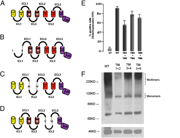
A–D, Truncations of the TSHR-TMD. The eight helices of the TSHR transmembrane domain are depicted by cylindrical structures connected to each other by their extra- and intracellular loops. The WT TSHR is shown in panel A, whereas panels B–D are structures with the paired truncations used in subsequent studies. The dotted line shows the actual connection of the ECLs and ICLs in the various truncated constructs. ECL, extracellular loop. E, Expression of pair-wise deleted mutants. WT TSHR and the truncated and paired TSHR mutants (TM1+TM2; TM3+TM4; and TM5+TM6) had a YFP tag at their carboxyl tail and were transfected into HEK293 cells and checked for total expression of YFP by flow cytometry in unfixed cells 48 hours after the transfection. The y-axis indicates the percentage of YFP-positive cells. Significant expression (from 50% to 90%) of truncated receptors was observed in these transiently transfected HEK293 cells. UT, untruncated. F, Western blotting of double TM truncated TSHRs. This figure illustrates the TSHR species identified in a nonreducing immunoblot of total membranes (100 μg/lane) prepared from the cells described in panel E after probing with the TSHR antibody (M1 at 2 μg/mL). There was a substantial decrease in TSHR oligomers (bands of ≥ to 220 kDa) with the TM1+TM2 deletion construct compared with WT TSHRs or other truncated pairs. Monomeric receptors of the expected 100–120 kDa were seen with the WT construct.
Individual TM1 and TM2 truncated TSHRs
Truncation of the TM1 alone showed a significant decrease in oligomer formation as compared with a TM2 truncated receptor and the wild-type receptor (Figure 4, A and B). Using CFP-tagged (donor) and YFP-tagged TSHRs and FRET analysis as detailed in Materials and Methods, we further established that the TM1 truncated receptors showed a significant decrease in FRET efficiency compared with WT receptors (Figure 4, C and D). Yet again, however, a significant degree of oligomerization continued to be present.
Figure 4.

A, Individual TM1 and TM2 truncated TSHRs. Total membranes prepared from constructs transfected into HEK293 cells with either TM1 deleted or TM2 deleted were immunoblotted using a TSHR antibody. There was a marked decrease in oligomers seen with the TM1 deleted mutant compared with WT TSHR and the TM2 deleted receptors. B, TSHR monomer to oligomer ratios. Densitometry of panel A yielded monomer to oligomer ratios of 1.0 compared with 1.4 for WT and thus emphasized the decreased formation of oligomers with the TM1 mutant compared with WT receptors. C, FRET analysis of the TM1 truncated construct. As detailed in Materials and Methods, FRET studies were carried out using CFP-tagged and YFP-tagged WT and TM1-deleted TSHR constructs after cotransfecting these tagged receptors into HEK293 cells. As controls we used CTV and C5V constructs tagged with CFP and Venus reporter genes. CTV is a construct in which the CFP (donor) is tagged to Venus (reporter) by a fixed set of amino acids that gives a positive FRET, whereas C5V is a truncated version of this arrangement in which the FRET efficiency is very low (56). FRET efficiency was measured for these different constructs after donor spectral bleed-through and acceptor spectral bleed-through subtraction using the pFRET software as described. The data indicate a significant decrease (75%) in FRET efficiency with the TM1-deleted receptors over that of WT TSHRs, illustrating decreased dimer or oligomer formation in TM1 truncated receptors. D, FRET images. WT (left panel) and TM1 and TM2 truncated (right panel) TSHR FRET images, after spectral bleed-through correction, showing the different fluorescent intensity obtained in the FRET analyses. The graphs below show the pixels measured across the images with their intensity (gray values) in the y-axis. Note the different scales needed to illustrate the decreased intensity obtained with the TM1 truncated receptor compared with that of the WT TSHR.
Restoring oligomerization by homologous replacement of region
We also replaced the missing TM1 helix with the TM1 helix from the LH receptor, which had 14 of 28 conserved residues (Figure 5A) and which caused the loss of BD predicted contact residue TM1 V433. The quantitation of the Western blot from the total cell membrane lysis showed no difference between WT TSHRs (Figure 5C) and the LH receptor TM1-substituted constructs (LHRsb), whereas the monomer to oligomer ratio remained significantly decreased in the TM1 truncated cells. The surface expression of the resulting constructs by flow cytometery was indistinguishable from the WT TSHR (Figure 5B). These data therefore allowed us to conclude that the loss of oligomer formation caused by the truncation of the TSHR TM1 helix was significant but not complete and that restoration of this interaction was achieved by a substitution with the LH receptor TM1, which was devoid of contact residue TM1 V433, indicating the presence of additional interacting residues.
Figure 5.

A, Aligned protein sequence of LHR and TSHR TM1 region. The protein sequences of the TSHR and LHR TM1 region were aligned to show the 54% conservation. The residues marked in red are the nonconserved substitutions found only in the LH receptor TM1 region. The residues of interest in TM1 for oligomer formation is marked in blue and underlined. LHR, LH receptor. B, Surface expression of the LH receptor substituted construct. Fluorescence-activated cell sorter analyses were performed with cells expressing WT-TSHR (upper right box) and TSHRs substituted with the LH receptor TM1 sequence (LHRsb) (lower right box) and TSHR truncated for TM1 (lower left box). These constructs had YFP on their carboxyl end and the unfixed cells were stained for surface TSHR expression with anti-TSHR (M1), which was subsequently detected with an antimouse PE as indicated in the x-axis. The upper left box shows the background binding with the second antibody alone. Some (29.2%) of the LH receptor TM1 substituted cells had surface expression of this chimeric receptor. C, The LH receptor TM1 restores TSHR TM1δ oligomerization. The densitometry of the Western blotting of lysates were prepared from WT-, LHRsb-, and TM1δ TSHR-expressing cells and probed for receptor forms with anti-TSHR (M1). As shown here, the TSHR TM1δ construct showed reduced monomer to oligomer ratio, but the substitution of LH receptor residues into the TSHR TM1δ truncated construct restored the oligomeric forms almost to the original levels seen with WT TSHR (gray filled bar). PE, Phycoerythrin.
Discussion
As seen with several well-characterized GPCRs, the TSHR exists as constitutive dimers and oligomers in native thyroid cells and overexpressed heterologous cells (14, 15, 36). These higher-order TSHR forms have been shown to have a role in negative cooperativity (36, 37), in regulating early events during receptor maturation and intracellular trafficking (1, 38), and recently in Gq11 signaling (39). We have previously shown that these monomeric and higher-order complexes can also bind autoantibodies (40) and may be regulated by TSH and stimulating TSHR antibodies within lipid rafts (16, 41). Furthermore, a direct relationship between the conformational changes associated with activation of the TMD and the allosteric behavior across glycoprotein hormone receptor dimers has been demonstrated experimentally (42).
Our biochemical observations showed that truncated TSHRs with β-subunits were still capable of forming dimers and oligomers like the holoreceptor (Figure 1A), showing that the TMD of the TSH receptor is a major site of interaction. This led us to map the interfacial contacts on the TMD. To proceed with mapping and the rediction of residues that may be involved in TSHR dimer/oligomer formation, we generated a model of the TSHR-TMD by constructing a TMD with extra- and intracellular loops (23). Our modeling data predicted that constitutive oligomerization of the TSHR would involve multiple residues in one or more helices, and the experimental data were also in favor of multiple interacting sites.
Despite recent advances in our understanding of the structural details and mode of oligomerization, predicting the exact dimerization interfaces for many GPCRs has been a challenge. Dimerization/oligomerization interfaces differ among highly related GPCRs. Correlated mutation analysis-based studies by Gouldson et al (43, 44) to identify dimerization interfaces of GPCRs predicted nearly every TM helix as a putative interface. Therefore, to experimentally identify these possible putative interacting TM helices, we used a reductionist approach by deleting the TSHR TM helices in pairs rather than a laborious cysteine-scanning experiment of the entire TMD. The truncated receptor constructs were then transiently expressed. By this analysis, the receptor protein pair having a TM1+TM2 deletion had a substantial decrease in the formation of oligomers, and the confirmation of this conclusion came from further examining TM1 and TM2 as individual truncations, both biochemically (immunoblotting) and by a biophysical method (FRET), in which we found that the TM1 truncated TSHRs showed a significant reduction in oligomer formation, thus demonstrating that TM1 played a more significant role in TSHR oligomerization compared with TM2. However, when we removed the TM1 from the TSHR and substituted the TM1 from the LH receptor with conserved residues but still lacking a leading interacting residue (V433), we were able to restore full oligomerization to the TSHR indicating the presence of a region of attraction rather than individual residue-dependent interactions.
The strong biochemical and biophysical evidence of TM1 as a potential regional interface for TSHR oligomerization was further supported by BD analysis based on our TMD structure developed from a rhodopsin-based model of the loop regions of the TSHR-TMD (23). Although both the homology model and the BD protocol used involved serious approximations, earlier work showed that this technique was capable of predicting actual dimerization surfaces. The major source of modeling errors includes the models for the loop regions, the fact that the proteins were held rigid, and the continuum representation of the membrane and aqueous environment. Because these are indeed significant approximations, it is not at all guaranteed that every application will produce a valid prediction and thus experimental confirmation is always necessary, as was done in this work. For example, it is quite possible that the residue pairs that we selected for cystein mutation change their side-chain conformation so that the cysteines are too far for forming the disulfide bond. It is also possible that the BD-predicted sites are widely spread out, which would mean that no real conclusion could be formed from the calculation. However, in our studies this was not the case: the BD predictions formed two well-defined, largely equivalent clusters, and the BD-suggested helix contacts were confirmed experimentally. Furthermore, the proposed contacts could explain not just dimerization but also oligomerization.
A proximity analysis of the contact residues of this central structure indicated three pairs of residues that were less than 4 Å apart in the two protomers. Interestingly, V433 in TM1 showed close proximity to L551 in TM4, whereas L467 and A470 in TM2 had interacting partners T588 and N590, respectively, in TM5. Indeed, rearrangement of aromatic clusters in response to ligand binding and the subsequent conformational changes have previously been shown to be a trigger for transition from an inactive state to an active state in GPCRs. These dynamic rearrangements may involve a series of TM helix movements (3, 45, 46). Furthermore, our truncation and substitution studies suggested that the oligomerization interfaces in the TSHR-TMD are not controlled by single residues but are formed cooperatively based on the cumulative effects of the interfacing residues. Our single-pair in silico mutations of the predicted residue pairs into cysteines did not show any significant alteration in the proximity distances as a result of these single mutations, which lead to mutating all six residues rather than as single pairs. Note, however, that due to the rigidity of the protein model used in the BD calculations, the analysis above is considered to be preliminary, especially the ones not confirmed by experiments. A definitive atomic-level description will require extensive molecular dynamics simulations with the TSHR-TMD dimer inserted in an explicitly represented lipid bilayer and solvated with water. Our dimer model can serve as a reasonable starting structure for such calculations.
Actual cysteine cross-linking experiments carried out using all six predicted contact residues were within interacting distances, demonstrating that residues in TM1, TM2, TM4, and TM5 are involved in the oligomerization. However, the presence of oligomers in the non-cross-linked Mut6 sample (Figure 2B) demonstrated that the six predicted mutants were not the only residues possibly involved in oligomerization, although these were within interacting distances. A correlated mutation analysis-based study (47) carried out with rhodopsin-like GPCRs also demonstrated that residues in TM1 and TM4 appeared most often as putative interfaces in these GPCRs. Similarly, experimental data have indicated a predominant role for these two TMHs in dimerization/oligomerization in rhodopsin and rhodopsin-like GPCRs (34, 48–50). In particular, atomic force microscopy analysis of rhodopsin (51) has suggested that TM1 and TM4 form distinct symmetrical interfaces in which TM4 was specifically implicated in intradimeric contacts between monomers, and TM1 was suggested to facilitate the formation of rhodopsin dimer rows. Calebiro et al (38) showed that a L467P mutation in TM2 of the TSHR resulted in a mutant receptor that was capable of holding the wild-type TSHR in the endoplasmic reticulum by virtue of dimerization, further suggesting a strong link to one of our BD predicted contact points in TM2. Furthermore, the crystal structure of the oligomeric β1-adrenergic receptor in the basal state has shown oligomerization interfaces mainly in TM1 as well as TM2, TM4, TM5, H8, and intracellular loop (ICL)-2 (52). Our BD studies, although not based on the β-adrenergic receptor, and our disulfide cross-linking experiments, also predicted possible dimerization/oligomerization contacts in these different TM helices. Hence, TM1 and TM2 appeared to contain some of the lead contact residues for TSH oligomerization but the absence of such a residue, V433 in TM1, did not prevent all the oligomers forming and indicated the cooperative nature of such interactions by additional residues able to substitute and achieve contact as evidenced by the cysteine mutation data.
These results in the existing lilterature therefore suggested that oligomer formation in the TSHR, and perhaps many GPCRs, can be the result of cooperative dimer interfaces. Yet this may not be the case for all GPCRs because I52 in TM1 and V150 in TM5 have been reported as the only key residues in the interaction surface between CCR5 molecules (53), although these data remain to be confirmed. In contrast, it is not uncommon for the structural space of protein-protein interfaces to be degenerate (54, 55), and this seems true for the TSHR because the restoration of the TM1-deleted residues by the LH receptor sequence did restore oligomer formation (Figure 5C). This may be explained for the TSHR as due to cooperative protein interactions (54, 55).
In conclusion, we present data obtained by computational methods and in vitro analyses that the TSHR-TMDs have oligomerization interfaces dominated by TM1 and TM5. Because TM1 and TM5 are at the opposite side of the helix bundle, such an arrangement allows the formation of larger oligomers. This resilient and unfailing system favoring receptor-receptor interaction strongly suggests the physiological importance of maintaining the TSHR oligomeric structures.
Acknowledgments
We thank Dr A. Periasamy (W. M. Keck Center for Cellular Imaging, University of Virginia, Charlottesville, Virginia) for providing the pFRET software and sensitized emission FRET methodology; Dr Steven S. Vogel (Section of Cellular Biophotonics, National Institutes of Health, Bethesda, Maryland) for providing us with the C5V and CTV plasmid constructs used as FRET standards; Dr B. Rees Smith (RSR Ltd, Cardiff, Wales) for the TSHR antibodies; and Dr Meng Cui (Virginia Commonwealth University School of Medicine, Richmond, Virginia) for helping us with the Brownian dynamics. We are also grateful to the members of our laboratory for critically reading this manuscript.
This work was supported by National Institutes of Health Grants DK069713 and DK052464 and the Veterans Affairs Merit Award Program (to T.F.D.). This work was also supported in part through the computational resources and staff expertise provided by the Department of Scientific Computing at the Icahn School of Medicine at Mount Sinai.
Disclosure Summary: The authors have nothing to disclose.
Footnotes
- BD
- brownian dynamics
- CHO
- Chinese hamster ovary
- CFP
- cyan fluorescent protein
- FRET
- Förster resonance energy transfer
- GPCR
- G protein-coupled receptor
- HEK
- human embryonic kidney
- ICL
- intracellular loop
- TM
- transmembrane
- TMD
- TM domain
- TSHR
- TSH receptor
- WT
- wild type
- YFP
- yellow fluorescent protein.
References
- 1. Latif R, Michalek K, Davies TF. Subunit interactions influence TSHR multimerization. Mol Endocrinol. 2010;24(10):2009–2018. [DOI] [PMC free article] [PubMed] [Google Scholar]
- 2. Ciruela F, Vallano A, Arnau JM, et al. G protein-coupled receptor oligomerization for what? J Recept Signal Transduct Res. 2010;30(5):322–330. [DOI] [PubMed] [Google Scholar]
- 3. Javitch JA. The ants go marching two by two: oligomeric structure of G-protein-coupled receptors. Mol Pharmacol. 2004;66(5):1077–1082. [DOI] [PubMed] [Google Scholar]
- 4. Whorton MR, Bokoch MP, Rasmussen SG, et al. A monomeric G protein-coupled receptor isolated in a high-density lipoprotein particle efficiently activates its G protein. Proc Natl Acad Sci USA. 2007;104(18):7682–7687. [DOI] [PMC free article] [PubMed] [Google Scholar]
- 5. Lee C, Ji I, Ryu K, Song Y, Conn PM, Ji TH. Two defective heterozygous luteinizing hormone receptors can rescue hormone action. J Biol Chem. 2002;277(18):15795–15800. [DOI] [PubMed] [Google Scholar]
- 6. Ferre S, Casado V, Devi LA, Filizola M, et al. G protein-coupled receptor oligomerization revisited: functional and pharmacological perspectives. Pharmacol Rev. 2014;66(2):413–434. [DOI] [PMC free article] [PubMed] [Google Scholar]
- 7. Rivero-Muller A, Chou YY, Ji I, et al. Rescue of defective G protein-coupled receptor function in vivo by intermolecular cooperation. Proc Natl Acad Sci USA. 2010;107(5):2319–2324. [DOI] [PMC free article] [PubMed] [Google Scholar]
- 8. Vassart G. An in vivo demonstration of functional G protein-coupled receptor dimers. Proc Natl Acad Sci USA. 2010;107(5):1819–1820. [DOI] [PMC free article] [PubMed] [Google Scholar]
- 9. De Felice M, Postiglione MP, Di Lauro R. Minireview: thyrotropin receptor signaling in development and differentiation of the thyroid gland: insights from mouse models and human diseases. Endocrinology. 2004;145(9):4062–4067. [DOI] [PubMed] [Google Scholar]
- 10. Rapoport B, Chazenbalk GD, Jaume JC, McLachlan SM. The thyrotropin (TSH) receptor: interaction with TSH and autoantibodies. Endocr Rev. 1998;19(6):673–716. [DOI] [PubMed] [Google Scholar]
- 11. Sanders J, Miguel RN, Furmaniak J, Smith BR. TSH receptor monoclonal antibodies with agonist, antagonist, and inverse agonist activities. Methods Enzymol. 2010;485:393–420. [DOI] [PubMed] [Google Scholar]
- 12. Latif R, Morshed SA, Zaidi M, Davies TF. The thyroid-stimulating hormone receptor: impact of thyroid-stimulating hormone and thyroid-stimulating hormone receptor antibodies on multimerization, cleavage, and signaling. Endocrinol Metab Clin North Am. 2009;38(2):319–341, viii. [DOI] [PubMed] [Google Scholar]
- 13. Davies TF, Ando T, Lin RY, Tomer Y, Latif R. Thyrotropin receptor-associated diseases: from adenomata to Graves disease. J Clin Invest. 2005;115(8):1972–1983. [DOI] [PMC free article] [PubMed] [Google Scholar]
- 14. Graves PN, Vlase H, Bobovnikova Y, Davies TF. Multimeric complex formation by the thyrotropin receptor in solubilized thyroid membranes. Endocrinology. 1996;137(9):3915–3920. [DOI] [PubMed] [Google Scholar]
- 15. Latif R, Graves P, Davies TF. Oligomerization of the human thyrotropin receptor: fluorescent protein-tagged hTSHR reveals post-translational complexes. J Biol Chem. 2001;276(48):45217–45224. [DOI] [PubMed] [Google Scholar]
- 16. Latif R, Graves P, Davies TF. Ligand-dependent inhibition of oligomerization at the human thyrotropin receptor. J Biol Chem. 2002;277(47):45059–45067. [DOI] [PubMed] [Google Scholar]
- 17. Latif R, Michalek K, Morshed SA, Davies TF. A tyrosine residue on the TSH receptor stabilizes multimer formation. PLoS One. 2010;5(2):e9449. [DOI] [PMC free article] [PubMed] [Google Scholar]
- 18. Fogolari F, Brigo A, Molinari H. The Poisson-Boltzmann equation for biomolecular electrostatics: a tool for structural biology. J Mol Recognit. 2002;15(6):377–392. [DOI] [PubMed] [Google Scholar]
- 19. Cui M, Mezei M, Osman R. Modeling dimerizations of transmembrane proteins using Brownian dynamics simulations. J Compur Aided Mol Des. 2008;22(8):553–561. [DOI] [PubMed] [Google Scholar]
- 20. Lowe SL, Adrian C, Ouporov IV, Waingeh VF, Thomasson KA. Brownian dynamics simulations of glycolytic enzyme subsets with F-actin. Biopolymers. 2003;70(4):456–470. [DOI] [PubMed] [Google Scholar]
- 21. Goldfeld DA, Zhu K, Beuming T, Friesner RA. Successful prediction of the intra- and extracellular loops of four G-protein-coupled receptors. Proc Natl Acad Sci USA. 2011;108(20):8275–8280. [DOI] [PMC free article] [PubMed] [Google Scholar]
- 22. Mustafi D, Palczewski K. Topology of class A G protein-coupled receptors: insights gained from crystal structures of rhodopsins, adrenergic and adenosine receptors. Mol Pharmacol. 2009;75(1):1–12. [DOI] [PMC free article] [PubMed] [Google Scholar]
- 23. Ali MR1, Latif R, Davies TF, Mezei M. Monte Carlo loop refinement and virtual screening of the thyroid-stimulating hormone receptor transmembrane domain. J Biomol Struct Dyn. 2014;July11:1–13. [DOI] [PMC free article] [PubMed] [Google Scholar]
- 24. Sali A, Blundell TL. Comparative protein modelling by satisfaction of spatial restraints. J Mol Biol. 1993;234(3):779–815. [DOI] [PubMed] [Google Scholar]
- 25. Northrup SH, Erickson HP. Kinetics of protein-protein association explained by Brownian dynamics computer simulation. Proc Natl Acad Sci USA. 1992;89(8):3338–3342. [DOI] [PMC free article] [PubMed] [Google Scholar]
- 26. Northrup SH, Boles JO, Reynolds JCL. Electrostatic effects in the Brownian dynamics of association and orientation of heme proteins. J Phys Chem. 1987;91(23):5991–5998. [Google Scholar]
- 27. Ciullo I, Latif R, Graves P, Davies TF. Functional assessment of the thyrotropin receptor-beta subunit. Endocrinology. 2003;144(7):3176–3181. [DOI] [PubMed] [Google Scholar]
- 28. Van Durme J, Horn F, Costagliola S, Vriend G, Vassart G. GRIS: glycoprotein-hormone receptor information system. Mol Endocrinol. 2006;20(9):2247–2255. [DOI] [PubMed] [Google Scholar]
- 29. Fiser A, Do RK, Sali A. Modeling of loops in protein structures. Protein Sci. 2000;9(9):1753–1773. [DOI] [PMC free article] [PubMed] [Google Scholar]
- 30. Laskowski RA, Moss DS, Thornton JM. Main-chain bond lengths and bond angles in protein structures. J Mol Biol. 1993;231(4):1049–1067. [DOI] [PubMed] [Google Scholar]
- 31. Mezei M. Simulaid: a simulation facilitator and analysis program. J Comp Chem. 2010;31(14):2658–2668. [DOI] [PubMed] [Google Scholar]
- 32. Downs GM. Clustering methods and their uses in computational chemistry. Rev Comp Chem. 2002;18:1–40. [Google Scholar]
- 33. Barnard JM, Downs GM, von Scholley-Pfab A, Brown RD. Use of Markush structure analysis techniques for descriptor generation and clustering of large combinatorial libraries. J Mol Graph Model. 2000;18(4–5):452–463. [PubMed] [Google Scholar]
- 34. Guo W, Shi L, Filizola M, Weinstein H, Javitch JA. Cross talk in G protein-coupled receptors: changes at the transmembrane homodimer interface determine activation. Proc Natl Acad Sci USA. 2005;102(48):17495–17500. [DOI] [PMC free article] [PubMed] [Google Scholar]
- 35. Hu J, Thor D, Zhou Y, et al. Structural aspects of M(3) muscarinic acetylcholine receptor dimer formation and activation. FASEB J. 2012;26(2):604–616. [DOI] [PMC free article] [PubMed] [Google Scholar]
- 36. Urizar E, Montanelli L, Loy T, et al. Glycoprotein hormone receptors: link between receptor homodimerization and negative cooperativity. EMBO J. 2005;24(11):1954–1964. [DOI] [PMC free article] [PubMed] [Google Scholar]
- 37. Chazenbalk GD, Kakinuma A, Jaume JC, McLachlan SM, Rapoport B. Evidence for negative cooperativity among human thyrotropin receptors overexpressed in mammalian cells. Endocrinology. 1996;137(11):4586–4591. [DOI] [PubMed] [Google Scholar]
- 38. Calebiro D, de Filippis T, Lucchi S, et al. Intracellular entrapment of wild-type TSH receptor by oligomerization with mutants linked to dominant TSH resistance. Hum Mol Genet. 2005;14(20):2991–3002. [DOI] [PubMed] [Google Scholar]
- 39. Allen MD, Neumann S, Gershengorn MC. Occupancy of both sites on the thyrotropin (TSH) receptor dimer is necessary for phosphoinositide signaling. FASEB J. 2011;25(10):3687–3694. [DOI] [PMC free article] [PubMed] [Google Scholar]
- 40. Graves PN, Vlase H, Davies TF. Folding of the recombinant human thyrotropin (TSH) receptor extracellular domain: identification of folded monomeric and tetrameric complexes that bind TSH receptor autoantibodies. Endocrinology. 1995;136(2):521–527. [DOI] [PubMed] [Google Scholar]
- 41. Latif R, Ando T, Davies TF. Lipid rafts are triage centers for multimeric and monomeric thyrotropin receptor regulation. Endocrinology. 2007;148(7):3164–3175. [DOI] [PubMed] [Google Scholar]
- 42. Zoenen M, Urizar E, Swillens S, Vassart G, Costagliola S. Evidence for activity-regulated hormone-binding cooperativity across glycoprotein hormone receptor homomers. Nat Commun. 2012;3:1007. [DOI] [PubMed] [Google Scholar]
- 43. Gouldson PR, Dean MK, Snell CR, Bywater RP, Gkoutos G, Reynolds CA. Lipid-facing correlated mutations and dimerization in G-protein coupled receptors. Protein Eng. 2001;14(10):759–767. [DOI] [PubMed] [Google Scholar]
- 44. Gouldson PR, Snell CR, Bywater RP, Higgs C, Reynolds CA. Domain swapping in G-protein coupled receptor dimers. Protein Eng. 1998;11(12):1181–1193. [DOI] [PubMed] [Google Scholar]
- 45. Hlavackova V, Zabel U, Frankova D, et al. Sequential inter- and intrasubunit rearrangements during activation of dimeric metabotropic glutamate receptor 1. Sci Signal. 2012;5(237):ra59. [DOI] [PubMed] [Google Scholar]
- 46. Pin JP, Comps-Agrar L, Maurel D, et al. G-protein-coupled receptor oligomers: two or more for what? Lessons from mGlu and GABAB receptors. J Physiol. 2009;587(Pt 22):5337–5344. [DOI] [PMC free article] [PubMed] [Google Scholar]
- 47. Filizola M, Olmea O, Weinstein H. Prediction of heterodimerization interfaces of G-protein coupled receptors with a new subtractive correlated mutation method. Protein Eng. 2002;15(11):881–885. [DOI] [PubMed] [Google Scholar]
- 48. Carrillo JJ, Pediani J, Milligan G. Dimers of class A G protein-coupled receptors function via agonist-mediated trans-activation of associated G proteins. J Biol Chem. 2003;278(43):42578–42587. [DOI] [PubMed] [Google Scholar]
- 49. Klco JM, Lassere TB, Baranski TJ. C5a receptor oligomerization. I. Disulfide trapping reveals oligomers and potential contact surfaces in a G protein-coupled receptor. J Biol Chem. 2003;278(37):35345–35353. [DOI] [PubMed] [Google Scholar]
- 50. Liang Y, Fotiadis D, Filipek S, Saperstein DA, Palczewski K, Engel A. Organization of the G protein-coupled receptors rhodopsin and opsin in native membranes. J Biol Chem. 2003;278(24):21655–21662. [DOI] [PMC free article] [PubMed] [Google Scholar]
- 51. Fotiadis D, Liang Y, Filipek S, Saperstein DA, Engel A, Palczewski K. Atomic-force microscopy: rhodopsin dimers in native disc membranes. Nature. 2003;421(6919):127–128. [DOI] [PubMed] [Google Scholar]
- 52. Huang J, Chen S, Zhang JJ, Huang XY. Crystal structure of oligomeric β1-adrenergic G protein-coupled receptors in ligand-free basal state. Nat Struct Mol Biol. 2013;20(4):419–425. [DOI] [PMC free article] [PubMed] [Google Scholar]
- 53. Hernanz-Falcon P, Rodriguez-Frade JM, Serrano A, et al. Identification of amino acid residues crucial for chemokine receptor dimerization. Nat Immunol. 2004;5(2):216–223. [DOI] [PubMed] [Google Scholar]
- 54. Escher C, Cymer F, Schneider D. Two GxxxG-like motifs facilitate promiscuous interactions of the human ErbB transmembrane domains. J Mol Biol. 2009;389(1):10–16. [DOI] [PubMed] [Google Scholar]
- 55. Gao M, Skolnick J. Structural space of protein-protein interfaces is degenerate, close to complete, and highly connected. Proc Natl Acad Sci USA. 2010;107(52):22517–22522. [DOI] [PMC free article] [PubMed] [Google Scholar]
- 56. Koushik SV, Chen H, Thaler C, Puhl HL, 3rd, Vogel SS. Cerulean, Venus, and VenusY67C FRET reference standards. Biophys J. 2006;91(12):L99–L101. [DOI] [PMC free article] [PubMed] [Google Scholar]
