Abstract
In vivo staining reveals cytological details that might otherwise not be apparent. The aim of the study was to test the utility of toluidine blue test in detecting various types of malignant and premalignant lesions in early stage. Fifty patients with lesion in oral cavity having suspicion of malignancy clinically were selected. After subjecting the patients to clinical examination, the suspicious lesions were stained with 1 % toluidine blue. The biopsy site was selected on the basis of clinical appearance and dye retention and in the sites where no retention of the stain occurred, clinical judgment directed the biopsy site. The sensitivity of toluidine blue in detecting premalignant or malignant lesions was found to be 97.8 % and the over all specificity was found to be 100 %. The positive predictive value, negative predictive value and diagnostic accuracy was reported to be 100, 80 and 90 % respectively. Toluidine Blue staining is highly a reliable source for the detection of insitu and invasive carcinomas. Staining with this stains is an adjunct to clinical judgment, assist in the choice of biopsy site, follow up of premalignant lesions and marginal demarcation of the malignant lesions enabling an intervention method to be adopted earlier for the disease, which carries a high rate of morbidity and mortality.
Keywords: Toluidine blue, In vivo staining, Intra-oral malignancies, Early detection
Introduction
Cancer is one among the most dreadful disease human race is suffering. Although oral cancer accounts for 3.6 % of all the cancers there are approximately 27,000 new cases and 9,000 deaths from oral cancer each year [1]. Oral cancer when caught at an early stage is often curable, inexpensive to treat and affords a better quality of life.
In vivo staining has been used extensively in gynaecologic practice for detection of malignant changes of the cervix during colposcopy and this technique has been applied in the oral setting for over 30 years by means of the dyes like toluidine blue. Toluidine blue by its property of retaining in the increased DNA and RNA cellular activity areas, aids in delineating the suspicious areas [2, 3].
Thus, this study was done to assess the utility of toluidine blue in assessing and detecting intra-oral malignancies at an early stage.
Materials and Method
The present study was carried out in Department of Otorhinolaryngology and Head and Neck Surgery, Netaji Subhash Chandra Bose Medical College and Hospital, Jabalpur, selecting 50 patients with lesion in oral cavity having suspicion of malignancy. Duration of study was 1 year (September 2011–August 2012). After subjecting the patients to clinical examination, the suspicious lesions were stained with toluidine blue. The patient was asked to rinse the mouth with 1 % acetic acid for 20 s. 1 % toluidine blue(w/w) was then applied with the help of a swab stick and retained for 20 s. Then again rinse with 1 % acetic acid for 20 s to eliminate mechanically retained stain. A punch biopsy was taken from the site after applying topical anaesthetic agent (xylocain 4 %). The biopsy site was selected on the basis of clinical appearance and dye retention and in the sites where no retention of the stain occurred, clinical judgment directed the biopsy site.
A dark blue (royal or navy) stain is considered positive if either the entire lesion being stained or a portion of it is stained or stippled (Figs. 1, 2). A light blue staining is considered doubtful. If there is no colour absorbed by the lesion, it is taken as a negative stain.
Fig. 1.
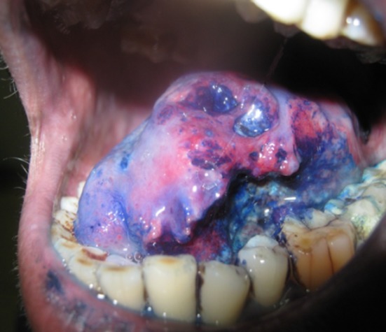
Ulcerative lesion tongue stained with toluidine
Fig. 2.
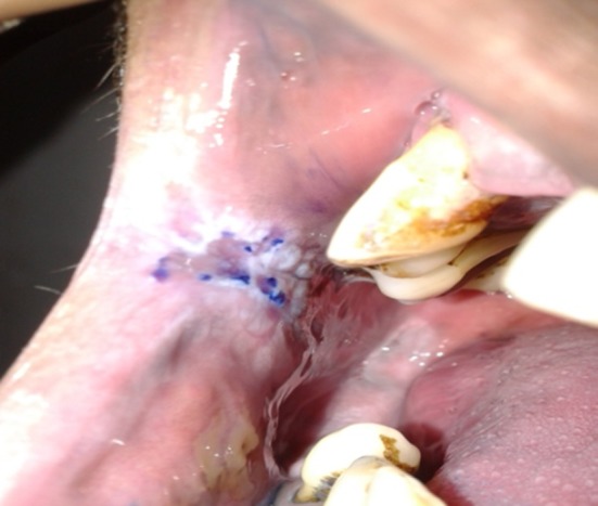
Lesion rt. Buccal mucosa stained with toluidine
Formultion of 1 % toluidine blue : (as described by Mashberg) [4, 5].
1 gm toluidine blue powder
10 ml of 1 % acetic acid
4.19 ml absolute alcohol
86 ml distilled water
pH = 4.5
Results
Most common age group was found to be 41–60 years. (24 cases 48 %) with mean age to be 49.2 years.
Male preponderance with male to female ratio of 8:5.
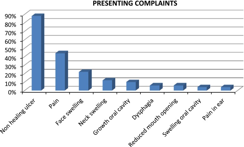
Most common presenting complaint was non healing ulcer in oral cavity (88 %) followed by pain (44 %), face swelling (22 %), neck swelling (12 %), growth (10 %), difficulty in swallowing (6 %), reduced mouth opening (6 %), pain in ear (4 %), swelling in oral cavity (4 %) (Fig. 3).
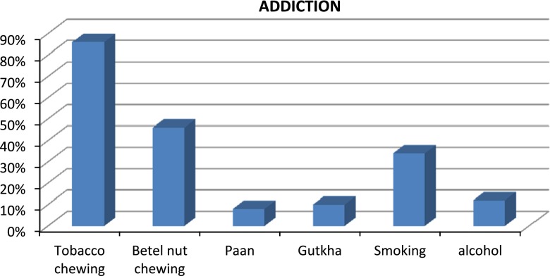
Majority of the cases had multiple risk factors but maximum had tobacco consumption in some or other form (Fig. 4).
Most common site of lesion was found to be buccal mucosa (40 %) followed by tongue (26 %), lower alveolus (14 %), palate (6 %), retero molar trigone (6 %), lips (4 %), flore of mouth (2 %), tonsil (2 %).
Clinically 48 (96 %) cases were diagnosed as malignant lesion and 2 (4 %) cases were suspected as benign lesions.
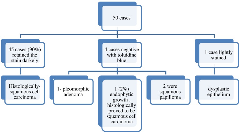
Of the 50 cases, 45 (90 %) cases took dark blue, one case (2 %) took light blue stain and four cases (8 %) did not pick any stain.
- Of the 45 cases stained darkely with toluidine blue, all (100 %) were histlogically proved as having squamous cell carcinoma. four cases negative with toluidine blue,1 (2 %) was an endophytic growth, histologically proved to be squamous cell carcinoma, one was pleomorphic adenoma, two were squamous papilloma. One case,lightly stained with toluidine blue, was proved to be dysplastic epithelium (Fig. 5; Table1).
- Senstivity: 45/46 = 97.8 %.
- Specificity: 4/4 = 100 %.
- PPW: 45/45 = 100 %.
- NPW: 4/5 = 80 %.
- % False Positive: 00 %.
- % False Negative: 20 %.
Fig. 3.
Presenting complaints of patients
Fig. 4.
Addiction present in patients
Fig. 5.
Result of toluidine blue staining and histopathology
Table 1.
Result of toluidine blue staining and histopathology
| Clinical diagnosis | Cases (n = 50) | Staining | Histology | ||
|---|---|---|---|---|---|
| Positive % | Negative % | Malignant % | Benign % | ||
| Suspicious lesion for malignancy | 48 | 45 | 03 | 46 | 02 |
| 93.75 | 06.2 | 95.8 | 4.16 | ||
| Premalignant or non malignant lesion | 02 | 00 | 02 | 00 | 02 |
| 00 | 100 | 00 | 100 | ||
Discussion
In vivo staining reveals cytological details that might otherwise not be apparent, thus aid in accelerating biopsies, diagnosis, and treatment. Toluidine blue, an acidophilic metachromatic dye of thiazine group selectively stains acidic tissue components (sulfates, carboxylates and phosphate radicals) thus staining DNA and RNA. It is used as an in vivo stain based on the fact that dysplastic and anaplastic cells may contain quantitatively more nucleic acids than normal tissues. Also malignant epithelium may contain intracellular canals that are wider than normal epithelium, which may facilitate penetration of the dye [2].
In our present study the sensitivity of toluidine blue in detecting premalignant or malignant lesions was 97.8 % and the results were in accordance of the findings of Mashberg [4] who found the sensitivity to be 98 % and also Hegde et al. [6], who found it to the 97.2 % (Table 2).
Table 2.
Result from different studies about the efficacy of TB staining
| Study | No. of pts. | Sensitivity (%) | Specificity (%) | False positive (%) | False negative (%) |
|---|---|---|---|---|---|
| Present study | 50 | 97.8 | 100 | 0 | 20 |
| Waruakulasuriya [8] | 145 | 100 | 62 | 20.50 | |
| Mashberg [4] | 235 | 98 | 92 | 8.5 | 6.7 |
| Epstein [2] | 59 | 92.5 | 63.2 | ||
| Neibel, Chomet [11] | 20 | 100 | 100 | ||
| Hegde et al. [6] | 90 | 97.2 | 100 | 7.69 | 16.67 |
| Nagaraju et al. [9] | 60 | 100 | 40 | 5.172 | 0 |
The reason for sensitivity not being 100 % in our study is attributed to fact that toluidine blue is unable to stain lesions with intact mucosa i.e. endophytic growth. Out of 50 cases studied, one was an endophytic growth tongue which was negative with toluidine blue stain, while came out to the squamous cell carcinoma Grade I in histopathological reporting.
In our study the over all specificity of TB was found to be 100 % and our results were in accordance with Neibel, Chomet [11] and Hegde et al. [6]. All cases staining positive with the stain were also positive in the histopathological slides.
However our results differed from findings of Waruakularuriya (62 %) [8]; Mashberg (92 %) [4], Epstein (63.2 %) [2], and Nagaraju et al. [9]. This difference can be attributed to interindividual differences applied for the selection criteria of patients for the study.
In our present study the PPV, NPV and diagnostic accuracy (DA) of TB stain was reported to be 100, 80 and 90 % respectively. The findings of the same reported by Silverman et al. [10] were 90 % PPV, 92 % NPV and 90 % DA. Another study conducted by Epstein et al. [2] reported 84 % PPV and 83 DA % while NPV showed significant difference of about 20 %. This can be attributed to the false negative results of the results of the stain i.e. failure of dye to retain in dysplastic/malignant lesions and these results have significantly reduced the NPV in the authors study.
Conclusion
In vivo stains are the prompt resources in diagnosing the molecular changes or some specific chemical reactions taking place within cells or tissues during the process of carcinogenesis. Toluidine Blue staining is highly reliable, cost effective, non invasive and easy source for the detection of insitu and invasive carcinomas. Staining should be routinely used as a method to assist in the choice of biopsy site and in the follow up of premalignant lesions and in the experienced hands marginal demarcation of the malignant lesions enables an intervention method to be adopted earlier for the diseases, which carries a high rate of morbidity and mortality.
Contributor Information
Deepti Singh, Email: drdeepti0810@gmail.com.
R. K. Shukla, Email: dr_r_k_shukla@hotmail.com
References
- 1.Rosenberg D, Cretin S. Use of meta-analysis to evaluate tolonium chloride in oral cancer screening. Oral Surg Oral Med Oral Pathol. 1989;67:621–627. doi: 10.1016/0030-4220(89)90286-7. [DOI] [PubMed] [Google Scholar]
- 2.Epstein JB, Scully C, Spinelli JJ. Toluidine blue and Lugol’s iodine application in the assessment of oral malignant disease and lesions at risk of malignancy. J Oral Pathol Med. 1992;21:160–163. doi: 10.1111/j.1600-0714.1992.tb00094.x. [DOI] [PubMed] [Google Scholar]
- 3.Yokoo K, Noma H, Inoue T, Hashimoto S, Shimono M. Cell proliferation and tumour suppressor gene expression in iodine-unstained area surrounding oral squamous cell carcinoma. Int J Oral Maxillofacial Surg. 2004;33:75–83. doi: 10.1054/ijom.2002.0457. [DOI] [PubMed] [Google Scholar]
- 4.Mashberg A. Re-evaluation of toluidine blue application as a diagnostic adjunct in the detection of asymptomatic oral squamous carcinoma. Cancer. 1980;46:758–763. doi: 10.1002/1097-0142(19800815)46:4<758::AID-CNCR2820460420>3.0.CO;2-8. [DOI] [PubMed] [Google Scholar]
- 5.Mashberg A. Tolonium (toluidine blue) rinse a screening method for recognition of squamous carcinoma—a continuing study of Oral Cancer IV. JAMA. 1981;245:2408–2410. doi: 10.1001/jama.1981.03310480024019. [DOI] [PubMed] [Google Scholar]
- 6.Hegde MC, Kamath PM, Shridharan S, Dannama NK, Raju RM. Supravital staining: its role in detecting early malignancies. Indian J Otorhinolaryngol Head Neck Surg. 2006;58(1):31–34. doi: 10.1007/BF02907735. [DOI] [PMC free article] [PubMed] [Google Scholar]
- 7.Martin IC, Kerawala CJ, Reed M. The application of toluidine blue as a diagnostic adjunct in the detection of epithelial dysplasia. Oral Surg Oral Med Oral Pathol. 1998;85:444–446. doi: 10.1016/S1079-2104(98)90071-3. [DOI] [PubMed] [Google Scholar]
- 8.Warnakulasuriya KAAS, Johnson NW. Sensitivity and specificity of orascan toluidine blue mouth rinse in the detection of oral cancer and precancer. J Oral Pathol Med. 1996;25:97–103. doi: 10.1111/j.1600-0714.1996.tb00201.x. [DOI] [PubMed] [Google Scholar]
- 9.Nagaraju K, Prasad S, Ashok L. Diagnostic efficiency of toluidine blue with Lugol’s iodine in oral premalignant and malignant lesions. Indian J Dent Res. 2010;21(2):218–223. doi: 10.4103/0970-9290.66633. [DOI] [PubMed] [Google Scholar]
- 10.Silverman S, Jr, Migliorati C, Barbosa J. Toluidine blue staining in the detection of oral precancerous and malignant lesions. Oral Surg Oral Med Oral Pathol. 1984;57:379–382. doi: 10.1016/0030-4220(84)90154-3. [DOI] [PubMed] [Google Scholar]
- 11.Niebel HH, Chomet B (1964) In vivo staining test for delineation of oral intraepithelial neoplastic change: preliminary report. J Am Dent Assoc 68:801–806 [DOI] [PubMed]


