CASE SUMMARY
Fifty-seven years old male patient, non-addict, unemployed, was referred to Pulmonary Medicine OPD by anesthesiologist, in view of right paratracheal shadow, detected as an incidental finding during routine preanesthetic check up. The patient was posted for left malleolar bursa excision surgery.
The patient did not have any respiratory complains, when he visited Pulmonary Medicine OPD. The patient had undergone angiography 7 months ago which did not reveal any abnormality. He was operated for fracture neck femur 13 years ago.
On examination the patient was afebrile, with a pulse rate of 80 beats per minute, regular, and had blood pressure of 140/90 mmHg, respiratory rate of 20/min. Rest of general examination and respiratory system examination did not reveal any abnormality. Breath sounds were equal on both sides.
INVESTIGATIONS
Hb: 9.6 gm%, WBC: 11,000, N: 70, L: 20, E: 03, M: 05, ESR: 79, Platelets: 2,80000, BUN: 18 mg, Serum creatinine: 1.0 mg/dl, Total proteins: 7.5 gm/dl Albumin: 3.6 gm/dl, Globulin: 3.9 gm/dl, Total Bilirubin: 0.6 mg/dl, Direct Bilirubin: 0.2 mg/dl, SGPT: 13 IU, SGOT: 19 IU, Alkaline phosphatase: 77 KA, FBS: 93 mg%, PLBS: 155 mg%, Spirometry was within normal limits.
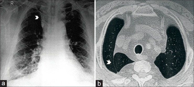
CXR: Right paratracheal shadow
Figure 1.

(a) CXR: Right paratracheal shadow. (b) CT thorax image
QUESTIONS
What is the diagnosis?
Q1. Enlarged thymus
Q2. Substernal goitre
Q3. Pleural Bands
Q4. Azygos lobe
ANSWER
Right azygos lobe
HRCT chest: Right azygos lobe
DISCUSSION
An azygos lobe is a rare anomaly of lung and the incidence of the same varies from 0.4% to 1%.[1]. Azygos lobe was first mentioned by Wrisberg in1778 from anatomical studies, described as “Lobus Wrisbergi.” An azygos lobe is formed when a precursor of the azygos vein fails to migrate over the apex of the lung during fetal life, instead of course through the lung dragging lung with it, the parietal and visceral pleura [Figure 2]. The four layers of pleura are known as “Azygos Fissure.” Since there is no corresponding alteration in segmental architecture, “Lobe” is a misnomer.[1] Embryologically, a part of upper lobe of the right lung may come to lie medial to the azygos vein. This part is called the azygos lobe. In this condition, the azygos vein is suspended from the wall of the thorax by a fold of parietal pleura (mesoazygous).[2] It is commonly seen in males and has also been reported on the left side.[3] An azygos lobe is not susceptible to disease.[1] However, multiple authors have reported spontaneous pneumothorax associated within the azyos lobe in adult and pediatric patients.[4,5,6] Clinically, the knowledge of azygos lobe anatomy is important during thoracic surgical approaches.[3]
Figure 2.

Formation of an azygos lobe
Footnotes
Source of Support: Nil
Conflict of Interest: None declared.
REFERENCES
- 1.Patil SJ. Azygos lobe A review. Int J Clin Surg Adv. 2013;1:17 9. [Google Scholar]
- 2.Singh I, Pal GP. Liver, pancreas, spleen, respiratory system, body cavities. In: Singh I, Pal GP, editors. Human Embryology. 9th ed. New Delhi: Macmillan Publishers India Limited; 2012. p. 190. [Google Scholar]
- 3.Cimen M, Erdil H, Karatepe T. A cadaver with azygos lobe and its clinical significance. Anat Sci Int. 2005;80:235 7. doi: 10.1111/j.1447-073X.2005.00116.x. [DOI] [PubMed] [Google Scholar]
- 4.Monaco M, Barone M, Barresi P, Carditello A, Pavia R, Mondello B. Azygos lobe and spontaneous pneumothorax. G Chir. 2000;21:457 8. [PubMed] [Google Scholar]
- 5.Betschart T, Goerres GW. Azygos lobe without azygos vein as a sign of previous iatrogenic pneumothorax: Two case reports. Surg Radiol Anat. 2009;31:559 62. doi: 10.1007/s00276-009-0479-x. [DOI] [PubMed] [Google Scholar]
- 6.Asai K, Urabe N, Takeichi H. Sponteneous pneumothorax and a coexistent azygos lobe. Jpn J Thorac Cardiovasc Surg. 2005;53:604 6. doi: 10.1007/s11748-005-0147-y. [DOI] [PubMed] [Google Scholar]


