Abstract
Objective
The Cavalieri principle was applied to consecutive pathology sections that were photographed at the same magnification and used to estimate tissue volumes via superimposing a point counting grid on these images. The goal of this study was to perform the Cavalieri method quickly and practically.
Materials and Methods
In this study, 10 adult female Sprague Dawley rats were used. Brain tissue was removed and sampled both systematically and randomly. Brain volumes were estimated using two different methods. First, all brain slices were scanned with an HP ScanJet 3400C scanner, and their images were shown on a PC monitor. Brain volume was then calculated based on these images. Second, all brain slices were photographed in 10× magnification with a microscope camera, and brain volumes were estimated based on these micrographs.
Results
There was no statistically significant difference between the volume measurements of the two techniques (P>0.05; Paired Samples t Test).
Conclusion
This study demonstrates that personal computer scanning of serial tissue sections allows for easy and reliable volume determination based on the Cavalieri method.
Keywords: Light microscopy, Cavalieri principle, Stereology, Histology
Özet
Amaç
Bu çalışmada aynı büyütmede fotoğraflanmış ışık mikroskobik seri kesitler üzerinde Cavalieri prensibi uygulandı. Daha sonra bu kesitler üzerinde noktalı ölçüm gridi kullanılarak hacim hesaplanması yapıldı ve böylece Cavalieri prensibinin hızlı ve etkin olarak kullanılması amaçlandı.
Gereç ve Yöntem
Bu çalışmada 10 adet sağlıklı ve erişkin Sprague Dawley cinsi sıçan kullanıldı. Bu sıçanlardan çıkarılan beyinler sistematik rastgele olarak örneklendi ve seri olarak kesildi. Beyin hacimleri ise iki farklı şekilde hesaplandı. İlk olarak beyin preparatları HP ScanJet 3400C marka scanner ile tarandı ve tarama görüntüleri bilgisayar monitörüne yansıtıldı. Daha sonra bu görüntüler üzerinde beyin hacimleri hesaplandı. İkinci olarak tüm beyin kesitleri x 10 büyütmede kamera ataçmanlı mikroskop ile fotoğraflandı ve beyin hacimleri bu mikrograflar üzerinde hesaplandı.
Bulgular
Bu çalışmanın sonucunda her iki farklı yöntemle elde edilen beyin hacmi bulguları birbiriyle kıyaslandı ve bu sonuçlar arasında istatistiksel açıdan herhangi bir fark olmadığı görüldü (P>0,05; Eşleştirilmiş Örnekler t Testi).
Sonuç
Neticede, seri kesitlerin scanner ile taranarak elde edilen görüntülerin bilgisayar monitörüne yansıtılmasının Cavalieri yönteminin uygulanabilirliği açısından kolay ve güvenilir olabileceğini düşünmekteyiz.
Introduction
Organ volumes can be estimated using the Cavalieri principle and consecutive serial tissue sections [1,2]. Our study demonstrated that the Cavalieri method is a simple, accurate, quick and inexpensive stereological approach for volume calculation using standard histological slides or magnetic images of any organ such as the brain [3].
The conventional method of applying the Cavalieri principle is based on histological slides under light microscopy and a set of serial consecutive sections. Volumes are estimated by using a superimposed point counting grid on these slides. Our method takes these histological slides and projects them onto a computer screen for volume determination. This technique is limited because some histological samples are too large to be completely projected on the screen. Thus, additional calibrations are required to obtain more reliable results. The goal of this study was to perform the Cavalieri method quickly and practically.
Materials and Methods
In this study, 10 adult female Sprague Dawley rats were used. The brain of each animal was removed and both systematically and randomly sampled. Then samples were prepared with routine histological methods and were cut (5 µm) consecutively. Based on a previous study, every 5th brain piece from all samples was selected for analysis. Also, every 5th brain section in a set of consecutive sections from each selected brain piece was selected. Choosing the first section was done randomly. Fifteen to twenty sections were sampled from each brain in a systematic random manner [1,3].
A point counting grid was used to estimate the area of each section of brain. The point density of the point counting grid was designed to obtain an appropriate coefficient of error (CE) for the serial sections [4]. The coefficient of error and coefficient of variation (CV) were estimated according to the Gundersen and Jensen formula [5] (Table 1).
Table 1.
Volumetric results, obtained from brain photomicrographs and scanned brain slices.
| Animals | Volume of brains (ml) | SD | |
|---|---|---|---|
| Brain slices, scanned with pc scanner | Brain photomicrographs (at ×10 magnification) | ||
| 1 | 2.23 | 2.27 | 0.028284 |
| 2 | 3.2 | 3.32 | 0.084853 |
| 3 | 2.95 | 3.04 | 0.06364 |
| 4 | 4.2 | 4.17 | 0.021213 |
| 5 | 2.4 | 2.38 | 0.014142 |
| 6 | 2.95 | 3.05 | 0.070711 |
| 7 | 3.3 | 3.21 | 0.06364 |
| 8 | 3.25 | 3.34 | 0.06364 |
| 9 | 3.98 | 3.86 | 0.084853 |
| 10 | 2.94 | 3.06 | 0.084853 |
| Means | 3.14 | 3.17 | 0.057983 |
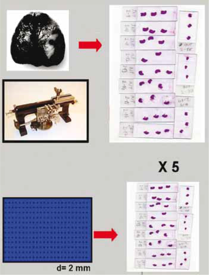
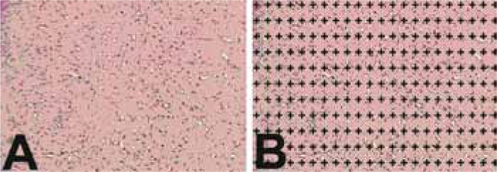
Brain volume was estimated using the Cavalieri method in two different ways. First, all brain slices and a ruler (for scale) were scanned with an HP ScanJet 3400C scanner, and their images were projected onto a PC monitor. Then the point counting volume estimation grid was superimposed on these images, and volume was calculated (Figure 1). Second all brain slices were photographed under ×10 magnification with a microscope camera (Olympus BH-2, Tokyo; Japan), and the volumes of the brains were estimated on these micrographs (Figure 2).
Fig. 1.

The application of the Cavalieri Principle on scanned slides.
Fig. 2.

The Application of the Cavalieri Principle on photomicrographs. (Magnification: ×10; d=2 mm).
Brain volume in each method was estimated using the following formula: Volume= t × a/p × P, where ‘t’ is the section thickness, ‘a/p’ is the representing area of each point on the point counting grid, and ‘P’ is the total number of points touching the sections’ surface areas.
Finally, all results obtained from the two different applications were subjected to statistical analysis using SPSS for Windows 13.0 (Paired Samples t Test). A value of p < 0.005 was considered to be statistically significant.
Results
The mean brain volumes estimated using the two different methods are summarized in Table 1. Analysis of the two volumetric calculations showed no statistically significant differences between the two methods (P>0.05; Paired Samples t Test).
Discussion
It is useful to examine structures that require assessment of changes in volume over time as an indicator of therapeutic effectiveness [6,7]. Change in brain volume is important to determine for the evaluation of neurodegenerative diseases, monitoring the organ by computer systems, and other applications [8,9]. Stereological procedures are efficient for quantitative analyses involving volumes of objects in any biological tissue or organ. Although conventional morphometric techniques have been used in most of the studies that have evaluated brain morphometry, a few stereological studies have been performed in order to estimate the volume of brain using the Cavalieri method [3]. These methods were used in our study. There was no statistically significant difference between the volume estimation based on PC images and photomicrographs.
Using consecutive sections scanned into a PC is beneficial because it prevents the time that is needed to photograph all of the slides. Also, it provides a chance to evaluate the whole section area without microscope magnification.
Footnotes
Conflict interest statement The authors declare that they have no conflict of interest to the publication of this article.
References
- 1.Cruz-Orive LM. Stereology of single objects. J Microsc. 1997;186:93–107. [Google Scholar]
- 2.Roberts N, Cruz-Orive LM, Reid NMK, Brodie DA, Bourne M, Edwards HT. Unbiased estimation of human body composition by the Cavalieri method using magnetic resonance imaging. J Microsc. 1992;171:239–53. doi: 10.1111/j.1365-2818.1993.tb03381.x. [DOI] [PubMed] [Google Scholar]
- 3.Bas O, Acer N, Mas N, Karabekir HS, Kusbeci OY, Sahin B. Stereologicalevaluation of the volume and volume fraction of intracranial structures in magnetic resonance images of patients with Alzheimer’s disease. Ann Anat. 2009;191:186–95. doi: 10.1016/j.aanat.2008.12.003. [DOI] [PubMed] [Google Scholar]
- 4.Odaci E, Sahin B, Sonmez OF, et al. Rapid estimation of the vertebral body volume: a combination of the Cavalieri principle and computed tomography images. Eur J Radiol. 2003;48:316–26. doi: 10.1016/s0720-048x(03)00077-9. [DOI] [PubMed] [Google Scholar]
- 5.Gundersen HJ, Jensen EB. The efficiency of systematic sampling in stereology and its prediction. Journal of Microscopy. 1987;147:229–63. doi: 10.1111/j.1365-2818.1987.tb02837.x. [DOI] [PubMed] [Google Scholar]
- 6.Schiano TD, Bodian C, Schwartz ME, Glajchen N, Min AD. Accuracy and significance of computed tomographic scan assessment of hepatic volume in patients undergoing brain transplantation. Transplantation. 2000;69:545–50. doi: 10.1097/00007890-200002270-00014. [DOI] [PubMed] [Google Scholar]
- 7.Kubota K, Makuuchi M, Kusaka K, et al. Measurement of brain volume and hepatic functional reserve as a guide to decision-making in resectional surgery for hepatic tumors. Hepatology. 1997;26:1176–81. doi: 10.1053/jhep.1997.v26.pm0009362359. [DOI] [PubMed] [Google Scholar]
- 8.Fernández-Viadero C, González-Mandly A, Verduga R, Crespo D, Cruz-Orive LM. Stereology as a tool to estimate brain volume and cortical atrophy in elders with dementia. Rev Esp Geriatr Gerontol. 2008;43:32–43. doi: 10.1016/s0211-139x(08)71147-8. [DOI] [PubMed] [Google Scholar]
- 9.Ekinci N, Acer N, Akkaya A, Sankur S, Kabadayi T, Sahin B. Volumetric evaluation of the relations among the cerebrum, cerebellum and brain stem in young subjects: a combination of stereology and magnetic resonance imaging. Surg Radiol Anat. 2008;30:489–94. doi: 10.1007/s00276-008-0356-z. [DOI] [PubMed] [Google Scholar]


