Abstract
Obturator hernia is a rare hernia in the world, diagnosed late since it has no specific symptoms and findings and generally occur in thin and old women with comorbidity.For this reason obturator hernia has high morbidity and mortality rates. In this study, we present an obturator hernia case that Howship-Romberg sign is positive and has typical appearance in computerized tomography. Laparotomy was performed on 89 years old female patient with body mass index 18.08 kg/m2 by low middle line incision. Following the segmentectomy to the strangulated small bowel loop, obturator canal is repaired by retroperitoneal application. No complication occurred in the postoperative period. Obturator hernia should be taken into consideration in old and thin female patients with intestinal obstruction. Computerized tomography should be performed for early diagnosis of the obturator hernia.
Keywords: Computerized tomography, Howship-Romberg sign, obturator hernia, small bowel obstruction
Özet
Obturator herni tüm dünyada nadir görülen, spesifik semptom ve bulgularının olmaması nedeni ile tanısı geciken, daha çok komorbiditesi olan yaşlı ve zayıf kadınlarda ortaya çıkan ve bu nedenlerle morbiditesi ve mortalitesi yüksek olan bir hernidir. Bu çalışmada Howship-Romberg işareti pozitif olan ve bilgisayarlı tomografide tipik görüntünün olduğu obturator herni vakasını sunuyoruz. 89 yaşında, body mass indeksi 18.08 kg/m2 olan kadın hastaya düşük orta hat insizyon ile laparotomi yapıldı. Strangüle olan ince barsak ansına segmentektomi yapıldıktan sonra obturator kanal retroperitoneal mesh tatbiki ile onarıldı. Postoperatif dönemde komplikasyon gelişmedi. İntestinal obstrüksiyonla gelen yaşlı ve zayıf kadın hastalarda obturator herni akılda tutulmalıdır. Erken tanı için bilgisayarlı tomografi çekilmelidir.
Introduction
Obturator hernia is a rare hernia with high mortality and morbidity. Since there are not any specific symptoms and findings of obturator hernia which occur commonly in old and thin female patients with comorbidity, this causes delays in diagnosis. As a result of this, due to strangulation of small bowel, perforation and high mortality rates occur [1]. Recently, computerized tomography (CT) became an important common diagnostic tool for the rapid diagnosis and early surgical treatment of these diseases [1, 2]. In the present study, we want to present a strangulated obturator hernia case that we suspected with the existence of Howship-Romberg sign and clarified the diagnosis by abdominal and pelvic CT.
Case Report
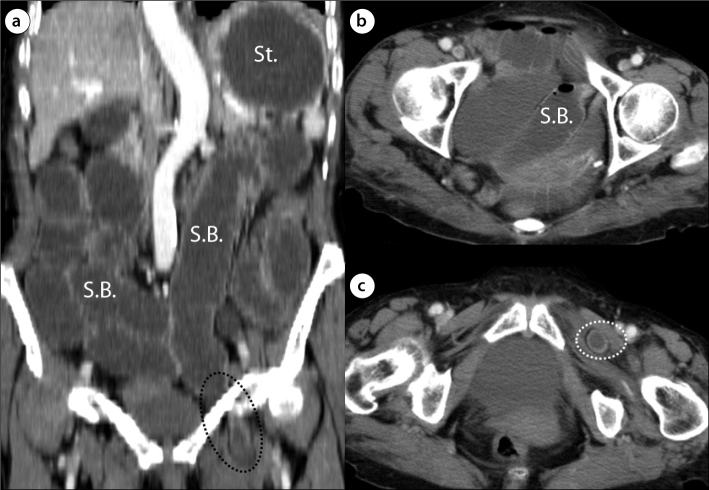
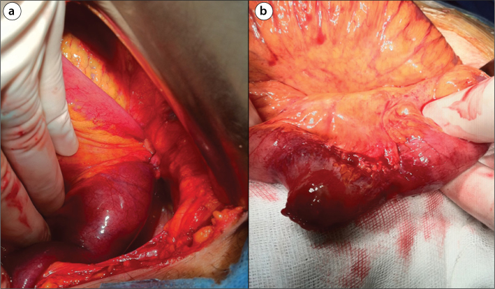
Eighty-nine years old female patient is hospitalized in our clinic due to nausea, vomiting and hip pain continuing for ten days. The patient described that her complaints were increased for the last few days because of hypertension and chronic obstructive pulmonary disease. The patient having no previous abdominal surgery had given birth to 5 kids. Body weight was 44 kg and body mass index (BMI) was 18.08 kg/m2. In her physical examination, the patient was dehydrated and abdomen was distended however there were no abdominal sensitivity and muscular defence. There was no significance in inguinal and rectal examination. Air-fluid levels were observed in the supine abdominal radiography. Laboratory tests were normal. The patient suffered pain in her left hip and thigh, the pain was detected to increase with the extension and abduction of the thigh. This finding was thought to be Howship-Romberg sign and CT was performed. Definitive diagnosis of obturator hernia was confirmed by the observation of dilated small bowel loops together with the increased content and herniated small bowel loop inside the obturator foramen (Figure 1) in CT. Emergency laparotomy by low middle line incision was made. 10 cm of small bowel loop at the 150 cm proximal of the ileocecal valve was observed as herniated to left obturator canal and strangulated (Figure 2). Proximal small bowel was dilated. Strangulated bowel loop has been resected. Obturator canal was repaired by the application of retroperitoneal mesh. There was not any complication occurred in the post-operative period. The patient had obvious regression of left hip and thigh pain and discharged at the 7th day with cure.
Figure 1. a–c.
Coronal (a) and axial (b and c) computed tomography scans reveal the left obturator hernia sac containing a distal ileal small bowel loop. In proximal of the left obturator hernia sac shows multiple dilated fluid-filled small bowel loops (S.B.) and dilated stomach (St.).
Figure 2. a, b.
In laparotomy, herniation of a small bowel loop inside obturator channel (a) and strangulation of this bowel loop (b) are seen.
Discussion
Obturator hernia is seen with a rate of 0.4% among bowel obstructions and 0.07–1.0% among hernias [3]. Obturator hernia was first described in 1724 by Ronsil. It occurs with the passing of the hernia sac down from the obturator canal where the obturator nerve and muscle pass [2]. It is seen more commonly in old and thin multiparous women with large and big pelvic bones and with a relatively horizontal obturator channel. Chronic obstructive pulmonary diseases, malnutrition, chronic constipation and diseases that cause increased intra-abdominal pressure are among the other pre-disposing factors. Symptoms of the obturator hernia are non-specific. The most common finding is an acute obstruction of the bowel [1]. However, John Howship in 1840 and Heinrich Romberg in 1845 have described that the occurrence of the acute pain by the incarceration of obturator nerve at the medial side of the thigh and exacerbation of this pain with the extension, abduction and medial rotation of the thigh are the cardinal signs of obturator hernia [4]. In the literature, Howship-Romberg sign was reported to exist in 13–65% of the obturator hernia patients [1, 5]. In our case, in the physical examination of the patient with symptoms of nausea, vomiting and left thigh pain, Howship-Romberg sign was also noticed as positive, CT performed and the presence of obturator hernia was seen together with small bowel obstruction.
In the periods that CT was not used, the diagnosis of obturator hernia was generally made intra-operatively. With the developments of imaging techniques, although imaging methods such as herniography, ultrasonography, barium enema and magnetic resonance imaging for the diagnosis of this disease, with a diagnostic value of 78–100% CT has become a golden standard [4, 5]. Although it is reported that the use of CT preoperatively in patients with obturator hernia decreases the intestinal resection rates and mortality [5], Nasir et al [1] have reported that preoperative CT is useful for the definitive and early diagnosis of the disease and predicting some of the post-operative complications but do not increase the post-operative survival rate. Chang et al. [6] have reported that one of the important reasons affecting the bowel resection in these patients is the duration of the symptoms. In the literature it is reported that; the strangulation rate can increase from 25% to 100% in the case of delayed surgical treatment [7]. In our case, although the diagnosis of obturator hernia was made preoperatively by CT, as a result of the continuing complaints of the patient for 10 days when admitted, resection of the small bowel has become unavoidable. Because of the delay in the diagnosis, intestinal ischemia and high incidence of perforation, post-operative mortality rate of these patients was reported as about 70% [8].
Although, a non-operative approach for the treatment of obturator hernia defined in literature [7], the only treatment of this disease is surgical. Retro pubic and trans-peritoneal inguinal approach, laparotomy with low midline incision and laparoscopic surgery preferred more common recently, are among the favourite surgical methods [2]. For the repair of the obturator canal, primary suture technique, closing with the neighbour tissue and closure with mesh methods are preferred [9]. Some authors have preferred to close by the primary suture technique in their cases that they performed a small bowel resection [2]. We repaired our case successfully by the application of retroperitoneal mesh following a small bowel resection with low midline incision.
In conclusion, the reason of the intestinal obstruction in thin and old female patients may be obturator hernia. The presence of Howship-Romberg sign should be controlled in suspicious cases. CT that was performed in the preoperative period is essential for early diagnosis and emergency surgical treatment.
Footnotes
Informed Consent: Written informed consent was obtained from patient who participated in this study.
Peer-review: Externally peer-reviewed.
Author Contributions: Concept - A.K., B.O.; Design - A.K., A.B.; Supervision - B.O., S.S.A.; Funding - A.K., I.Y.; Materials - A.B., İ.Y.; Data Collection and/or Processing - A.K.; S.S.A.; Analysis and/or Interpretation - A.B., I.Y.; Literature Review - B.O., S.S.A.; Writing - A.K., I.Y.; Critical Review - A.B., S.S.A.; Other - B.O., I.Y.
Conflict of Interest: No conflict of interest was declared by the authors.
Financial Disclosure: The authors declared that this study has received no financial support.
References
- 1.Nasir BS, Zendejas B, Ali SM, et al. Obturator hernia: the Mayo Clinic experience. Hernia. 2012;16:315–9. doi: 10.1007/s10029-011-0895-9. http://dx.doi.org/10.1007/s10029-011-0895-9. [DOI] [PubMed] [Google Scholar]
- 2.Cai X, Song X, Cai X. Strangulated intestinal obstruction secondary to a typical obturator hernia: a case report with literature review. Int J Med Sci. 2012;9:213–5. doi: 10.7150/ijms.3894. http://dx.doi.org/10.1007/s10029-011-0895-9. [DOI] [PMC free article] [PubMed] [Google Scholar]
- 3.Yokoyama Y, Yamaguchi A, Isogai M, et al. Thirty-six cases of obturator hernia: does computed tomography contribute to postoperative outcome? World J Surg. 1999;23:214–6. doi: 10.1007/pl00013176. http://dx.doi.org/10.1007/PL00013176. [DOI] [PubMed] [Google Scholar]
- 4.Karasaki T, Nakagawa T, Tanaka N. Obturator hernia: the relationship between anatomical classification and the Howship-Romberg sign. Hernia. 2014;18:413–6. doi: 10.1007/s10029-013-1068-9. http://dx.doi.org/10.1007/s10029-013-1068-9. [DOI] [PubMed] [Google Scholar]
- 5.Kammori M, Mafune K, Hirashima T, et al. Forty-three cases of obturator hernia. Am J Surg. 2004;187:549–52. doi: 10.1016/j.amjsurg.2003.12.041. http://dx.doi.org/10.1016/j.amjsurg.2003.12.041. [DOI] [PubMed] [Google Scholar]
- 6.Chang SS, Shan YS, Lin YJ, et al. A review of obturator hernia and a proposed algorithm for its diagnosis and treatment. World J Surg. 2005;29:450–4. doi: 10.1007/s00268-004-7664-1. http://dx.doi.org/10.1007/s00268-004-7664-1. [DOI] [PubMed] [Google Scholar]
- 7.Leow JJ, How KY, Goh MH, et al. Non-operative management of obturator hernia in an elderly female. Hernia. 2014;18:431–3. doi: 10.1007/s10029-012-1036-9. http://dx.doi.org/10.1007/s10029-012-1036-9. [DOI] [PubMed] [Google Scholar]
- 8.Rodríguez-Hermosa JI, Codina-Cazador A, Maroto-Genover A, et al. Obturator hernia: clinical analysis of 16 cases and algorithm for its diagnosis and treatment. Hernia. 2008;12:289–97. doi: 10.1007/s10029-007-0328-y. http://dx.doi.org/10.1007/s10029-007-0328-y. [DOI] [PubMed] [Google Scholar]
- 9.Pandey R, Maqbool A, Jayachandran N. Obturator hernia: a diagnostic challenge. Hernia. 2009;13:97–9. doi: 10.1007/s10029-008-0406-9. http://dx.doi.org/10.1007/s10029-008-0406-9. [DOI] [PubMed] [Google Scholar]


