Abstract
The present study investigated the seroprevalence of Toxoplasma gondii (T. gondii) antibodies by ELISA in horses reared in Korea. Serum samples were collected from 2009 through 2013 from 816 horses reared in Korea. Analysis was performed using a commercial toxoplasmosis ELISA kit to detect anti-T. gondii antibodies. Overall, 24 out of 816 horses (2.9%) were seropositive for T. gondii. The result was analyzed by age, gender, breed and region. Significant differences were observed according to breed and region (P<0.05). This is the first nationwide serological investigation of T. gondii in horses reared in Korea. The study results reveal that T. gondii occurs nationwide in Korean horses.
Keywords: ELISA, equine, Korea, prevalence, toxoplasmosis
Toxoplasma gondii (T. gondii) is an obligate intracellular parasite involved in the pathogenesis of toxoplasmosis. This pathogen can infect most warm-blooded animals, including humans, whereas felids are the only spreader as well as the definitive host. The two main routes of transmission to humans are ingestion of food or water that is contaminated by T. gondii oocysts and ingestion of undercooked meat with tissue cysts of T. gondii[2]. In humans, toxoplasmosis in immunocompetent adults is usually asymptomatic. However, T. gondii is known as one of the opportunistic pathogens, and T. gondii infection can be lethal in immunodeficient people, such as AIDS patients, and may cause abortion, stillbirth and congenital abnormalities if infection occurs during pregnancy in women [11].
T. gondii infection in the horse generally causes subclinical infection; however, fever, ataxia, retinal degeneration and encephalomyelitis may occasionally occur, as well as abortion or stillbirth in pregnant equids [9]. Although there are several reports on toxoplasmosis in other animals [3, 5, 8] in the Republic of Korea (ROK), information on equine toxoplasmosis is limited in ROK as well as worldwide. Thus, the purpose of the present study was to investigate the seroprevalence of T. gondii on a nationwide scale in horses raised in ROK.
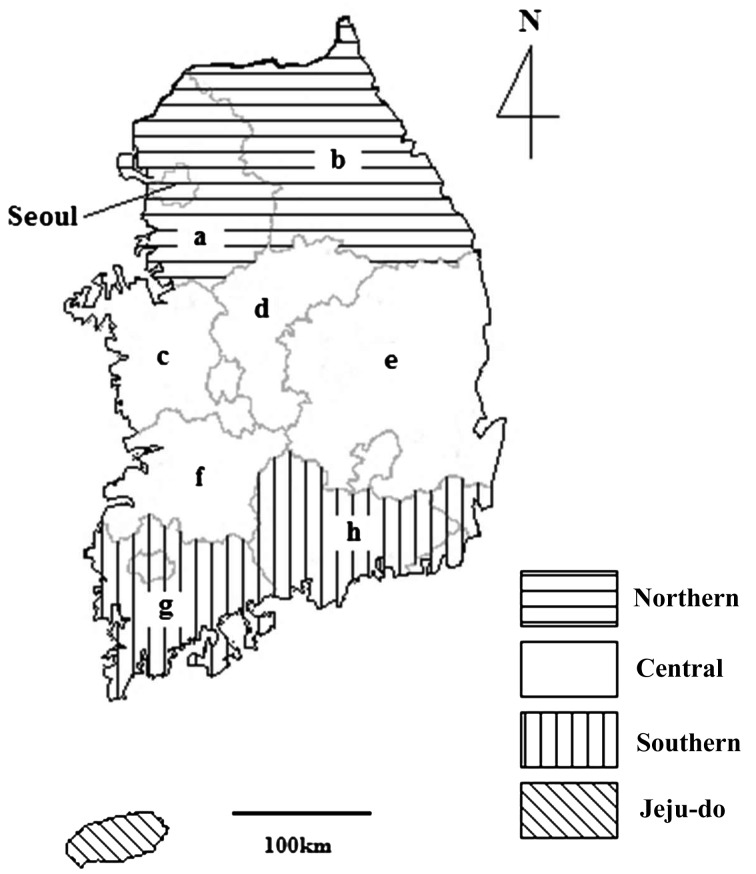
Blood samples were collected from 2009 through 2013 from the jugular vein of 816 horses reared in Korea. The use of experimental animals was approved by the Kyungpook National University Institutional Animal Care and Use Committee (Approval #KNU-200900501). The breeds of horses included the Thoroughbred (69.5%, 567/816), Korean native pony (13.4%, 109/816), warmblood (7.5%, 61/816) and mixed breed (9.7%, 79/816). The study area included all parts of the country and horse farms in each province and was divided into four regions (northern, central, southern and Jeju-do) (Fig. 1). Data regarding age, gender, breed and region of origin of each animal were collected; data were classified as “unknown” in cases of insufficient data. Serum was separated by centrifugation and stored at −20°C until use.
Fig. 1.
Regional map of the Republic of Korea showing the four study regions in which horse serum samples were collected for detection of the presence of anti-Toxoplasma gondii antibodies: northern (Seoul, Gyeonggi-do [a] and Gangwon-do [b]), central (Chungcheongnam-do [c], Chungcheongbuk-do [d], Gyeongsangbuk-do [e] and Jeollabuk-do [f]), southern (Jeollanam-do [g] and Gyeongsangnam-do [h]) and Jeju-do.
The anti-T. gondii antibodies in each serum sample were detected using a commercial toxoplasmosis multi-species ELISA kit (IDvet, Montpellier, France). All experimental steps were conducted according to the manufacturer’s instructions. Specificity and sensitivity of the multi-species kit are 99.9% and 84%, respectively, in the pig [1]. To determine seroprevalence, optical density (OD) values were measured at 450 nm, and the positive percentage of the sample (S/P) was calculated according to the manufacturer’s instructions as follows: S/P=100 × [(ODsample − ODnegative control) / (ODpositive control − ODnegative control)]. S/P was interpreted as follows: negative (<40%), doubtful (40–50%) and positive (>50%). In this study, doubtful results were considered negative.
For statistical analysis, chi-square and Fisher’s exact tests were used. The data of the “unknown” group were disregarded in the chi-square and Fisher’s exact tests. Statistical analyses were carried out using SPSS 21.0 (IBM Corporation, Armonk, NY, U.S.A.), and P-values of <0.05 were regarded as significant. A 95% confidence interval for all estimates was calculated.
Among the 816 tested horses, 24 (2.9%) were found to be seropositive for anti-T. gondii antibodies by ELISA (Table 1). With regards to age, the seroprevalence was 1.9% (5/269), 3.6% (9/251), 1.0% (2/202) and 8.5% (8/94) for horses aged <5 years, 5–10 years, >10 years and of unknown age, respectively. Regarding gender, 1.3% (2/160), 2.8% (8/282), 2.1% (6/280) and 8.5% (8/94) of the samples were seropositive in the male, female, castrated and unknown groups, respectively. There was no significant difference among the groups in age and gender.
Table 1. Seroprevalence of Toxoplasma gondii in 816 horses according to age, gender, breed and region.
| Group | No. positive / total (%) | 95% CIa) | |
|---|---|---|---|
| Age | <5 y | 5/269 (1.9) | 0.2–3.5 |
| 5–10 y | 9/251 (3.6) | 1.3–5.9 | |
| >10 y | 2/202 (1.0) | 0–2.4 | |
| Unknown | 8/94 (8.5) | 2.9–14.1 | |
| Gender | Male | 2/160 (1.3) | 0–3.0 |
| Female | 8/282 (2.8) | 0.9–4.8 | |
| Castrated | 6/280 (2.1) | 0.5–3.8 | |
| Unknown | 8/94 (8.5) | 2.9–14.2 | |
| Breedb) | Thoroughbred | 11/567 (1.9) | 0.8–3.1 |
| Korean native pony | 8/109 (7.3) | 2.4–12.2 | |
| Warmblood | 3/61 (4.9) | 0–10.3 | |
| Mixed | 2/79 (2.5) | 0–6.0 | |
| Regionb) | Northern | 8/301 (2.7) | 0.8–4.5 |
| Central | 4/178 (2.2) | 0.1–4.4 | |
| Southern | 4/243 (1.6) | 0.1–3.3 | |
| Jeju-do | 8/94 (8.5) | 2.9–14.2 | |
| Total | 24/816 (2.9) | 1.8–4.1 | |
a) CI, confidence interval, b) P<0.05.
Regarding breed, 1.9% (11/567), 7.3% (8/109), 4.9% (3/61) and 2.5% (2/79) of the samples were seropositive in the Thoroughbred, Korean native pony, warmblood and mixed breed, respectively. According to region, 2.7% (8/301), 2.2% (4/178), 1.6% (4/243) and 8.5% (8/94) of the samples were seropositive in the northern, central, southern and Jeju-do regions, respectively. There were significant differences according to breed and region (P<0.05). In this study, breed of horse was associated with region of collection; most of the samples (94/109) from Korean native ponies were collected in Jeju-do, and the highest seroprevalence (8.5%, 8/94) occurred in Jeju-do in regional analysis. In addition, all of the Korean native pony samples that were seropositive were collected in Jeju-do (data not shown). Although additional investigation is needed that takes into account environmental conditions, genetic factors and food preferences of the residents, an association may exist between seroprevalence and region, particularly with respect to Jeju-do, for T. gondii infection.
The higher prevalence in Jeju-do may be explained by two factors. First, horses in Jeju-do are allowed to graze freely, whereas mainland horses are reared semi-intensively, i.e., horses are reared in a confined system with limited grazing time. Thus, horses in Jeju-do are likely to more easily acquire parasites from the natural environment. Second, the infection status of the definitive host, i.e., cats, in Jeju-do appears higher than that on the mainland. Although the studies were not conducted in the same year, the seroprevalence of T. gondii among cats in Jeju-do was 37.0% despite a very small population [6]; this is in contrast to 15.8% (69/456) in Seoul [8], 16.1% in Gyeonggi-do [4] and 13.1% in Jinju [10], which are located on the mainland. Thus, the higher prevalence in horses could be related to the higher prevalence in stray cats. However, a further epidemiological comparative study between horses and cats and investigation of distribution according to that of T. gondii in the natural environment are needed.
This is the first nationwide large-scale serological study on the prevalence of T. gondii in horses raised in ROK. The overall positive rate of T. gondii antibodies in horses is low, and there is higher prevalence of T. gondii among horses in Jeju-do than mainland regions. The high prevalence in Jeju-do is significant, because most of the production and consumption of horse meat in ROK occur in Jeju-do [7]. Our findings provide an update on the status of T. gondii, particularly in the horse and may serve as the basis of future investigations on the significance of this parasite in ROK. In addition, continuous monitoring and implementation of integrated control strategies of T. gondii infections are needed to prevent human infections and public health considerations.
Acknowledgments
This research was supported by grants from the Korea Centers for Disease Control and Prevention (4847-302-210-13, 2013) and the Basic Science Research Program through the National Research Foundation of Korea (NRF) funded by the Ministry of Education (NRF-2013R1A1A2013102).
REFERENCES
- 1.Bokken G. C., Bergwerff A. A., van Knapen F.2012. A novel bead-based assay to detect specific antibody responses against Toxoplasma gondii and Trichinella spiralis simultaneously in sera of experimentally infected swine. BMC Vet. Res. 8: 36. doi: 10.1186/1746-6148-8-36 [DOI] [PMC free article] [PubMed] [Google Scholar]
- 2.Dubey J. P.2009. History of the discovery of the life cycle of Toxoplasma gondii. Int. J. Parasitol. 39: 877–882. doi: 10.1016/j.ijpara.2009.01.005 [DOI] [PubMed] [Google Scholar]
- 3.Jung B. Y., Gebeyehu E. B., Lee S. H., Seo M. G., Byun J. W., Oem J. K., Kim H. Y., Kwak D.2014. Detection and determination of Toxoplasma gondii seroprevalence in native Korean goats (Capra hircus coreanae). Vector Borne Zoonotic Dis. 14: 374–377. doi: 10.1089/vbz.2013.1452 [DOI] [PubMed] [Google Scholar]
- 4.Kim H. Y., Kim Y. A., Kang S., Lee H. S., Rhie H. G., Ahn H. J., Nam H. W., Lee S. E.2008. Prevalence of Toxoplasma gondii in stray cats of Gyeonggi-do, Korea. Korean J. Parasitol. 46: 199–201. doi: 10.3347/kjp.2008.46.3.199 [DOI] [PMC free article] [PubMed] [Google Scholar]
- 5.Kim J. H., Kang K. I., Kang W. C., Sohn H. J., Jean Y. H., Park B. K., Kim Y., Kim D. Y.2009. Porcine abortion outbreak associated with Toxoplasma gondii in Jeju Island, Korea. J. Vet. Sci. 10: 147–151. doi: 10.4142/jvs.2009.10.2.147 [DOI] [PMC free article] [PubMed] [Google Scholar]
- 6.Kim S., Kim Y.1989. On the distribution of Toxoplasma antibodies in Cheju-Do 1. Distribution of Toxoplasma antibodies in swine, cats and butchers. Korean J. Vet. Res. 29: 333–342 (in Korean with English abstract). [Google Scholar]
- 7.KOSIS. 2013. http://kosis.kr/statisticsList/statisticsList_01List.jsp?vwcd=MT_ZTITLE&parentId=F.
- 8.Lee S. E., Kim N. H., Chae H. S., Cho S. H., Nam H. W., Lee W. J., Kim S. H., Lee J. H.2011. Prevalence of Toxoplasma gondii infection in feral cats in Seoul, Korea. J. Parasitol. 97: 153–155. doi: 10.1645/GE-2455.1 [DOI] [PubMed] [Google Scholar]
- 9.Miao Q., Wang X., She L. N., Fan Y. T., Yuan F. Z., Yang J. F., Zhu X. Q., Zou F. C.2013. Seroprevalence of Toxoplasma gondii in horses and donkeys in Yunnan Province, Southwestern China. Parasit. Vectors 6: 168. doi: 10.1186/1756-3305-6-168 [DOI] [PMC free article] [PubMed] [Google Scholar]
- 10.Sohn W. M., Nam H. W.1999. Western blot analysis of stray cat sera against Toxoplasma gondii and the diagnostic availability of monoclonal antibodies in sandwich-ELISA. Korean J. Parasitol. 37: 249–256. doi: 10.3347/kjp.1999.37.4.249 [DOI] [PMC free article] [PubMed] [Google Scholar]
- 11.Tenter A. M., Heckeroth A. R., Weiss L. M.2000. Toxoplasma gondii: from animals to humans. Int. J. Parasitol. 30: 1217–1258. doi: 10.1016/S0020-7519(00)00124-7 [DOI] [PMC free article] [PubMed] [Google Scholar]


