ABSTRACT
Coxsackievirus B3 (CVB3) is trophic for cardiac tissue and is a major causative agent for viral myocarditis, where local viral replication in the heart may lead to heart failure or even death. Recent studies show that inserting microRNA target sequences into the genomes of certain viruses can eradicate these viruses within local host tissues that specifically express the cognate microRNA. Here, we demonstrated both in vitro and in vivo that incorporating target sequences for miRNA-133 and -206 into the 5′ untranslated region of the CVB3 genome ameliorated CVB3 virulence in skeletal muscle and myocardial cells that specifically expressed the cognate cellular microRNAs. Compared to wild-type CVB3, viral replication of the engineered CVB3 was attenuated in human TE671 (rhabdomyosarcoma) and L6 (skeletal muscle) cell lines in vitro that expressed high levels of miRNA-206. In the in vivo murine CVB3-infection model, viral replication of the engineered CVB3 was attenuated specifically in the heart that expressed high levels of both miRNAs, but not in certain tissues, which allowed the host to retain the ability to induce a strong and protective humoral immune response against CVB3. The results of this study suggest that a microRNA-targeting strategy to control CVB3 tissue tropism and pathogenesis may be useful for viral attenuation and vaccine development.
IMPORTANCE Coxsackievirus B3 (CVB3) is a major causative agent for viral myocarditis, and viral replication in the heart may lead to heart failure or even death. Limiting CVB3 replication within the heart may be a promising strategy to decrease CVB3 pathogenicity. miRNAs are ∼21-nucleotide-long, tissue-specific endogenous small RNA molecules that posttranscriptionally regulate gene expression by imperfectly binding to the 3′ untranslated region (UTR), the 5′ UTR, or the coding region within a gene. In our study, muscle-specific miRNA targets (miRT) were incorporated into the CVB3 genome. Replication of the engineered viruses was restricted in the important heart tissue of infected mice, which reduced cardiac pathology and increased mouse survival. Meanwhile, replication ability was retained in other tissues, thus inducing a strong humoral immune response and providing long-term protection against CVB3 rechallenge. This study suggests that a microRNA-targeting strategy can potentially control CVB3 tissue tropism and pathogenesis and may be useful for viral attenuation and vaccine development.
INTRODUCTION
Coxsackievirus B3 (CVB3) is a clinically relevant infectious agent that causes viral myocarditis, pancreatitis, and aseptic meningitis. CVB3 is cardiotropic and efficiently replicates in the heart. We and others have shown in various animal models that CVB3 replication induces a direct cytopathic effect on infected cells, leading to tissue damage (1, 2). At present, there is no effective therapy against CVB3, and strategies to develop such a therapy are greatly desired (3, 4). Limiting CVB3 replication within the heart may be a promising strategy to decrease CVB3 pathogenicity, and one relatively recent method to eliminate tissue-tropic viral infection takes advantage of factors specifically expressed in the tropic tissue, including cellular microRNAs (miRNAs) (5–7). In addition, specific antiviral vaccines are widely used and effectively function to immunize human hosts against pathogens such as HBV, H1N1, and poliomyelitis (8–11), and ways to generate effective memory immune responses against CVB3 will be important for protection against future infections.
miRNAs are a class of ∼21-nucleotide (nt)-long endogenous small RNA molecules that posttranscriptionally regulate gene expression by imperfectly binding to the 3′ untranslated region (UTR), the 5′ UTR, or the coding region within a gene. Other well-described characteristics of miRNAs are their tissue- and process-specific expression pattern and relative conservation across different species. For instance, miRNA-133 (miR-133), miR-1, and miR-206 are expressed mainly in myogenic tissues such as skeletal- and cardiac-muscle tissues in human, rats, and mice, and similarly participate in cellular differentiation and proliferation; they are also involved in metabolic control and development in cardiac myocytes (12, 13). Thus, these miRNAs are considered to be tissue-specific factors with restricted expression in the heart.
Previous studies showed that endogenous miRNAs could successfully recognize and regulate the expression of transgenes incorporated into a genome via treatment with gene-therapy vectors (5). We therefore hypothesized that muscle-specific miRNAs could regulate the tissue tropism of pathogenic CVB3 engineered to contain the specific sequences targeted by these miRNAs. To test our hypothesis, the cognate target sequences for muscle-specific miR-206 and miR-133 were incorporated into the 5′ UTR of the CVB3 genome, and the replicative ability of the engineered CVB3 was evaluated in miR-206-expressing host cell lines in vitro, as well as in mouse hearts in vivo. The engineered virus was unable to replicate and had an attenuated phenotype in cells expressing the corresponding miRNAs but not HeLa cells that had little miR-133 or miR-206 expression. In a mouse model of CVB3 infection, the virulence of the engineered CVB3 was attenuated in cardiac tissue, with decreased CVB3-mediated pathology and increased survival compared to infection with wild-type (WT) CVB3 (CVB3-WT). Importantly, the ability to provide long-lasting protective immunity against challenge with a pathogenic CVB3 strain was maintained after infection with the engineered CVB3, with the host exhibiting a high survival rate.
MATERIALS AND METHODS
Molecular biology and viruses.
The full-length CVB3 infectious clone pCVB3M/CMV (a generous gift from Decheng Yang at the University of British Columbia, British Columbia, Canada) was used as the starting material for genetic manipulation (14). The CVB3 mutants were constructed by inserting an miRNA target (miRT) sequence, followed by a 10-bp KOZAK-like sequence at bp positions 731 to 740 (ATACAGCAAA) of the CVB3 genome, which maintained the integrity of the initiation codon, into the location between the 5′-UTR and the coding sequences; either one copy of the miR-206 target sequence (miR-206T) or two copies of miR-206T plus one copy of miR-133T in tandem (miR-133T–miR-206T–miR-133T) were inserted into the CVB3 genome to create CVB-206T and CVB-3*T, respectively. We also constructed control viruses containing miRNA target sequences corresponding to a Caenorhabditis elegans-derived miR-39 that had no identified target in mammalian cells, called CVB-CON (Table 1). The progeny were passaged twice at a high multiplicity of infection (multiplicity of infection [MOI] = 10) at 37°C in HeLa cells, and viruses from the second passage were used for experiments. To confirm the stability of the mutants, supernatants containing mutant viral particles were harvested at different time points for RNA extraction, reverse transcription-PCR (RT-PCR) was performed as previously described (15), and the sequence was determined.
TABLE 1.
Sequence of inserted miRNA targets for CVB-206T, CVB-3*T, and CVB-CON
| miRT | Inserted sequence (5′–3′)a |
|---|---|
| miR-206T | CCACACACTTCCTTACATTCCA |
| miR-3*T | ACAGCTGGTTGAAGGGGACCAACGATCCACACACTTCCTTACATTCCAACCGGTACAGCTGGTTGAAGGGGACCAA |
| miR-CON | CAAGCTGATTTACACCCGGTGA |
Spacer elements are indicated in italicized text.
For transfecting cells with a small interfering RNA (siRNA) to silence the endonuclease Argonaute2 (Ago2) and the RNase III enzyme Drosha, 20 pmol of Ago2-siRNA or Drosha-siRNA was transfected into TE671 cells using Lipofectamine 2000 (Invitrogen, Carlsbad, CA) according to the manufacturer's instructions. The medium was replaced after 12 h, and viral infections were carried out 24 h later. The siRNA sense sequences against Ago2 and Drosha were 5′-GGAAAAUGAUGCUGAAUAUUU-3′ and 5′-GAGUAGGCUUCGUGACUUAUU-3′, respectively; the scramble siRNA (5′-UUCUCCGAACGUGUCACGU-3′), designated a non-siRNA, was used as the negative control. Ago2, Drosha, and miRNA expression were analyzed using the real-time RT-PCR method as previously described (15). The following primers were used: Drosha forward (5′-CTCCCCGCCTACGAACTTTTTA-3′) and reverse (5′-TTCTCCGGCATCATCTCTTCAAT-3′) and Ago2 forward (5′-GACAGTGCTGAAGGAAGCCATA-3′) and reverse (5′-GCCATCTGTTGGTCTGAGTGTAG-3′). β-Actin was used as the internal control. The specific forward primers for detecting miR-133 and miR-206 were 5′-ACACTCCAGCTGGGTTTGGTCCCCTTCAACCA-3′ and 5′-ACACTCCAGCTGGGTGGAATGTAAGGAAGTGT-3′, respectively, and the universal reverse primer was 5′-TGGTGTCGTGGAGTCG-3′. For absolute quantification, double-strand miRNA sequences were chemically synthesized and used for generating standard curves of miRNAs (Invitrogen). The miRNA concentration was represented as copies/104 cells; for miRNA relative quantification, U6 was used as the internal control.
miR-133 and miR-206 mimics were purchased from Qiagen (Valencia, CA). We transfected the miRNA mimic with Lipofectamine 2000 at 200 nM into HeLa cells. Four hours after transfecting in the mimics, cells were infected with virus at an MOI of 1. At 24 h postinfection, the cells were harvested for use in the 3-(4,5-dimethylthiazol-2-yl)-5-(3-carboxymethoxyphenyl)-2-(4-sulfophenyl)-2H-tetrazolium salt (MTS) cell viability assay (described below), and the supernatant was harvested for titration.
One-step growth curve.
One-step growth analysis was performed on HeLa cell monolayers in six-well plates at 37°C. Cells were infected with CVB3-WT, CVB-206T, CVB-3*T, or CVB-CON at an MOI of 1 for 60 min at room temperature, washed three times with phosphate-buffered saline (PBS), and then fed with MEM plus 10% newborn calf serum at 37°C. Supernatants were harvested at 2, 4, 6, 8, 10, 12, 18, and 24 h postinfection, and the number of virus titers at each time point was determined by viral plaque assay (described below).
Viral plaque assay.
Virus titers were determined by a plaque assay. Briefly, HeLa cells (8 × 10⁵ cells/well) were seeded into six-well plates and incubated at 37°C for 24 h. When cell confluence reached ca. 90%, the cells were washed twice with PBS and then overlaid with 200 μl of 1:10 diluted virus-containing supernatant. Cell cultures were incubated at 37°C for 60 min, the supernatant was removed, and the cells were washed with PBS. Finally, the cells were overlaid with 2 ml of sterilized soft Bacto agar-MEM (2× MEM–1.5% Bacto agar [1:1]). The cells were incubated at 37°C for 72 h, fixed with Carnoy's fixative for 30 min, and then stained with 1% crystal violet. The plaques were counted, and the viral PFU/ml values were calculated.
Cell viability assay.
Cell viability was measured using an MTS assay kit (Promega, Madison, WI) according to the manufacturer's instructions. The cells were incubated with MTS solution for 2 h, and the absorbance was measured at 492 nm using a microplate reader (Thermo Scientific, Waltham, MA). The survival values for the absorbance measured from the control mock-infected cells were defined as 100%, and the values obtained from the other infected cells were converted into a ratio based on the value of the mock-infected sample.
Infection of susceptible mice.
The animal study was approved by the Review Board of the Capital Institute of Pediatrics (Beijing, China). Male BALB/c mice (6 weeks old) were purchased from the Institute of Laboratory Animal Sciences of China (Beijing, China). CVB3-WT and engineered viruses were inoculated into mice by the intraperitoneal (i.p.) route. Mice were monitored daily for the onset of paralysis and survival. For measuring the 50% lethal dose (LD50) of CVB3-WT and engineered viruses, mice (n = 20) were infected i.p. with four doses of each recombinant virus (104, 105, 106, and 107 PFU/mouse, five mice/group). Animals were monitored until no new deaths were observed. The LD50 was determined by the Reed and Muench method (16). To analyze viral tissue tropism and histopathology, we harvested the whole heart from mice in each group on the indicated days, and the hearts were dissected into two sections. One half of the heart was weighed and homogenized in 0.5 ml of MEM and centrifuged for 10 min at 800 × g/min; infectious virus titers in the supernatants were then analyzed by the viral plaque assay, and the virus titers in infected tissue were expressed as PFU/g. The other half of the heart was examined histopathologically for evidence of inflammation and necrosis, as previously described (14, 17). Neutralization assays were performed as previously described by Barnes et al. (7) on sera collected from mice 1 month after i.p. immunization with PBS (mock), 104 PFU UV-inactivated CVB3-WT, CVB-206T, or CVB-3*T virus. The assay was performed three times, and the reciprocal of the neutralizing dilution was determined as the neutralizing antibody titer. Another group of mice immunized with equal amounts of the indicated virus was challenged with 100× the LD50 of the CVB3-WT and monitored for survival.
Statistical analysis.
All statistical analyses were performed using the SPSS 11.5 computer software program (SPSS, Inc., Chicago, IL). Survival was analyzed using the log-rank (Mantel-Cox) method. The significance of variability among the experimental groups was determined by the Mann-Whitney U test. All differences were considered statistically significant at a P of <0.05.
RESULTS
Engineered CVB3 retains infectious and stable replication activity.
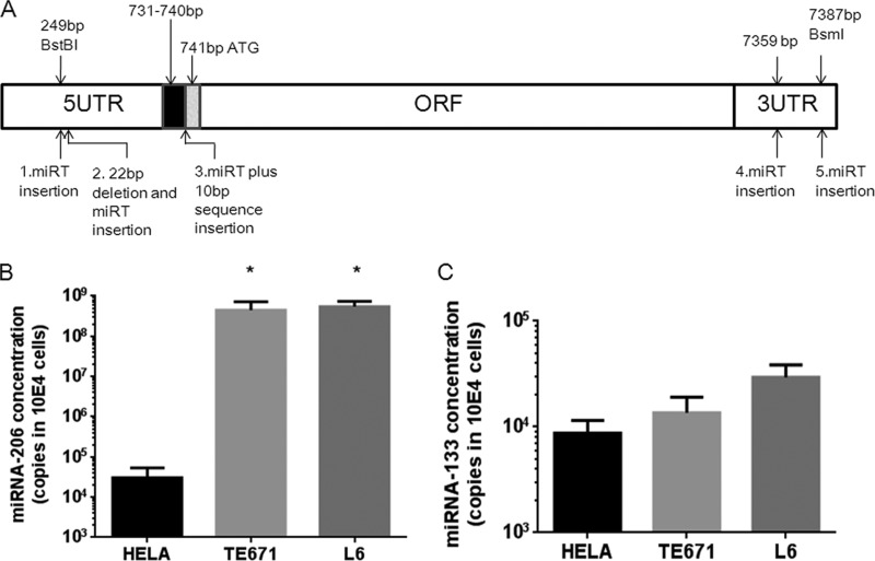
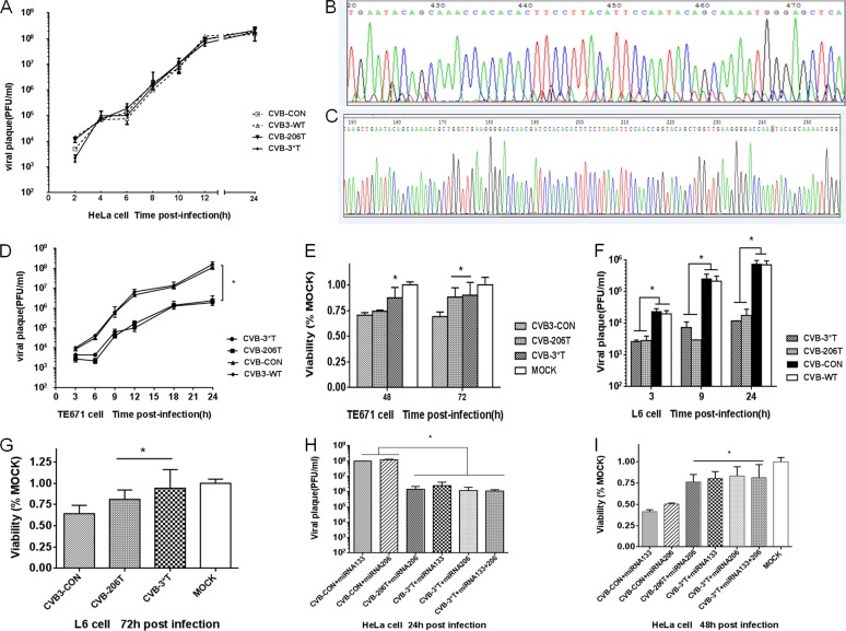
To begin testing our hypothesis that endogenous cellular miRNAs specifically expressed in cardiac tissues could suppress the pathogenicity of miRNA target-containing CVB3, we engineered a pathogenic CVB3-WT strain to contain one or several miRNA target sequences followed by a repeating KOZAK-like sequence into the 5′ UTR at a location immediately upstream of the coding sequences (Fig. 1A and Table 1). We first evaluated the ability of these miRT-containing viruses to replicate in vitro by infecting and evaluating the one-step growth curves in HeLa cells, which expressed little of either miRNA (Fig. 1B and C). Both the WT and the engineered CVB3 exhibited identical growth kinetics and had comparable final virus titers (Fig. 2A), indicating that the insertion of the miRT sequences did not impair viral replication. Sequence analysis also revealed that all recombinant miRT-containing viruses were stable in HeLa cells after four cell passages and still contained the desired miRNA target insertions (Fig. 2B and C).
FIG 1.
Construction of recombinant CVB3 and detection of endogenous specific miRNA. (A) Cognate target sequences for miR-133 and/or miR-206 were inserted into different locations of the CVB3 genome; location 3 in the figure was chosen. (B and C) Endogenous muscle-specific miR-206 and miR-133 expression in TE671, L6, and HeLa cells. The average concentrations of miR-206 in TE671 and L6 cells were 4.49 × 10⁸ and 5.46 × 10⁸ copies in 104 cells, values significantly higher than those observed in HeLa cells, i.e., 3.06 × 10⁴ copies in 104 cells; the differences in the miR-133 average concentrations in TE671, L6, and HeLa cells were not significant, i.e., 1.36 × 10⁴, 2.93 × 10⁴, and 8.7 × 10³ in 104 cells, respectively. *, P < 0.01 for miR-206 concentrations in TE671 and L6 cells compared to HeLa cells.
FIG 2.
Engineered CVB3-miRTs exhibit attenuated replication kinetics due to endogenous miR-206– and miR-133–mediated repression. (A) Replication kinetics of CVB3-WT, CVB-206T, CVB-3*T, and CVB-CON in HeLa cells, which express low levels of either miR-206 or miR-133. Virus titer values represent the means ± the standard deviations (SD) of three independent experiments. CVB-206T (B) and CVB-3*T (C) virus in the supernatants of HeLa cells after four passages were sequenced. (D) Replication kinetics of CVB-206T, CVB-3*T, CVB3-WT, and CVB-CON in TE671 cells that had high levels of endogenous miR-206 expression. Virus titer values represent the means ± the SD of three independent experiments. (E) TE671 cell viability in cells infected with the CVB-miRTs compared to cells infected with the control viruses, as determined by MTS assay. (F) Virus titers of CVB3-miRTs and CVB-CON in L6 cells that express a high level of endogenous miR-206. Virus titer values represent the means ± the SD of three independent experiments. (G) L6 cell viability in cells at 72 h postinfection with the CVB-miRTs compared to cells infected with the control viruses, as determined by MTS assay. (H) Virus titers of CVB3-miRTs and CVB-CON in HeLa cells transfected with miR-133 and/or miR-206 mimics 4 h before infection. (I) HeLa cell viability in cells transfected with miR-133 and/or miR-206 mimics. *, P < 0.01 for the CVB-206T and CVB-3*T groups compared to the CVB-CON group. Values represent the means ± the SD.
Muscle-specific miRNAs inhibit miRT-containing viral replication and increase viability of infected host cells.
To test whether miRNAs had an effect on CVB-206T and CVB-3*T viral growth, TE671 cells expressing high levels of miR-206 (Fig. 1B and C) were infected with CVB-206T, CVB-3*T, or CVB-CON (MOI = 1). The viral plaque assay performed on the supernatants showed that the replication capacity of both CVB-206T and CVB-3*T was decreased in host cells compared to that of the CVB-CON (Fig. 2D). The final titers of the engineered CVB3 viruses were 100-fold lower than in the control groups at 24 h postinfection, indicating that the combination of cellularly expressed miR-206 with the target sequences within CVB-206T and CVB-3*T successfully degraded the virus sequences. Based on these results showing that host cells infected with the miRT-containing CVB3 harbored lower titers of the pathogenic CVB3 due to decreased viral replication, we predicted that host cell viability might also improve. As expected, TE671 cells infected with CVB-206T and CVB-3*T showed increased cell viability compared to the CVB-CON (Fig. 2E). Similar results were obtained from CVB-206T- and CVB-3*T-infected L6 cells, which also expressed high miR-206 levels (Fig. 1B, 1C, 2F, and 2G).
To determine whether muscle-specific miRNAs could control replication of the recombinant engineered CVB3 viruses, we next infected HeLa cells with the engineered or control viruses (MOI = 1) after transfection with miRNA mimics corresponding to miR-133 or miR-206, or with a control mimic (miRNA-mimic–con) that had no identified target in mammalian cells. miR-206 and/or miR-133 mimics partially inhibited viral replication (Fig. 2H) and significantly protected HeLa cells from viral lysis by CVB3 (Fig. 2I). As expected, cells transfected with miRNA-mimic–con had no effect on inhibiting viral replication or on improving cell viability (data not shown). These data suggest that inhibition effect on viral replication was attributed to high levels of relevant miRNAs.
miRNA machinery in host cells mediates the attenuation of miRT-containing CVB3 viral replication.
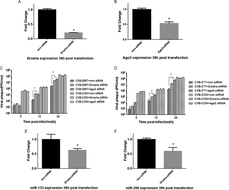
Since our data strongly suggested that viral replication was inhibited through the miRNA degradation pathway, we next determined whether core components of the miRNA machinery were required for controlling replication of the miRT-containing CVB3 mutants. To address this, two core components of the miRNA machinery, Ago2 and Drosha, were specifically depleted by siRNA in TE671 cells. Ago2 acts as an endonuclease within the RNA-induced silencing complex (RISC) machinery, and Drosha—a double-stranded RNA-specific RNase III-type endonuclease—functions to process nascent miRNA transcripts to pre-miRNA precursors in the nucleus. We transfected TE671 cells with Ago2 siRNA or Drosha siRNA (Fig. 3A and B) and then infected these cells with the miRT-containing CVB3 mutants or CVB-CON (MOI = 1). A viral plaque assay of the supernatant showed that downregulation of both Ago2 and Drosha partially rescued replication of the miRT-containing CVB3 mutants with a >10-fold increase in titers compared to those of control groups transfected with a control siRNA; moreover, transfection of Ago2 siRNA and Drosha siRNA had no effect on CVB3-WT replication (Fig. 3C and D). To illustrate the effect of Drosha and Ago2 siRNA on miRNA expression, we measured miR-133 and miR-206 expression in siRNA-treated cells and found that both miRNAs were reduced in the Drosha siRNA-treated group compared to cells treated with the control siRNA (Fig. 3E and F), although these miRNAs were less affected in the Ago2 siRNA-treated groups (data not shown). Together, these data establish that an interaction between cellular miR-133 and miR-206 with the target sequences in the CVB3 mutants can account for the potent inhibition of viral replication. These data further indicate that Ago2, the core component of RISC, exerts its enzymatic “slicing” function during the cleavage process in CVB3 mutants through endogenous miRNAs.
FIG 3.
The miRNA machinery mediates attenuation of viral replication. (A and B) Ago2 and Drosha were knocked down in TE671 cells 36 h after siRNA transfection. Viral replication was assessed for CVB-206T (C) and CVB-3*T (D) posttransfection; non-siRNA was used as a control. (E and F) miR-206 and miR-133 expression was analyzed after Drosha-siRNA transfection. *, P < 0.01 for the CVB-206T- and CVB-3*T-infected groups compared to the CVB-CON group. Virus titer values represent the means ± the SD of three independent experiments.
miRNAs modulate tissue tropism of the miRT-containing CVB3 mutants in vivo.
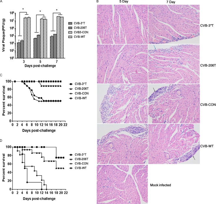
Based on the in vitro experiments, we hypothesized that miR-206 and miR-133 could alleviate the CVB3 pathogenesis in an animal model of infection. We first determined the LD50 for CVB-WT, CVB-CON, CVB-206T, and CVB-3*T in mice according to the Reed and Muench method. For 20 mice i.p. infected with serial dilutions of the viruses, the LD50 of CVB-WT was determined to be 105 PFU, whereas the LD50 values for CVB-206T and CVB-3*T were greater than for CVB-WT at 106 PFU and 5 × 10⁶ PFU, respectively (Table 2). In other words, i.p. injection of 105 PFU of CVB-WT into mice resulted in 50% death within 14 days, whereas injection of the engineered viruses led to no death (for CVB-3*T) or only a few deaths (for CVB-206T). CVB-CON displayed LD50 values similar to those of CVB-WT, as expected. We next determined viral replication of the miRT-containing CVB3 mutants within tissues that express these miRNAs in vivo. Mice in each group were inoculated i.p. with equivalent amounts (105 PFU) of CVB-206T, CVB-3*T, CVB3-WT, or CVB-CON. Evaluating homogenates of the hearts harvested from infected mice at 3, 5, or 7 days after infection, CVB-206T and CVB-3*T replication was more than 2 orders of magnitude lower than CVB-CON (Fig. 4A), suggesting that replication of miRT-containing CVB3 mutants was significantly reduced in the heart, which highly expressed miR-206 and miR-133 (18). Cardiac histopathology revealed that mice infected with CVB-3*T and CVB-206T showed only minor dropsy and hemorrhage, while mice in the CVB-CON group showed significant necrosis and signs of mononuclear cell infiltration on days 5 and 7 after infection (Fig. 4B), further suggesting that miR-206 and miR-133 expression specifically in the heart attenuated CVB-3*T and CVB-206T infection.
TABLE 2.
Engineered viruses are less virulent than CVB3-WT and CVB-CON
| Virus | LD50 (PFU)a |
|---|---|
| CVB3-WT | 1 × 10⁵ |
| CVB-CON | 1 × 10⁵ |
| CVB-206T | 1 × 10⁶ |
| CVB-3*T | 5 × 10⁶ |
LD50 values were calculated for each virus in BALB/c mice using the Reed and Muench method.
FIG 4.
Tissue tropism and protective effect on mouse survival of CVB3-miRTs are controlled by miRNA machinery. (A) BALB/c mice were infected i.p. with the engineered or control CVB3. Viruses isolated from the heart were analyzed by a standard plaque assay at 3, 5, and 7 days postinfection. Virus titers (PFU/g) were normalized to tissue mass. Virus titer values represent the means ± the SD of three independent experiments (n = 5 mice/group). (B) Histological analysis of CVB3 infection in the heart tissues of mock-infected mice or mice i.p. infected with 105 PFU of CVB3-WT or the CVB3-miRTs. Heart sections were prepared 5 and 7 days postinfection. Examples of necrosis, calcification (black arrows), and infiltration (blue arrow) are shown. Original magnification, ×200. (C and D) Survival of BALB/c mice infected with CVB-206T, CVB-3*T, or control viruses (n = 20 mice/group). (C) Survival of BALB/c mice after infection with 105 PFU—the LD50 for CVB3-WT—of the indicated virus. (D) Survival of BALB/c mice infected with 106 PFU—the LD100 for CVB3-WT—of the indicated virus.
To next test whether the attenuated replication and lower pathology of the miRT-containing CVB3 mutants in the heart tissue protected mice against early death after CVB3 infection, we evaluated the survival rate of mice infected with the engineered CVB3 compared to CVB3-CON. The survival of mice infected with an LD50 or LD100 (106 PFU) dose of the control CVB-CON virus was indistinguishable from that of mice infected with the CVB3-WT, while both CVB-206T and CVB-3*T significantly improved the life span of mice (Fig. 4C and D). Collectively, these results indicate that the replication and virulence of CVB3 engineered to carry miRNA target sites were highly attenuated in an in vivo murine experimental CVB3 model.
miRNA-targeted CVB3 are promising vaccine candidates.
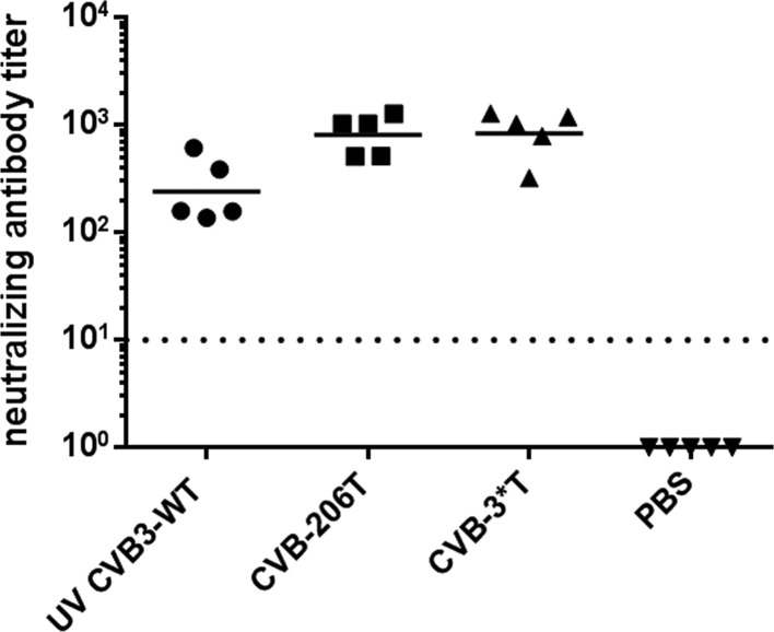
The generation of an effective antiviral memory response is essential for host protection against reinfection. To determine whether a host infected with the miRT-containing CVB3 mutants were still capable of generating immune protection against CVB3 despite attenuated replication in the heart, we next examined the immunogenic potential of CVB-3*T and CVB-206T in mice inoculated i.p. with the engineered viruses by evaluating the levels of neutralizing antibodies—considered a key correlate of vaccine efficacy against viruses—in the infected hosts 4 weeks after immunization. CVB-3*T and CVB-206T induced higher levels of CVB3-specific neutralizing antibodies than that of mice immunized with UV-inactivated CVB3-WT (Fig. 5); as a control, antibody titers for mock-immunized (PBS) mice were below the detection level of the assay. These data suggest that engineered viruses may be candidates for developing effective CVB3 vaccines. To verify that the engineered CVB3 genomes remained stable in vivo, viruses were isolated from the hearts of moribund mice until no new deaths appeared in the groups treated with attenuated viruses (∼18 days postinfection). RT-PCR and direct sequencing of the viral RNA revealed that the miRT sequences inserted into CVB-206T and CVB-3*T remained unchanged, indicating that the viral genome within the engineered viruses was stable over time.
FIG 5.
Infection with CVB-miRTs induces high levels of neutralizing antibody. Neutralizing antibody titers (i.e., the reciprocal of the serum dilution able to neutralize 100× the 50% tissue culture infectious dose [50% tissue culture infective dose of CVB3-WT]) were determined from sera collected from immunized mice. The data for individual mice and the average titers for each experimental group are shown. Titers for PBS-immunized mice were below the detection level of the assay, indicated by a dashed line.
To formally test whether the high titers of neutralizing antibody generated in mice after infection with the engineered CVB3 viruses could protect mice against a normally lethal challenge of the pathogenic CVB3-WT virus, mice infected with miRT-containing CVB3 or control CVB3 viruses were challenged i.p. with a lethal dose (100× the LD50) of CVB3-WT 4 weeks after i.p. immunization. Compared to the PBS-immunized group in which 0/15 animals survived after a lethal challenge of CVB3-WT, 12/15 CVB-206T-immunized mice and 13/15 CVB-3*T-immunized mice exhibited complete protection and remained alive, while only 4/15 mice immunized with the control UV-inactivated CVB3-WT were protected (Table 3). Furthermore, the death of mice infected with either of the engineered viruses occurred at a later date than mice in the control groups (∼14 days postinfection for the engineered viruses versus 2 days for the PBS group and 5 days for the UV-inactivated group) (data not shown). These results indicated that CVB-206T and CVB-3*T conferred protective immunity to viral reinfection, and the production of neutralizing antibodies of immunized mice correlated with the percentage of mice surviving a lethal dose of CVB3.
TABLE 3.
Infection with CVB-miRTs confers immunity against a lethal challenge with CVB3-WTa
| Immunogen | 100× the LD50 |
|---|---|
| PBS | 0/15 |
| UV-inactivated CVB3-WT | 4/15 |
| Live CVB-206T | 12/15 |
| Live CVB-3*T | 13/15 |
Mice were immunized with PBS or 104 PFU (0.1 LD50 for CVB3-WT) of UV-inactivated CVB3-WT, live CVB-206T, or live CVB-133*T. Four weeks later, mice were challenged with 102 LD50 of CVB3-WT via i.p. injection. The numbers of surviving mice are indicated in the table.
DISCUSSION
The incorporation of cognate miRNA target sequences into viral genomes is an approach that has been successfully applied to several different viruses in order to attenuate viral tropism and pathogenesis. For example, an engineered oncolytic virus can inhibit tumor growth while restricting viral replication in normal tissues (11). miRNA-targeted viruses have also been shown to exhibit increased genetic stability without compromising replication robustness in nonlethal organs, providing an alternative method to rationally design live attenuated vaccines (7). In the present study, we report that miRTs corresponding to muscle-specific miR-133 and/or miR-206 inserted into the 5′ UTR of the CVB3 genome can attenuate the cardiotropic virulence phenotype and control cardiac pathogenesis after CVB3 infection while retaining the immunogenicity of the virus.
The cardiotropic CVB3 can efficiently replicate in the heart and is among the most common and significant causative agents of heart disease in humans. Similar to humans, mice inoculated with CVB3 can die from direct pathological effects on the heart. miR-206 and miR-133 are muscle-specific miRNAs, and their expression is broadly distributed throughout heart and skeletal muscle, as well as in muscle-derived cells. In the present study, a plasmid containing the CVB3m strain viral genome was reconstructed to contain miRNA targets. Although CVB3 engineered to contain targets for miR-206 alone or miR-206 plus miR-133 exhibited normal replication in HeLa cells, which had little expression of either miRNA, replication was distinctly reduced (100-fold lower) compared to control viruses in TE671 cells and L6 cells that expressed high levels of miR-206. Further, when miRNA mimics for miR-133 or miR-206 were transfected into HeLa cells, CVB-206T and CVB-3*T replication was significantly suppressed. Thus, insertion of the miRNA targets did not impair viral reproduction in the absence of miR-133 and miR-206, but their presence inhibited viral replication and importantly led to improved host cell viability. In vivo, CVB-206T and CVB-3*T viral replication was more than 2 orders of magnitude lower than that exhibited by the control CVB3 viruses in the hearts of infected mice. Importantly, this lower viral load was accompanied by attenuated pathological injury in the heart and increased survival of the mice, indicating that virulence of the engineered viruses was effectively restricted in the desired organ due to the expression of the specific miRTs. An important concern is that miRT-carrying CVB3 may act as sponges for endogenous miRNAs. However, previous reports found that some effects caused by miRNA misexpression might become masked by counterbalancing regulatory signals originating from other cell types in the tissue (19–21); thus, miRNA expression may be modulated and maintain a homeostatic state in vivo. Overall, our data indicate that insertion of miRNA targets into the CVB3 virus may provide an effective approach to restrict viral replication, reduce inflammatory responses, and control viral cardiotropism and pathogenesis.
Live attenuated vaccines remain the safest, most cost-effective intervention against viral infections. However, live attenuated vaccines may revert back to a virulent phenotype, causing pathology, along with regaining the ability to reproduce within the host, where reproduction of highly pathogenic viruses in major organs may also cause severe infection. Thus, limiting CVB3 viral reproduction in specific tissues may be a key factor to consider when developing effective live attenuated vaccines. In the present study, even though CVB-206T and CVB-3*T replication was notably attenuated in the heart, these CVB3 mutants retained the ability to replicate normally in peripheral tissues. In terms of providing immune protection, mice immunized with either the engineered CVB3 or the UV-inactivated CVB3-WT elicited similarly strong neutralizing antibody responses against CVB3 and were effectively protected against a lethal challenge of CVB3-WT; moreover, the life span of mice treated with these engineered viruses significantly improved over mice treated with the control viruses. Thus, while replication of the engineered CVB3 viruses was restricted in the important heart tissue, the normal replication maintained in other tissues was still able to elicit a complex and long-lasting immune response, which might be advantageous for CVB3 vaccine design.
Previous reports indicate that increasing the number of miRNA targets within a genome is the most efficacious way to silence gene expression (22, 23). In the present study, an enhanced effect on attenuating CVB3 virulence in vitro was not evident when inserting three target sequence copies (miR-206 plus miR-133) rather than just a single copy of miR-206. Both engineered viruses elicited comparable levels of neutralizing antibody. Although the protective effect of CVB-3*T appeared to be more effective than CVB-206T in controlling virus titers in the heart as well as in improving the survival of mice after infection or after CVB3-WT challenge after immunization, these differences were not significant. We thus consider that miR-206 may play a more important role in controlling viral replication than miR-133 or that the insertion of more miRNA targets may further increase the efficacy of the engineered virus.
Previous studies provided evidence to support that miRNAs could modulate viral translation through imperfect or perfect binding to the 3′ UTR (24), the 5′ UTR (25–27), or the sequences within the coding region (28–30). The 5′ UTR (7), 3′ UTR (11, 31, 32), or coding position locations (7) selected for miRT insertion differed among various viruses depending upon where insertion was tolerated within the particular virus. In the present study, several locations within the 5′ UTR or 3′ UTR of the CVB3 genome were selected for constructing the virus (see genomic map of tested sites in Fig. 1A). Liu et al. provided evidence to support that the core IRES sequence was located between 432 and 639 bp in the CVB3 genome and that deletion of less than 29 nt at bp 249 (the BstBI restriction site) using exonuclease Bal31 did not abolish CVB3 infectivity (14). However, viral infectivity was not observed in our hands after inserting the miRT sequence into the BstBI site, transfecting the reconstructed plasmid into HeLa cells, and passaging the progeny three times. We also tried 22-bp substitutions—equivalent to the length of one copy of the miRNA target sequence—at the BstBI site, but viral infectivity was again abolished. Insertion of the miRTs in the 3′ UTR of the CVB3 genome was also tested: miR-133T or miR-206T was incorporated into either the BsmI restriction site at bp 7387 or between bp 7359 and 7360, where insertion of exogenous sequence could not disturb the dominant hairpin structure of the 3′ UTR (33), but both locations in the 3′ UTR did not tolerate sequence insertion. One possible explanation for these results is that the higher-order RNA structure of the 5′ UTR and 3′ UTR may have been disturbed by the insertion. Once we inserted the KOZAK-like site alongside the miRTs, viral infectivity and replicability were retained (34, 35), and the altered genome remained stable in host cells, even to passage 5.
In conclusion, we demonstrated that modifying CVB3 by inserting a single copy or three tandem copies of miRNA target sequences could not only attenuate cellular virulence in cells that express the specific miRNAs in vitro but also nearly abolished the cardiovirulent phenotype in vivo. These findings support the rationale for using this miRNA-targeting approach to control CVB3 cardiotropism and to develop live attenuated virus vaccines. Since pancreatitis and meningitis may also occur after CVB3 infection, the insertion of miRNA targets for pancreas-specific miRNAs, such as let-7, miR-200, or the central nervous system-specific miR-124a/let-7b, into the viral genome may provide even more effective protection against CVB3 infection.
ACKNOWLEDGMENTS
We thank Qinghua Zhang and Liying Huang at the Capital Institute of Pediatrics (Beijing, China) for construction of the CVB3 mutants. We also thank Jie Deng at the Capital Institute of Pediatrics (Beijing, China) for technical support with the neutralization assays.
This study was supported by the National Natural Science Foundation of China (81271809), the Beijing Natural Science Foundation (7102023), and the Beijing Training Programme Foundation for Talents (2011A003034000032).
We do not have any conflict of financial or other interest.
REFERENCES
- 1.Fairweather D, Rose NR. 2007. Coxsackievirus-induced myocarditis in mice: a model of autoimmune disease for studying immunotoxicity. Methods 41:118–122. doi: 10.1016/j.ymeth.2006.07.009. [DOI] [PMC free article] [PubMed] [Google Scholar]
- 2.Gauntt CJ, Tracy SM, Chapman N, Wood HJ, Kolbeck PC, Karaganis AG, Winfrey CL, Cunningham MW. 1995. Coxsackievirus-induced chronic myocarditis in murine models. Eur Heart J 16(Suppl O):56–58. doi: 10.1093/eurheartj/16.suppl_O.56. [DOI] [PubMed] [Google Scholar]
- 3.Esfandiarei M, McManus BM. 2008. Molecular biology and pathogenesis of viral myocarditis. Annu Rev Pathol 3:127–155. doi: 10.1146/annurev.pathmechdis.3.121806.151534. [DOI] [PubMed] [Google Scholar]
- 4.Tam PE. 2006. Coxsackievirus myocarditis: interplay between virus and host in the pathogenesis of heart disease. Viral Immunol 19:133–146. doi: 10.1089/vim.2006.19.133. [DOI] [PubMed] [Google Scholar]
- 5.Brown BD, Venneri MA, Zingale A, Sergi SL, Naldini L. 2006. Endogenous microRNA regulation suppresses transgene expression in hematopoietic lineages and enables stable gene transfer. Nat Med 12:585–591. doi: 10.1038/nm1398. [DOI] [PubMed] [Google Scholar]
- 6.Kelly EJ, Nace R, Barber GN, Russell SJ. 2010. Attenuation of vesicular stomatitis virus encephalitis through microRNA targeting. J Virol 84:1550–1562. doi: 10.1128/JVI.01788-09. [DOI] [PMC free article] [PubMed] [Google Scholar]
- 7.Barnes D, Kunitomi M, Vignuzzi M, Saksela K, Andino R. 2008. Harnessing endogenous miRNAs to control virus tissue tropism as a strategy for developing attenuated virus vaccines. Cell Host Microbe 4:239–248. doi: 10.1016/j.chom.2008.08.003. [DOI] [PMC free article] [PubMed] [Google Scholar]
- 8.Centers for Disease Control and Prevention. 1999. Impact of vaccines universally recommended for children–United States, 1900-1998. JAMA 281:1482–1483. doi: 10.1001/jama.281.16.1482. [DOI] [PubMed] [Google Scholar]
- 9.Cova L. 2014. Advances and challenges in the development of therapeutic DNA vaccines against hepatitis B virus infection. Curr Gene Ther 14:149–160. doi: 10.2174/1566523214666140509102644. [DOI] [PubMed] [Google Scholar]
- 10.Jang YH, Byun YH, Lee YJ, Lee YH, Lee KH, Seong BL. 2012. Cold-adapted pandemic 2009 H1N1 influenza virus live vaccine elicits cross-reactive immune responses against seasonal and H5 influenza A viruses. J Virol 86:5953–5958. doi: 10.1128/JVI.07149-11. [DOI] [PMC free article] [PubMed] [Google Scholar]
- 11.Kelly EJ, Hadac EM, Greiner S, Russell SJ. 2008. Engineering microRNA responsiveness to decrease virus pathogenicity. Nat Med 14:1278–1283. doi: 10.1038/nm.1776. [DOI] [PubMed] [Google Scholar]
- 12.Townley-Tilson WH, Callis TE, Wang D. 2010. MicroRNAs 1, 133, and 206: critical factors of skeletal and cardiac muscle development, function, and disease. Int J Biochem Cell Biol 42:1252–1255. doi: 10.1016/j.biocel.2009.03.002. [DOI] [PMC free article] [PubMed] [Google Scholar]
- 13.Care A, Catalucci D, Felicetti F, Bonci D, Addario A, Gallo P, Bang ML, Segnalini P, Gu Y, Dalton ND, et al. . 2007. MicroRNA-133 controls cardiac hypertrophy. Nat Med 13:613–618. doi: 10.1038/nm1582. [DOI] [PubMed] [Google Scholar]
- 14.Liu Z, Carthy CM, Cheung P, Bohunek L, Wilson JE, McManus BM, Yang D. 1999. Structural and functional analysis of the 5′ untranslated region of coxsackievirus B3 RNA: in vivo translational and infectivity studies of full-length mutants. Virology 265:206–217. doi: 10.1006/viro.1999.0048. [DOI] [PubMed] [Google Scholar]
- 15.Yao H, Zhang Y, He F, Wang C, Xiao Z, Zou J, Wang F, Liu Z. 2012. Short hairpin RNA targeting 2B gene of coxsackievirus B3 exhibits potential antiviral effects both in vitro and in vivo. BMC Infect Dis 12:177. doi: 10.1186/1471-2334-12-177. [DOI] [PMC free article] [PubMed] [Google Scholar]
- 16.Reed LJ, Muench H. 1938. A simple method of estimating fifty percent endpoints. Am J Hyg 27:493–497. [Google Scholar]
- 17.He F, Yao H, Xiao Z, Han J, Zou J, Liu Z. 2014. Inhibition of IL-2 inducible T-cell kinase alleviates T-cell activation and murine myocardial inflammation associated with CVB3 infection. Mol Immunol 59:30–38. doi: 10.1016/j.molimm.2013.12.004. [DOI] [PubMed] [Google Scholar]
- 18.Zhang Q, Xiao Z, He F, Zou J, Wu S, Liu Z. 2013. MicroRNAs regulate the pathogenesis of CVB3-induced viral myocarditis. Intervirology 56:104–113. doi: 10.1159/000343750. [DOI] [PubMed] [Google Scholar]
- 19.Elia L, Quintavalle M, Zhang J, Contu R, Cossu L, Latronico MV, Peterson KL, Indolfi C, Catalucci D, Chen J, Courtneidge SA, Condorelli G. 2009. The knockout of miR-143 and -145 alters smooth muscle cell maintenance and vascular homeostasis in mice: correlates with human disease. Cell Death Differ 16:1590–1598. doi: 10.1038/cdd.2009.153. [DOI] [PMC free article] [PubMed] [Google Scholar]
- 20.Liu N, Bezprozvannaya S, Williams AH, Qi X, Richardson JA, Bassel-Duby R, Olson EN. 2008. microRNA-133a regulates cardiomyocyte proliferation and suppresses smooth muscle gene expression in the heart. Genes Dev 22:3242–3254. doi: 10.1101/gad.1738708. [DOI] [PMC free article] [PubMed] [Google Scholar]
- 21.Diederichs S, Haber DA. 2007. Dual role for argonautes in microRNA processing and posttranscriptional regulation of microRNA expression. Cell 131:1097–1108. doi: 10.1016/j.cell.2007.10.032. [DOI] [PubMed] [Google Scholar]
- 22.Brown BD, Gentner B, Cantore A, Colleoni S, Amendola M, Zingale A, Baccarini A, Lazzari G, Galli C, Naldini L. 2007. Endogenous microRNA can be broadly exploited to regulate transgene expression according to tissue, lineage and differentiation state. Nat Biotechnol 25:1457–1467. doi: 10.1038/nbt1372. [DOI] [PubMed] [Google Scholar]
- 23.Heiss BL, Maximova OA, Thach DC, Speicher JM, Pletnev AG. 2012. MicroRNA targeting of neurotropic flavivirus: effective control of virus escape and reversion to neurovirulent phenotype. J Virol 86:5647–5659. doi: 10.1128/JVI.07125-11. [DOI] [PMC free article] [PubMed] [Google Scholar]
- 24.Sisk JM, Witwer KW, Tarwater PM, Clements JE. 2013. SIV replication is directly downregulated by four antiviral miRNAs. Retrovirology 10:95. doi: 10.1186/1742-4690-10-95. [DOI] [PMC free article] [PubMed] [Google Scholar]
- 25.Jopling CL, Yi M, Lancaster AM, Lemon SM, Sarnow P. 2005. Modulation of hepatitis C virus RNA abundance by a liver-specific microRNA. Science 309:1577–1581. doi: 10.1126/science.1113329. [DOI] [PubMed] [Google Scholar]
- 26.Norman KL, Sarnow P. 2010. Modulation of hepatitis C virus RNA abundance and the isoprenoid biosynthesis pathway by microRNA miR-122 involves distinct mechanisms. J Virol 84:666–670. doi: 10.1128/JVI.01156-09. [DOI] [PMC free article] [PubMed] [Google Scholar]
- 27.Roberts AP, Lewis AP, Jopling CL. 2011. miR-122 activates hepatitis C virus translation by a specialized mechanism requiring particular RNA components. Nucleic Acids Res 39:7716–7729. doi: 10.1093/nar/gkr426. [DOI] [PMC free article] [PubMed] [Google Scholar]
- 28.Tong L, Lin L, Wu S, Guo Z, Wang T, Qin Y, Wang R, Zhong X, Wu X, Wang Y, et al. . 2013. MiR-10a* upregulates coxsackievirus B3 biosynthesis by targeting the 3D-coding sequence. Nucleic Acids Res 41:3760–3771. doi: 10.1093/nar/gkt058. [DOI] [PMC free article] [PubMed] [Google Scholar]
- 29.Wang L, Qin Y, Tong L, Wu S, Wang Q, Jiao Q, Guo Z, Lin L, Wang R, Zhao W, et al. . 2012. MiR-342-5p suppresses coxsackievirus B3 biosynthesis by targeting the 2C-coding region. Antivir Res 93:270–279. doi: 10.1016/j.antiviral.2011.12.004. [DOI] [PubMed] [Google Scholar]
- 30.Ye X, Liu Z, Hemida MG, Yang D. 2011. Targeted delivery of mutant tolerant anti-coxsackievirus artificial microRNAs using folate conjugated bacteriophage phi29 pRNA. PLoS One 6:e21215. doi: 10.1371/journal.pone.0021215. [DOI] [PMC free article] [PubMed] [Google Scholar]
- 31.Yen LC, Lin YL, Sung HH, Liao JT, Tsao CH, Su CM, Lin CK, Liao CL. 2013. Neurovirulent flavivirus can be attenuated in mice by incorporation of neuron-specific microRNA recognition elements into viral genome. Vaccine 31:5915–5922. doi: 10.1016/j.vaccine.2011.09.102. [DOI] [PubMed] [Google Scholar]
- 32.Jin H, Lv S, Yang J, Wang X, Hu H, Su C, Zhou C, Li J, Huang Y, Li L, et al. . 2011. Use of microRNA Let-7 to control the replication specificity of oncolytic adenovirus in hepatocellular carcinoma cells. PLoS One 6:e21307. doi: 10.1371/journal.pone.0021307. [DOI] [PMC free article] [PubMed] [Google Scholar]
- 33.Melchers WJ, Hoenderop JG, Bruins SH, Pleij CW, Pilipenko EV, Agol VI, Galama JM. 1997. Kissing of the two predominant hairpin loops in the coxsackie B virus 3′ untranslated region is the essential structural feature of the origin of replication required for negative-strand RNA synthesis. J Virol 71:686–696. [DOI] [PMC free article] [PubMed] [Google Scholar]
- 34.Kozak M. 1981. Possible role of flanking nucleotides in recognition of the AUG initiator codon by eukaryotic ribosomes. Nucleic Acids Res 9:5233–5252. doi: 10.1093/nar/9.20.5233. [DOI] [PMC free article] [PubMed] [Google Scholar]
- 35.Harkins S, Cornell CT, Whitton JL. 2005. Analysis of translational initiation in coxsackievirus B3 suggests an alternative explanation for the high frequency of R+4 in the eukaryotic consensus motif. J Virol 79:987–996. doi: 10.1128/JVI.79.2.987-996.2005. [DOI] [PMC free article] [PubMed] [Google Scholar]


