ABSTRACT
The determinants of the maintenance of chronic hepadnaviral infection are yet to be fully understood. A long-standing unresolved argument in the hepatitis B virus (HBV) research field suggests that during chronic hepadnaviral infection, cell-to-cell spread of hepadnavirus is at least very inefficient (if it occurs at all), virus superinfection is an unlikely event, and chronic hepadnavirus infection can be maintained exclusively via division of infected hepatocytes in the absence of virus spread. Superinfection exclusion was previously shown for duck HBV, but it was not demonstrated for HBV or HBV-related woodchuck hepatitis virus (WHV). Three woodchucks, which were chronically infected with the strain WHV7 and already developed WHV-induced hepatocellular carcinomas (HCCs), were superinfected with another WHV strain, WHVNY. Six weeks after the superinfection, the woodchucks were sacrificed and tissues of the livers and HCCs were examined. The WHVNY superinfection was demonstrated by using WHV strain-specific PCR assays and (i) finding WHVNY relaxed circular DNA in the serum samples collected from all superinfected animals during weeks one through six after the superinfection, (ii) detecting replication-derived WHVNY RNA in the tissue samples of the livers and HCCs collected from three superinfected woodchucks, and (iii) finding WHVNY DNA replication intermediates in tissues harvested after the superinfection. The results are consistent with the occurrence of continuous but inefficient hepadnavirus cell-to-cell spread and superinfection during chronic infection and suggest that the replication space occupied by the superinfecting hepadnavirus in chronically infected livers is limited. The findings are discussed in the context of the mechanism of chronic hepadnavirus infection.
IMPORTANCE This study aimed to better understand the determinants of the maintenance of chronic hepadnavirus infection. The generated data suggest that in the livers chronically infected with woodchuck hepatitis virus, (i) hepadnavirus superinfection and cell-to-cell spread likely continue to occur and (ii) the virus spread is apparently inefficient, which is consistent with the interpretation that a limited number of cells in the livers facilitates the spread of hepadnavirus. The limitations of the cell-to-cell virus spread most likely are mediated at the level of the cells and do not reflect the properties of the virus. Our results further advance the understanding of the mechanism of chronic hepadnavirus infection. The significance of the continuous but limited hepadnavirus spread and superinfection for the maintenance of the chronic state of infection should be further evaluated in follow-up studies in order to determine whether blocking the virus spread would facilitate the suppression of chronic hepadnavirus infection.
INTRODUCTION
Worldwide, approximately four hundred million people are chronic carriers of human hepatitis B virus (HBV), which is a prototype hepadnavirus. Chronic HBV infection is a main risk factor of hepatocellular carcinoma (HCC), and it is associated with more than 50% of all known cases of HCC (1–14). During the early stage of hepadnaviral infection, virtually the entire liver becomes infected with a hepadnavirus within several weeks. However, during chronic infection, areas of apparently hepadnavirus-free hepatocytes were observed in HBV carrier humans and chimpanzees and also in woodchucks chronically infected with woodchuck hepatitis virus (WHV), which is closely related to HBV. The hepadnavirus-free hepatocytes tested negative for (i) the hepadnavirus core antigen (by immunostaining) and (ii) the viral DNA (by in situ hybridization) regardless of the ongoing viremia. In HBV carrier humans, up to 90% of all hepatocytes could appear to be HBV free. Some of the virus-free hepatocytes form foci of altered hepatocytes (FAH) with morphology similar to that of liver cancer cells and are considered premalignant. In addition, the cells of hepadnavirus-induced HCCs often were reported to be virus free as well (1, 4, 15–23). In contrast, several previous studies reported the presence of hepadnaviral DNA and RNA in HCC cells (24–26), making the issue still controversial. The observations of apparent hepadnavirus-free HCC cells and hepatocytes, along with the analysis of the kinetics of the emergence of drug-resistant HBV mutants, laid the foundation for a long-standing unresolved argument in the HBV field that during chronic infection, cell-to-cell spread of a hepadnavirus is at least very inefficient (if it occurs at all), and superinfection is an unlikely event. It was proposed (i) that the above-mentioned virus-free hepatocytes accumulate via the immune selection, because they are no longer targeted by the immune system and have a selective advantage for clonal expansion, and (ii) that HCCs develop from the virus-free hepatocytes of FAH. Superinfection exclusion was shown for the duck HBV and was suggested, but has never been demonstrated, for WHV and HBV. The cases of the occurrence of intergenotype HBV recombinants may argue against superinfection exclusion for HBV, although they have been challenged by researchers who favor the hypothesis that chronic hepadnavirus infection is maintained exclusively by the division of infected hepatocytes in the absence of virus spread and superinfection (3, 15–22, 27–30). We recently used three woodchucks that were chronic carriers of WHV and had already developed HCCs, and we superinfected them with serum-derived hepatitis delta virus (HDV) coated with the envelope proteins of WHV (wHDV). For all three superinfected animals, we demonstrated that wHDV was able to infect not only nonmalignant hepatocytes but also fractions of the cells in WHV-induced HCCs which were developed prior to wHDV superinfection. The efficiency of wHDV superinfection was comparable in most HCCs and matching nonmalignant liver tissues. These results indicated that in WHV-induced HCCs, at least a fraction of tumor cells expresses functional WHV receptors and support all of the steps of the attachment and entry that are regulated by the WHV envelope proteins. Taking into consideration that it was previously hypothesized that the cells of hepadnavirus-induced HCCs are resistant to reinfection with a hepadnavirus in vivo, our data suggested that if such resistance exists, it is mediated via a block at a postentry step. In addition, using quantitative real-time PCR assays (qPCRs), we were able to demonstrate the presence of pregenome/precore RNA (pgRNA) and covalently closed circular DNA (cccDNA) of WHV in harvested samples of WHV-induced HCC tissues. However, the amounts of pgRNA and cccDNA in HCC tissues were reduced, often significantly, compared to those in corresponding nonmalignant liver tissues (31). In the current study, we also used three WHV carrier woodchucks with already formed WHV-induced HCCs. These WHV carrier woodchucks were generated by infection with the strain WHV7 (32). We superinfected these WHV7 carriers with a different WHV strain, WHVNY (33), and then monitored them for a period of 6 weeks after the superinfection. In a nutshell, using WHV strain-specific PCR assays and examining the sera, livers, and matching HCC tissue samples, we obtained evidence of superinfection with strain WHVNY for all three superinfected woodchucks. Our data, however, indicated that the replication space occupied by WHVNY in the livers 6 weeks after the superinfection was limited.
MATERIALS AND METHODS
Superinfection with the strain WHVNY of woodchucks chronically infected with WHV7 strain.
In the current study, we used three adult WHV carrier woodchucks, F7808, F7806, and M7761, which originally were generated by neonatal infection with the strain WHV7 performed at 3 days of age. The initial inoculation of these woodchucks (as neonates) was done using the inoculum cWHV7P1 (32). Woodchucks F7808, F7806, and M7761 were bred, infected with WHV7, and maintained as chronic WHV7 carriers under contract N01AI05399 (College of Veterinary Medicine, Cornell University, Ithaca, NY). All experimental manipulations of animals have been performed under protocols approved by the Institutional Animal Care and Use Committee. The above-described three chronic WHV7 carrier woodchucks that already developed WHV-induced HCCs were superinfected with strain WHVNY. Each animal was inoculated with 1.66 × 10⁸ genome equivalents (GE) of serum-derived WHVNY, which was obtained from woodchuck 2761 (33). One week prior to superinfection, the animals underwent limited surgery (laparotomy) in order to verify the locations of the established HCCs in the infected livers, which were previously determined by ultrasound imaging. At this point, two biopsy samples were obtained from each animal. One sample was taken from nonmalignant liver tissue, and a second sample was harvested from one HCC located during the limited surgery. Serum samples were collected at 1 week prior to the superinfection (week −1), immediately before the superinfection (week 0), and during each week after the superinfection for a period of 6 weeks. Six weeks after the superinfection, the woodchucks were sacrificed, and from each animal, nonmalignant tissue samples were taken from the left medial liver lobe (LM), right lateral liver lobe (RL), and left liver lobe (LL). In addition, for each HCC that was found during necropsy, a sample of the center core of the tumor was harvested for subsequent analysis.
Isolation of rcDNA of WHV from serum samples.
The WHV relaxed circular DNA (rcDNA) was extracted using a modification of a previously described method (29). Each aliquot of 50 μl of woodchuck serum was incubated for 2 h at 37°C with 450 μl of lysis buffer containing 10 mM Tris-HCl, pH 8.0, 10 mM EDTA, 100 mM NaCl, and 2% SDS and supplemented with 2 mg/ml pronase (Fisher Scientific). The rcDNA then was extracted with one volume of Tris-saturated phenol, pH 7.8 to 8.2 (Acros Organics), followed by extraction of the obtained aqueous phase with one volume of a mixture of chloroform-isoamyl alcohol (24:1). DNA next was precipitated from the aqueous phase by adding 2 volumes of 100% ethanol, 0.1 volume of 3 M sodium acetate (pH 5.5), and 2.5 μl of 10 μg/μl dextran (MP Biomedicals) solution in DNase/RNase-free water per sample. Samples were placed at −80°C for at least 1 h, and then DNA was sedimented by centrifugation at 13,000 rpm at 4°C for 30 min. The resulting DNA pellet was redissolved in 20 μl of DNase/RNase-free water.
Molecular clones and consensus sequences of WHVNY and WHV7.
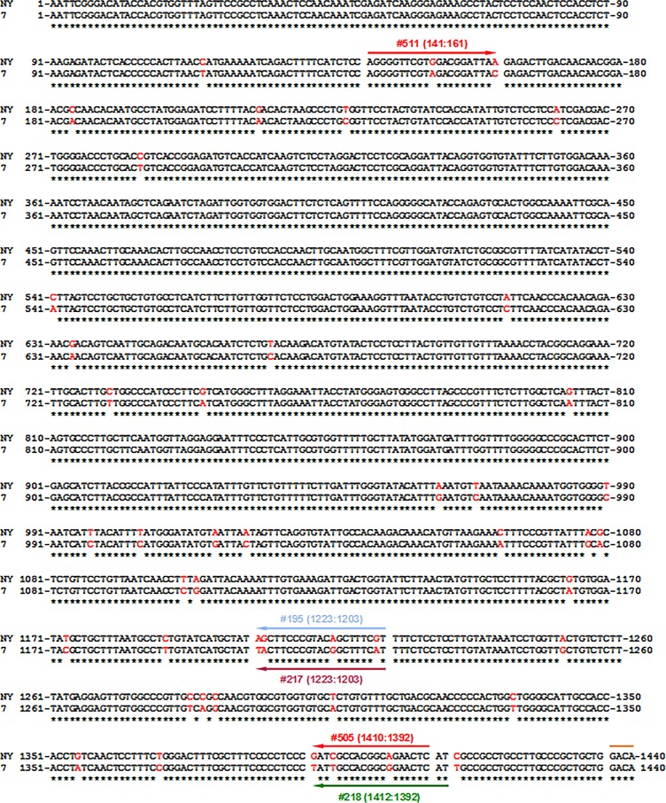
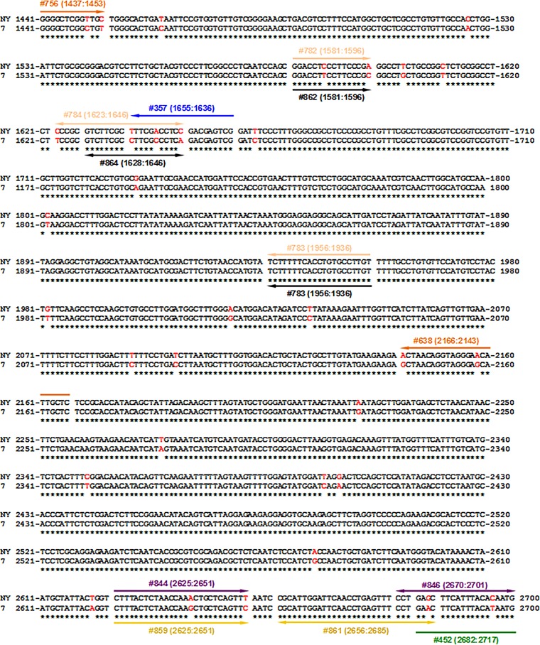
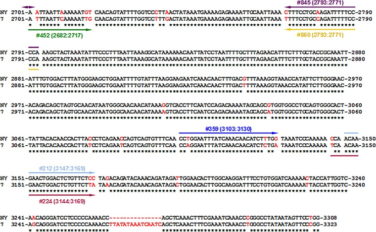
The rcDNA of WHVNY was extracted from serum of woodchuck 2761, which was monoinfected with WHVNY (33). The serum sample from woodchuck 7946 (monoinfected with WHV7) was used to isolate rcDNA of WHV7. A modification of the strategy described by Gunther et al. (34, 35) was used to obtain PCR products spanning the entire genomes of WHVNY and WHV7. The PCR mixture contained 2 μl of isolated rcDNA, 15 pmol of the primer 206A (GAT-1935-ATCTTTTTCACCTGTGCCTTGTTT-1958 [the created EcoRV site is underlined]), 15 pmol of the primer 231 (CTCGAG-1942-AAAAAGATACATGGTTACAG-1923 [the artificial XhoI site is underlined]), 10 nmol of each deoxynucleoside triphosphate (dNTP) (Fermentas), 5 μl of 10× Pfu buffer, and 2.5 U of PfuUltra high-fidelity DNA polymerase (Agilent Technologies) in a final volume of 50 μl. The PCR conditions were 95°C for 2 min; 30 cycles of 95°C for 30 s, 55°C for 30 s, and 72°C for 3.5 min; and a final extension step for 10 min at 72°C. The expected sizes of PCR products were 3,324 bp for WHVNY and 3,339 bp for WHV7. After PCR amplification, 3′A overhangs were added by adding 0.2 μl of Klentaq polymerase (AB Peptides) to the PCR mixture and incubating samples for an additional 10 min at 72°C. The PCR products were cloned into PCR IV Topo vector (for WHVNY) or into PCR II Topo vector (for WHV7). The resulting constructs were analyzed by the sequencing of overlapping fragments using M13 primers (Invitrogen) and WHV-specific primers 23 (2504-AGAAGACGCACTCCCTCTCCT-2524), 126 (2963-TTGGGAACACAGACAGCTAGTGC-2985), 170 (148-CGTAGACGGATTACGAGACTTGAC-171), 172 (891-CCGCACTTCTGAGCATCTTAC-911), and 174 (1729-CAGAATTGCGAACCATGGATTCC-1750). The obtained sequences of WHV genomic fragments then were aligned using ClustalW software (http://www.ch.embnet.org/software/clustalw.html) in order to produce the complete genome sequences for WHVNY and WHV7. Eight complete genome molecular clones were obtained for each of the WHV strains. Based on the molecular clones, the consensus sequences of WHVNY and WHV7 strains were generated using MacVector 11.1.1 software. The consensus sequences of WHVNY and WHV7 were aligned using ClustalW2 software (http://www.ebi.ac.uk/Tools/msa/clustalw2/), and the generated alignment is presented in Fig. 1. The alignment was used further for the design of the WHV strain-specific PCR assays. The numbering shown in Fig. 1 was used to indicate the positions of the WHV-specific primers and probes for real-time PCR.
FIG 1.
Alignment of the full-genome consensus sequences of the strains WHVNY and WHV7. As described in Materials and Methods, the rcDNA of WHVNY and WHV7 was isolated from the serum samples of monoinfected woodchucks. Eight full-genome molecular clones for each WHV strain were generated using a strategy based on the method described by Gunther et al. (34, 35). Using the molecular clones, the consensus sequences for WHVNY and WHVN7 were obtained using MacVector 11.1.1 software. The alignment of the consensus sequences generated using ClustalW2 software (http://www.ebi.ac.uk/Tools/msa/clustalw2/) is shown. The sequence of WHVNY is shown on the top row, while the sequence of WHV7 is on the bottom row. Identical nucleotides are indicated by asterisks underneath. The nonidentical nucleotides are shown in red. The genome of WHVNY contains a 15-nucleotide-long deletion which corresponds to the positions 3264 to 3278 of the WHV7 genome. The positions of primers (each primer is shown as a line with a single arrow) and probes (each probe is shown as a line with two arrows) used for the strain-specific conventional PCR and qPCR assays are indicated relative to the alignment. In the first round of nested PCR to detect serum WHVNY rcDNA, primers 359 (3103:3130) and 357 (1655:1636) (blue) were used. For the second round of PCR, primers 212 (3147:3169) and 195 (1223:1203) (light blue) were employed. For WHV7 rcDNA detection, primers 452 (2682:2717) and 218 (1412:1392) (green) were used for the first round of PCR. The second round of amplification was carried out using primers 224 (3144:3169) and 217 (1223:1203) (light purple). For WHVNY RNA detection, the cDNA synthesis was carried out using WHVNY-specific primer 357. Amplification was carried out by seminested PCR using primers 511 (141:161) and 505 (1410:1392) (in some cases, primer 357 was used instead of 505 [see Materials and Methods]) for the first round (red). The second round of amplification was carried out using primers 511 and 195. WHV7 cDNA synthesis was performed using WHV7-specific primer 218. Amplification was carried out by the same nested PCR method as that used for the rcDNA detection, employing primers 452 and 218 for the first round of amplification and primers 224 and 217 for the second round of PCR. The quantification of serum rcDNA and replicative intermediate DNA (RI-DNA) of WHVNY in tissue samples was done by qPCR using primers 844 (2625:2651) and 845 (2793:2771) and probe 846 (2670:2701) (purple). For WHV7 rcDNA and RI-DNA quantification, primers 859 (2625:2651) and 860 (2793:2771) and probe 861 (2656:2685) (yellow) were used. For the quantification of WHVNY cccDNA in tissue samples, a preamplification step (conventional PCR) was carried out using primers 756 (1437:1453) and 638 (2166:2143) (orange). Then, in the next step, qPCR was performed using primers 782 (1581:1596) and 783 (1956:1936) and probe 784 (1623:1646) (beige). The quantification of WHV7 cccDNA was carried out using primers 862 (1581:1596) and 783 and probe 864 (1623:1646) (black).
Plasmids and standards.
WHV strain-specific DNA standards were made using the plasmids pN4C4 (WHVNY-pCR IV colony 4) and p72C6 (WHV7-pCR II colony 6), which were obtained by cloning the WHVNY and WHV7 genomes using the serum rcDNA as the starting material as described above. Plasmid DNAs were digested with EcoRV and XhoI. The generated fragments, greater than WHV genome-length fragments and corresponding to the genomes of WHVNY and WHV7 (3,321 bp and 3,336 bp, respectively), were gel purified using a QIAEX II gel extraction kit (Qiagen) and cloned into pcDNA3 vector (Invitrogen), which was predigested with EcoRV and XhoI and then dephosphorylated using shrimp alkaline phosphatase (Fermentas). The resulting constructs were analyzed by sequencing using the primers 23, 126, 170, 172, 174, 735 (4673-CACGCTGCGCGTAACCAC-4656), and 740 (748-GTAACAACTCCGCCCCATTGA-768). The primers 735 and 740 spanned the indicated positions in the backbone of pcDNA3 and allowed sequencing of the ends of WHV-specific inserts. The final constructs pJSWHVNY-4C4 and pJSWHV7-2C6 were used as the standards for the analysis of WHV DNA and RNA by conventional PCR and qPCR assays. For the analysis of WHVNY cccDNA, a new DNA standard was generated based on the strategy described previously (36). The WHVNY rcDNA isolated from the serum of monoinfected woodchuck F6541 was used for the amplification of two fragments of the WHVNY genome. Fragment A was amplified using primers 382 (TTAGGTACC-1697-GTCCGGTCCGTGTTGCTT-1714 [the artificial Acc65I restriction site is underlined]) and 383 (14-GTATGTCCCGAATT-1-CCAGGAACTATTATAGG-3293 [the natural EcoRI site is underlined]). Fragment B was amplified using the primers 380 (3287-CCGGGCCTATAATAGTTCCTGG-3808-AATTCCTA [the natural EcoRI site is underlined]) and 381 (CTACTCGAG-2219-TAGTTAATTCATCCCAGCATACTAAAGCTTG-2189 [the artificial XhoI site is underlined]). The amplification was performed using the PfuUltra high-fidelity DNA polymerase kit (Agilent Technologies) as described above under the following cycling conditions: 95°C for 2 min, followed by 30 cycles of 95°C for 30 s, 59°C for 30 s, and 72°C for 2.5 min, and a final extension step for 10 min at 72°C. Fragment A and pcDNA3 vector then were digested with Acc65I and EcoRI. Digested DNAs were purified by a QIAquick PCR purification kit (Qiagen). The digested pcDNA3 vector then was treated with shrimp alkaline phosphatase (Fermentas). At the next step, the predigested fragment A and the larger fragment of pcDNA3 vector were gel purified as described above. The obtained DNA fragments were ligated using a rapid DNA ligation kit (Roche) according to the manufacturer's instructions, and an aliquot of the ligation mixture was used to transform One Shot TOP10 chemically competent bacterial cells (Invitrogen). Transformants were analyzed by digestion with HindIII, and one plasmid that displayed the expected pattern of restriction fragments was designated the pcDNA3+A construct. Fragment B and the pcDNA3+A construct were digested with EcoRI and XhoI. After that, digested pcDNA3+A was treated with shrimp alkaline phosphatase (Fermentas). The generated larger fragments of fragment B and pcDNA3+A then were gel purified and ligated, and an aliquot of the ligation mixture was transformed into bacterial cells as described above. The transformants were analyzed by digestion with HindIII. The plasmids that displayed the expected pattern of restriction were sequenced using the primers 23, 126, 170, 172, 174, 735, and 740. The construct that bore greater than the full-length genomic sequence of WHVNY was designated pLRWHVNY-001. The large fragment generated by digestion of pLRWHVNY-001 with MunI was gel purified and then used as the DNA standard for the quantification of WHVNY cccDNA by qPCR. On several occasions, the plasmid pUC-CMVWHV (37) was used as the standard/control for WHV7-specific assays. Thus, the plasmid pUC-CMVWHV was digested with two enzymes, Eco105I and XmaJI (Eco105I+XmaJI). After the digestion, the larger fragment of pUC-CMVWHV/(Eco105I+XmaJI) was gel purified and then used for generation of a standard curve for WHV7 cccDNA-specific qPCR. In addition, pUC-CMVWHV was used as the substitute for WHV7 cccDNA species during the evaluation of qPCR assays that were employed to quantify replicative intermediate DNA (RI-DNA) and cccDNA of WHV7. Although the plasmid pUC-CMVWHV bears the sequence of strain WHV8, strain WHV8 has a sufficiently high degree of sequence identity to WHV7 to allow the use of pUC-CMVWHV as the DNA template together with WHV7-specific primers and probes and to conduct WHV7-specific assays (31 and data not shown). The additional surrogates that represent double-stranded linear DNA (DSL) genomes of WHVNY and WHV7 also were used for validation of the qPCR assays that measured RI-DNA and cccDNA. Both surrogates were prepared using PCR with the primers 900 (1935-ATCTTTTTCACCTGTGCCTTGTTTTTG-1961) and 901 (1945-GTGAAAAAGATACATGGTTACAGAAGTCGC-1916). For the WHVNY DSL substitute, the plasmid pJSWHVNY-4C4 was employed as the template for PCR, while for the DSL mimic of WHV7, the PCR template was pJSWHV7-2C6. The amplification was performed as described above under the following cycling conditions: 95°C for 2 min; 30 cycles of 95°C for 30 s, 58°C for 30 s, and 72°C for 3 min; and a final extension step for 10 min at 72°C. The PCR products were gel purified as described above. The resulting DSL surrogates were double-stranded, linear, greater-than-genome-length DNA species (3,333 bp for WHV7 and 3,318 bp for WHVNY), which had their right-end position at nucleotide 1945 and left-end position at nucleotide 1935, as described for putative boundaries of natural WHV DSL (38).
The RNA standards for analysis of WHVNY and WHV7 RNA intermediates were made using an in vitro RNA transcription reaction followed by a gel purification step based on a previously described strategy (39, 40). The plasmids, pJSWHV7-2C6 and pJSWHVNY-4C4, each linearized with XhoI, were used as the templates for an RNA transcription reaction that employed a RiboMax large-scale RNA production system-T7 kit (Promega).
Detection of the rcDNA of WHV7 and WHVNY in sera from woodchucks.
The isolated rcDNA samples of WHVNY and WHV7 were analyzed using WHV strain-specific conventional nested PCR assays. Unless stated otherwise, all PCRs were carried out as follows: 5 μl of DNA was used as the template for amplification with 3.75 U of the Klentaq polymerase (AB Peptides) in a 30-μl reaction mixture containing 1× PC2 buffer, 0.2 mM each dNTP (Fermentas), and 15 pmol each primer. The cycling conditions were denaturation at 94°C for 2 min, followed by 30 cycles of amplification, which included denaturation at 94°C for 30 s, annealing for 30 s, and elongation at 72°C for 2 min. A final extension step was carried out for 10 min at 72°C. The number of cycles and annealing temperature varied for each combination of primers. In order to achieve the highest stringency possible and to detect as few as 2 copies of WHVNY in the presence of a huge excess of WHV7 (and vice versa), the primers were designed based on the presence of different polymorphisms in the two WHV strains (Fig. 1). Preferentially, primers were designed having a polymorphism on the terminal nucleotide at the 3′ end in order to maximize the specificity and selectivity for a particular WHV strain. In some cases, additional modifications were made to the primers. These modifications were the introductions of an artificial mismatched nucleotide upstream of the original mismatched 3′ nucleotide(s). This strategy aimed to further reduce the chances of the primer pairing with the undesired sequence. Originally this modification strategy was used for the simple allele-discriminating PCR (SAP); therefore, the modified primers were called SAP primers (41). The current study used the same convention in indicating the modified primers. For the detection of WHVNY rcDNA isolated from serum samples, the SAP primers 359 (3103-CCTGGAATTTATCAAACAACATCTTTGG-3130) and 357 (1655-CGACTCGTCGGAGGTCGACA-1636) were used for the first round of PCR, which consisted of 25 cycles of amplification and employed an annealing temperature of 57°C. The SAP modifications in the primers are underlined. Next, 3 μl of the reaction mixture of the first PCR was used as a template for a second round of amplification with the primers 212 (3147-ACAAGAACTGGACTCTGTTCTCC-3169) and 195 (1223-ACGAAAGCTGTACGGGAAGCT-1203). The second PCR consisted of 25 cycles of amplification and used an annealing temperature of 59°C. The expected size of the final WHVNY-specific PCR product was 1,384 bp. For WHV7, a 1:10 dilution of rcDNA extracted from serum was prepared, and 5 μl of diluted rcDNA preparation was used as a template for the first round of amplification, which employed the SAP primer 452 (2682-GAACTTCATTTACATAATGATTTAATTCAAAAACTG-2717) and primer 218 (1412-ATGAGTTCCGCCGTGGCAATA-1392). The first PCR consisted of 30 cycles of amplification and used an annealing temperature of 60°C. Two μl of the reaction mixture of the first PCR was used as a template for the second round of amplification, which employed the primers 224 (3144-TCAACAAGAACTGGACTCTGTTCTTA-3169) and 217 (1223-ATGAAAGCCGTACGGGAAGTA-1203), included 25 cycles, and used an annealing temperature of 60°C. The expected size of the final WHV7-specific PCR product was 1,402 bp. The positions of primers used are shown in Fig. 1.
Detection of WHV7 and WHVNY RNA in harvested nonmalignant liver tissues and HCC samples.
Approximately 50 to 100 mg of liver or HCC tissue sample (harvested at necropsy) was ground in a glass homogenizer in 2 ml of Tri Reagent (Molecular Research Center), and each sample was divided into 0.5-ml aliquots. Total RNA was extracted according to the instructions of the manufacturer. The RNA pellets were resuspended in 20 μl of DNase/RNase-free water each and then combined. Pooled RNA samples were extracted one more time with 0.5 ml Tri Reagent and resuspended in 50 μl of DNase/RNase-free water. After the isolation, 12 μg of RNA was treated with 8 U of Turbo DNase (Life Technologies) for 1 h at 37°C. An additional 2 U of Turbo DNase then was added to each sample, and the samples were incubated at 37°C for another hour. The DNase-treated RNA (dtRNA) then was reextracted with Tri Reagent. The resulting RNA pellet was resuspended in 12 μl of DNase/RNase-free water. The synthesis of WHVNY cDNA was performed by using 5 μg of reextracted dtRNA (assuming no RNA losses during reextraction) in the presence of 10 pmol of WHVNY-specific SAP primer 357 and dNTP mixture (the final concentration was 0.5 mM each deoxynucleoside triphosphate). The reaction mixture was denatured at 65°C for 5 min and immediately cooled to 4°C for 5 min. The cDNA synthesis reaction was started by adding 200 U of ProtoScript II reverse transcriptase (RT; New England BioLabs [NEB]), 2 μl of 10× ProtoScript II buffer, and dithiothreitol (DTT) (to a final concentration of 10 mM). The final reaction volume was 20 μl. After incubation for 1 h at 42°C, an additional 200 U of ProtoScript II reverse transcriptase was added to the reaction mixture. Samples then were incubated at 42°C for another hour. For the analysis of the total RNA samples from (i) the nonmalignant liver tissues of the right lateral liver lobe (RL), left liver lobe (LL), and tissue samples from HCC1 (T1) and HCC2 (T2) of woodchuck F7806 and (ii) all liver and HCC tissue samples of woodchuck F7808, 200 U of ProtoScript II reverse transcriptase was added to the cDNA synthesis mixture for the third time, and the samples were incubated for one more hour at 42°C. Finally, the samples were incubated for 15 min at 70°C. The synthesis of WHV7 cDNA was conducted mostly as described above using 1 μg of reextracted dtRNA as the template. However, the primer used for the reverse transcription was 218. The reaction mixture was incubated for only 1 h at 42°C in the presence of 200 U of ProtoScript II reverse transcriptase. No additional incubations with increased amounts of reverse transcriptase were performed. For WHVNY detection, 9 μl of cDNA synthesized with a WHVNY-specific primer (357) was used as a template for the first round of amplification, which employed the primers 511 (141-AGGGGTTCGTGGACGGATTAA-161) and 505 (1410-GAGTTCTGCCGTGGCGATC-1392). In some cases, an alternative version of the first PCR was conducted: primer 505 was replaced with primer 357. This was done for several samples that did not generate the final product of WHVNY-specific seminested PCR. The use of primer 357 facilitated the generation of the expected-size PCR product after the second PCR round. One explanation is that during WHVNY replication, there was a mutation(s) generated in the binding site for the primer 505, which precluded the primer's proper functioning during the first round of seminested PCR. The PCR was performed using the above-described conditions, with an annealing temperature of 58°C for 30 cycles for the first PCR round. At the next step, a 1:10 dilution of the reaction mixture of the first PCR was prepared when primer 505 was used. Alternatively, a 1:5 dilution of the reaction mixture was prepared when primer 357 was used. As a template for the second PCR, 5 μl of diluted reaction mixture from the first PCR was used. The amplification was performed using the primers 511 and 195. The second PCR consisted of 35 cycles and used an annealing temperature of 61°C. The expected size of the final WHVNY-specific PCR product was 1,082 bp. The detection of tissue RNA-derived WHV7 sequences was performed using the same primers and cycling conditions as those used for the detection of WHV7 rcDNA from serum. As a template for the first round of PCR amplification, 5 μl of cDNA synthesized with a WHV7-specific primer (218) was used. Five μl of the reaction mixture of the first PCR was then used as the template for the second round of PCR amplification. The primers' locations are indicated in Fig. 1.
Quantification of WHV7 and WHVNY rcDNA in woodchuck serum samples.
For each reaction mixture, 5 μl of isolated rcDNA was used as the template for the quantification of WHVNY viral titers by qPCR. Tenfold serial dilutions of the plasmid bearing the sequence of the WHVNY genome (pJSWHVNY-4C4) were prepared and used for the generation of a standard curve, ranging from 2.0 × 10⁶ to 2.0 GE of WHVNY. The analysis was performed using 20-μl reaction mixtures containing 1× TaqMan gene expression master mix (Life Technologies), 900 nM primer 844 (2625-CTTTACTCTAACCAAACTGCTCAGTTT-2651), 900 nM primer 845 (2793-TGGGGAAAAATCTTGCAGGAAAG-2771), and 250 nM probe 846 (2670-/6-FAM/CCTGAGTTTCCTGAGCTTCATTTACACAATGA/BHQ_1/-2701) (6-FAM stands for 6-carboxyfluorescein [at the 5′ end], and BHQ_1 stands for black hole quencher 1 [at the 3′ end]). The amplification was performed under standard cycling conditions: 50°C for 2 min, 95°C for 10 min, and then 40 cycles of 95°C for 15 s and 60°C for 1 min. The serum titers of WHV7 were measured in a similar fashion, with a few modifications. First, a 1:100 dilution of rcDNA was prepared for each sample, and 5 μl of diluted rcDNA was used as the template for qPCRs which employed the primers 859 (2625-CTTTACTCTAACCAAGCTGCTCAGTTC-2651) and 860 (2793-TGGGGAAAAATCTGGCAGGAAAA-2771) and the probe 861 (2656-/6-FAM/CGCATTGGATTCAACCTGAGTTTCCTGAAC/3BHQ_1/-2685). The plasmid that bears the WHV7 genome sequence (pJSWHV7-2C6) was used for generating the standard curve that ranged from 2.0 × 10⁷ to 2.0 GE of WHV7. The positions of primers and probes are indicated in Fig. 1.
Quantification of RI-DNA and cccDNA of WHV7 and WHVNY in nonmalignant liver tissues and HCC samples.
The isolation of total DNA was based on a previously described protocol (38). Approximately 50 to 100 mg of nonmalignant liver or HCC tissue sample (harvested at necropsy) was homogenized in 1.5 ml of TE (10:10) buffer (10 mM Tris-HCl, pH 7.6, 10 mM EDTA) using a glass homogenizer. Total DNA was extracted from 0.7 ml of the prepared tissue homogenate by adding 2 ml of lysis buffer, which contained 10 mM Tris-HCl, pH 7.6, 10 mM EDTA, 4 mg/ml pronase, and 0.2% SDS. Samples then were incubated for 2 h at 37°C and extracted twice with a mixture of phenol-chloroform (1:1). The upper phase produced during the extraction was collected. Two volumes of 100% ethanol, 0.1 volume of 3 M sodium acetate, pH 5.5, and 2.5 μl of 10 μg/μl dextran (MP Biomedicals) solution in DNase/RNase-free water then were added to each collected upper-phase fraction. DNA was precipitated by overnight incubation at −20°C. DNA was sedimented by centrifugation for 15 min at 13,000 rpm at 4°C, and the resulting pellets were washed two times using 100% ethanol. Pellets then were air dried and resuspended in 60 μl of DNase/RNase-free water each.
Quantification of RI-DNA in the isolated total DNA fraction was performed by qPCR using the same parameters, standards, primers, and probes as those used for the detection of rcDNA in serum samples. A volume of 5 μl of isolated total DNA was used as the template for WHVNY detection. A 1:100 dilution of total DNA was prepared, and 5 μl of diluted total DNA was used as the template for WHV7 detection. The concentration of DNA was quantified using the DNA-binding fluorescent dye Hoechst 33258. The measurements were conducted using a DyNA Quant 200 fluorimeter (Hoefer) with excitation at 360 nm and emission at 460 nm. The measured DNA concentration values were used for the normalization of the RI-DNA and cccDNA numbers obtained by qPCRs.
An aliquot of 0.7 ml of the remainder of the above-described tissue homogenate was used for producing a cccDNA-enriched DNA preparation (Hirt extract) using a previously described method (38). A volume of 3.1 ml of TE buffer (10:10) and 0.2 ml 10% SDS solution was added, and the mixture was vortexed vigorously. One ml of 2.5 M KCl solution then was added, and the mixture was vortexed again. The samples then were incubated for 30 min at room temperature and then centrifuged for 30 min at 13,000 rpm at 4°C. The supernatant was extracted with 1 volume of Tris-saturated phenol, pH 7.8 to 8.2 (Acros Organics), and then the aqueous phase was extracted with one volume of a mixture of phenol-chloroform (1:1). Two volumes of 100% ethanol were added to the final aqueous phase, and DNA was precipitated overnight at room temperature. The Hirt DNA was precipitated by centrifugation for 30 min at 13,000 rpm at 4°C. The DNA pellet was washed twice with 70% ethanol and then once with 100% ethanol. The pellet then was air dried and resuspended in DNase/RNase-free water as specified below. The RI-DNA quantification suggested that measurements of WHVNY cccDNA would be challenging because of the very low abundance of WHVNY DNA replication intermediates. Therefore, for the tissue samples harvested from animals F7808 and F7806, and also for T1 of M7761, the DNA precipitated after the extraction was resuspended in 40 μl of DNase/RNase-free water, and the measurements of WHVNY cccDNA were conducted using 5 μl Hirt DNA. Furthermore, for a number of tissues (all tissue samples collected from F7808, with the exception of HCC1 [T1], all tissue samples harvested from F7806, with the exception of RL and HCC3 [T3], and HCC1 [T1] of M7761), the procedure was modified additionally to facilitate the detection of very small amounts of WHVNY cccDNA. Thus, three Hirt DNA extractions were made separately for the same tissue (40 μl each), and 5 μl of each Hirt DNA sample was analyzed for the presence of WHVNY cccDNA. If they tested negative for WHVNY cccDNA, the three obtained Hirt DNA preparations were pooled and Hirt DNA was reprecipitated as described above by adding 2 volumes of 100% ethanol, 0.1 volume of 3 M sodium acetate, pH 5.5, and 2.5 μl of 10 μg/μl dextran/sample and incubating the obtained mixture at −80°C for at least 1 h. The resulting DNA was resuspended in 12 μl of DNase/RNase-free water, and then 5 μl of DNA solution was used for the detection of WHVNY cccDNA. For the tissue samples of woodchuck M7761 (with the exception of HCC1 [T1]), with a higher abundance of WHVNY DNA replication intermediates, the precipitated DNA was resuspended in 120 μl of DNase/RNase-free water, and the resulting Hirt DNA (100 μl) was subjected to additional enzymatic treatment (see the paragraph below), which produced the DNA preparation that was highly enriched in cccDNA content and was efficiently depleted from other WHV DNA intermediates.
Thus, in the case of tissue samples of M7761 (except HCC1), the obtained Hirt DNA (100 μl out of 120 μl [see above]) was treated with a cocktail of enzymes that contained (i) 300 U of Plasmid-Safe ATP-dependent DNase (PSD) (Epicentre Technologies), which digests noncircular DNA; (ii) 500 U SacII (NEB), which linearizes WHV7 DNA (and not WHVNY DNA), making it susceptible to PSD digestion; and (iii) 50 U HpaI (NEB), which breaks down genomic DNA, making it more susceptible to degradation by PSD. The treatment of Hirt DNA with the above-described enzymes was performed overnight at 37°C in 1× NEB buffer 4 supplemented with 1 mM ATP and 40 μg/ml RNase A (Roche). For the detection of WHV7 cccDNA, the remainder of the obtained Hirt DNA was subjected to additional enzymatic treatment on all occasions. Thus, 10 μl of Hirt DNA was treated with the same cocktail of the enzymes as that used for WHVNY, with the exception of SacII. For both WHVNY and WHV7 analysis, after the additional enzymatic treatment, the samples were extracted with 1 volume of Tris-saturated phenol, pH 7.8 to 8.2 (Acros Organics). The aqueous phase then was extracted with 1 volume of phenol-chloroform (1:1), and DNA was precipitated from the obtained upper phase using the addition of 2.5 volumes of 100% ethanol, 0.1 volume of 3 M sodium acetate, pH 5.5, and 2.5 μl of 10 μg/μl dextran per sample and subsequent incubation at −80°C for at least 1 h. Pellets were obtained by centrifugation at 13,000 rpm for 30 min at 4°C and then washed three times with 70% ethanol. DNA was resuspended in 300 μl of DNase/RNase-free water and reprecipitated again as described above. Each final pellet was resuspended in 40 μl of DNase/RNase-free water during incubation at 65°C for at least 30 min.
To measure the amounts of WHVNY cccDNA, which were anticipated to be very low based on the RI-DNA measurements (as mentioned above), a preamplification round using a conventional PCR was performed prior to conducting the qPCR. A volume of 5 μl of either Hirt DNA preparation (that did not undergo the additional enzymatic treatment, i.e., samples from F7808 and F7806 and HCC1 [T1] from M7761) or Hirt DNA plus PSD preparation (that resulted from the additional treatment of Hirt DNA with PSD plus SacII from animal M7761, with the exception of HCC1 [T1]) was used as the template for 25 cycles of PCR amplification, employing WHVNY-specific primers 756 (1437-GACAGGGGCTCGGTTGC-1453) and 638 (2166-GAGCAATGTTCCCTACCTGTTAGT-2143) that flanked the gap region of the positive strand of WHVNY rcDNA. The PCR was performed as described above and used an annealing temperature of 63°C. In order to determine the copy numbers of cccDNA that were present in the initial samples, 10-fold serial dilutions of the control DNA (gel-purified large fragment of plasmid pLRWHVNY-001 linearized with MunI [i.e., containing a single copy of the qPCR amplicon]) ranging from 2.0 × 10⁴ to 2.0 GE of WHVNY were prepared and used as the standards. The standards were preamplified and then diluted (prior to the qPCR step) the same way as the experimental samples. Dilutions (1:25) were prepared from the preamplified samples, and 5 μl of each diluted sample was used as the template for quantification by qPCR using the primers 782 (1581-GGACCTCCCTTCCCGA-1596) and 783 (1956-ACAAGGCACAGGTGAAAAAGA-1936) and probe 784 (1623-/6-FAM/CCCGCGTCTTCGCTTTCGACCTCC/BHQ_1/-1646).
For the quantification of WHV7 cccDNA, no preamplification step was necessary. Instead, a 1:100 dilution of the PSD-treated Hirt DNA was prepared, and 5 μl of the diluted DNA was used as the template for amplification by qPCR with the primers 862 (1581-GGACCTTCCTTCCCGC-1596) and 783 and the probe 864 (1628-/6-FAM/GTCTTCGCCTTCGCCCTCA/BHQ_1/-1646). The gel-purified larger fragment of the precut plasmid pUC-CMVWHV/(Eco105I+XmaJI) was used to generate a standard curve ranging between 2.0 × 10⁷ and 2.0 GE. The positions of primers and probes used in the strain-specific qPCR assays are depicted in Fig. 1.
Sequencing of serum-derived rcDNA and tissue-derived RNA of WHVNY.
The WHVNY-specific products of the above-described conventional PCR generated using as the templates the rcDNA isolated from serum samples harvested at weeks 1, 4, and 6 after the superinfection were further examined by sequencing. The PCR products were gel purified and cloned into PCR IV Topo vector as described above. The correct plasmids were identified using digestion with EcoRI and then subjected to sequencing. The origin of the obtained sequences was verified by comparison to the consensus sequence of WHVNY (Fig. 1). The selected WHVNY-specific PCR products derived from total RNA isolated from liver/HCC tissue samples also were analyzed by sequencing. The examined products were obtained using the tissues of (i) LL and HCC2 (T2) of woodchuck M7761, (ii) RL and HCC1 (T1) of animal F7806, and (iii) LM and HCC2 (T2) of woodchuck F7808. The selected PCR products were gel purified and cloned as described above. The correct plasmids were identified by digestion with EcoRI and then sequenced. The origin of the sequences was verified by alignment with the generated consensus sequence of WHVNY (Fig. 1).
Analysis of serum samples harvested from two WHV7 carrier woodchucks that were superinfected with wHDV and two woodchucks transiently infected with WHVNY.
Serum samples were collected from two WHV7 carrier woodchucks superinfected with wHDV, M6593 (at week 5 after wHDV superinfection) and F6438 (at week 4 after the superinfection), and two woodchucks transiently infected with WHVNY, F6678 (week 8 postinfection) and F6541 (week 9 postinfection). For each animal, the total RNA was extracted from 20 μl of serum using 500 μl of Tri Reagent (Molecular Research Center). The resulting RNA pellets were resuspended in 20 μl of DNase/RNase-free water and then treated with 2 U of Turbo DNase (Life Technologies) for 1 h at 37°C. Two U of Turbo DNase was then added to each sample, and the samples were incubated at 37°C for 2 additional hours. The reaction was stopped by adding 4 μl of inactivation buffer and incubating at 37°C for 5 min. The inactivation buffer was removed by centrifugation at 13,000 rpm for 2 min, and the supernatant containing the dtRNA was placed in a fresh tube. The synthesis of cDNA of WHVNY and cDNA of WHV7 and the subsequent PCR assays to detect the WHV-strain-specific RNAs were conducted basically as described above. The reverse transcription reaction used 200 U of ProtoScript II reverse transcriptase (NEB) and was conducted for 2 h at 42°C. For WHVNY detection, 5 μl of dtRNA was used as the template, whereas for WHV7 detection, 2 μl of dtRNA was used. In the case of HDV RNA analysis, 2 μl of dtRNA was mixed with 20 pmol of primer 335 (22-CTCGCTCGGAACTTGGCTCA-3) and 400 ng of total RNA isolated from Huh7 cells (RNA carrier), denatured at 65°C for 5 min, and immediately cooled to 4°C for 5 min. The cDNA synthesis was performed by using a high-capacity cDNA reverse transcription kit (Life Technologies). The reaction mixture containing 0.8 μl of 100 mM dNTPs, 1 μl of 50 U/μl of MultiScribe reverse transcriptase, and 2 μl of 10× RT buffer was added to dtRNA that underwent the annealing procedure in the presence of primer 335 (see above), and then DNase/RNase-free water was used to reach a final volume of 20 μl. The cDNA synthesis reaction mixture was incubated at 42°C for 1.5 h and then at 85°C for 5 min. HDV-specific PCR was performed using the primers 331 (751-CGGTAATGGCGAATGGGACC-770) and 334 (1664-CGAGTCCAGCAGTCTCCTCT-1645) for the first round of amplification, which consisted of 30 cycles, used an annealing temperature of 57°C, and had an elongation step at 72°C for 1.5 min. Five μl of the first PCR amplification mixture then was used as a template for the second round of amplification of 30 cycles. This round of PCR used (i) the primers 332 (811-GTGGCTCTCCCTTAGCCATC-830) and 333 (1630-AAGAGTACTGAGGACTGCCGC-1610), (ii) an annealing temperature of 57°C, and (iii) an elongation step at 72°C for 1 min. The numbering of HDV sequences was as previously described by Kuo et al. (42). The expected size of the final HDV-specific PCR product was 819 bp.
Histopathology.
The harvested tissue samples were examined by the pathologist (B. V. Kallakury) using the slides of paraffin sections of formalin-fixed tissues that were stained with hematoxylin-eosin (HE) as described previously (43).
RESULTS
Consensus sequence of the complete WHVNY genome.
The relaxed circular DNA (rcDNA) of the strains WHVNY and WHV7 was isolated from the serum samples collected from two woodchucks that were monoinfected with either WHVNY or WHV7 and then used for PCR amplification, which used a modification of the method of Gunther et al. (34, 35). For each strain of WHV, eight molecular clones were generated. For WHVNY, the characteristic deletion of 15 nucleotides (33) compared to the WHV7 sequence was observed in all molecular clones obtained. As expected, none of eight WHV7 molecular clones contained the above-mentioned WHVNY-specific deletion (spanning positions 3264 to 3278 of WHV7 sequence). The molecular clones were used to generate the consensus sequences for the entire genomes of WHVNY and WHV7 and to produce their alignment (Fig. 1). The presence of numerous unique nonidentical nucleotides throughout the genomic sequences of both strains facilitated the development of several WHV strain-specific PCR assays, which allowed us (i) to selectively detect WHVNY and WHV7 sequences in the serum, liver, and HCC tissue samples using conventional nonquantitative PCR assays and (ii) to quantify rcDNA in serum samples and viral replicative intermediate DNA (RI-DNA) and covalently closed circular DNA (cccDNA) of WHVNY and WHV7 in harvested tissue samples using real-time PCR assays (qPCRs) (Fig. 1).
Relaxed circular DNA of WHVNY in the serum samples of chronic WHV7 carrier woodchucks that were superinfected with the strain WHVNY.
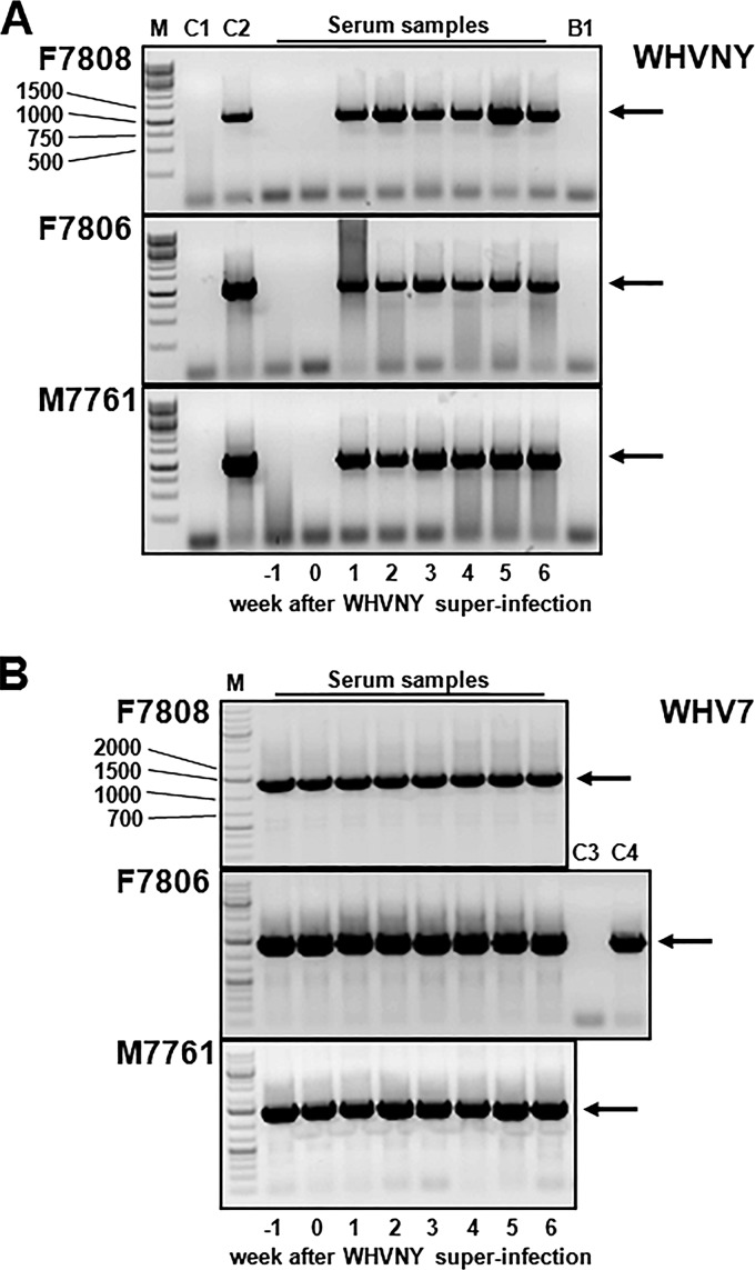
Each of three woodchucks that were WHV7 chronic carriers was superinfected with 1.66 × 10⁸ genome equivalents (GE) of WHVNY. Given that the liver of an adult woodchuck contains about 3 × 10¹⁰ hepatocytes (27), the multiplicity of WHVNY infection corresponded to approximately one WHVNY virion per 181 hepatocytes. The animals were monitored for a period of 6 weeks after the superinfection. The serum samples were collected at 1 week prior to the superinfection (week −1), on the day of the superinfection (week 0), and weekly after the superinfection with WHVNY. The results of the analysis of the serum samples harvested from the superinfected animals are summarized in Fig. 2. The analysis was carried out using two different WHV strain-specific nested PCR assays. The assays are described in detail in Materials and Methods. For all three superinfected woodchucks, the presence of the rcDNA of WHVNY was detected in the serum samples collected during weeks one through six after the superinfection with WHVNY. As shown in Fig. 2A, the WHVNY-specific PCR product (marked with an arrow) was observed only after the superinfection and was not detected in the samples collected prior to the superinfection (weeks −1 and 0). The WHVNY-specific PCR assay was able to detect 2 copies of WHVNY DNA and was not able to detect 2.0 × 10⁷ GE of WHV7 (Fig. 2A, controls C2 and C1, respectively). As expected, Fig. 2B demonstrates the presence of WHV7 rcDNA in all samples collected from all three animals at all time points, including weeks −1 and 0. It is shown that the WHV7-specific nested PCR assay produces the expected PCR product (indicated with arrow) for 2 × 10³ GE of WHV7, while it was unable to detect 2 × 10⁸ GE of WHVNY (Fig. 2B, controls C4 and C3, respectively). It should be noted that the amounts of isolated rcDNA used as templates for the detection of WHVNY were 100-fold higher than that for WHV7 (see Materials and Methods), indicating low titers of WHVNY in the sera. In addition, the presence of WHVNY in the sera was confirmed by sequencing of the cloned selected products of the WHVNY-specific nested PCR assay. The sequencing was conducted for the PCR products that were obtained for all three woodchucks, F7808, F7806, and M7761, and corresponded to weeks 1, 4, and 6 after the superinfection. The WHVNY sequences were verified using the unique WHVNY-specific nucleotides that were identified during the analysis of WHVNY molecular clones and were different from the WHV7 sequence (Fig. 1 and data not shown). Therefore, the generated data were consistent with the superinfection of all three WHV7 carrier woodchucks with a different strain, WHVNY.
FIG 2.
Detection of rcDNA of WHVNY and WHV7 in serum samples harvested from WHV7 chronic carrier woodchucks superinfected with strain WHVNY. The isolation of rcDNA and conventional nested WHV strain-specific PCR assays are described in Materials and Methods. Serum samples were collected at 1 week prior to the superinfection with WHVNY (week −1), immediately before the superinfection (week 0), and every week after the superinfection for the next 6 weeks (weeks 1 to 6). The time of the sample collection is shown at the bottom of each panel. The PCR products were analyzed in 1% agarose gels stained with ethidium bromide. Each gel image represents the analysis of serum samples collected from one woodchuck. The woodchucks are identified at the left. Results of WHVNY-specific PCR assay (A) and WHV7-specific PCR assay (B) are shown. The strain-specific PCR products are indicated with arrows on the right. The expected sizes of the final nested PCR products were 1,384 bp for WHVNY and 1,402 bp for WHV7. The serum sample-derived PCR products are indicated at the top. The control samples are the following: B1, no template; C1, 2.0 × 10⁷ GE of WHV7 DNA standard (plasmid pJSWHV7-2C6); C2, 2.0 GE of WHVNY DNA standard (plasmid pJSWHVNY-4C4); C3, 2.0 × 10⁸ GE of WHVNY DNA standard; and C4, 2.0 × 10³ GE of WHV7 DNA standard. The DNA size markers are shown on the left (lanes labeled M).
WHVNY RNA in the livers and matching HCCs of the superinfected woodchucks.
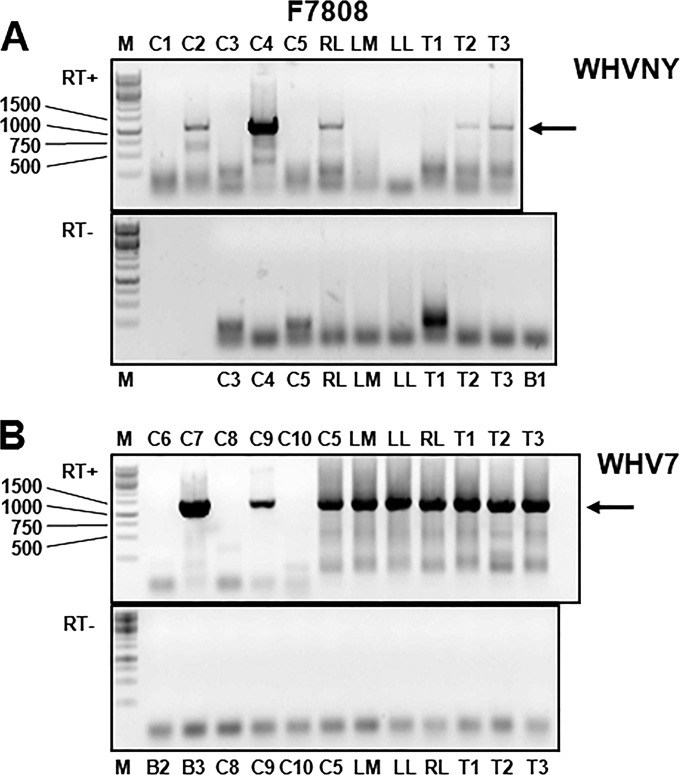
We next examined the tissue samples collected from the superinfected animals at the completion of the experiment. From each animal, the nonmalignant liver tissues and tissues of the matching HCCs were harvested. As nonmalignant liver tissues, the samples from the left medial liver lobe (LM), right lateral liver lobe (RL), and left liver lobe (LL) were collected. For each hepatocellular carcinoma (HCC) found in the livers upon necropsy, the center core of the tumor was excised and used for subsequent analysis. The isolation of RNA from tissue samples, preparation of cDNAs, and strain-specific conventional PCR assays that were employed for the detection of tissue-derived RNA of either WHVNY or WHV7 are detailed in Materials and Methods. It needs to be emphasized that the procedure for the detection of WHVNY RNA was modified, so it was possible to detect small amounts of RNA templates. No similar modifications were necessary for the detection of WHV7 RNA. These aspects of RNA detection in tissues indicated the relative abundance of WHV7 RNA and the presence of small amounts of WHVNY RNA intermediates. For all experiments assaying for WHV RNAs in tissues harvested at necropsy, the same set of control samples was used. The results obtained are shown in Fig. 3 to 5. Figure 3 represents the data generated during the analysis of tissues collected from woodchuck F7808. Briefly, the WHVNY-specific assay was shown to detect 4 copies of WHVNY DNA and 9 copies of WHVNY RNA (Fig. 3A, top image, lanes C2 and C4, respectively). However, 4.0 × 10⁷ GE of WHV7 DNA and 9.0 × 10⁷ GE of WHV7 RNA were not amplified by this assay (Fig. 3A, top image, lanes C1 and C3, respectively). In addition, no WHVNY-specific PCR product was observed when the total RNA from the woodchuck liver that was monoinfected with WHV7 was analyzed (Fig. 3A, top image, lane C5). These PCR results demonstrate that the assay employed is highly sensitive and strictly WHVNY specific. Using separate pieces of the same tissue sample, the total RNA extraction and subsequent analysis of isolated RNA was repeated for each tissue sample at least two times. Figure 3A represents typical results of the WHVNY-specific reverse transcription-seminested PCR analysis. The tissues that tested positive for WHVNY RNA on at least two separate occasions were RL, T2 (i.e., HCC2), and T3. For each of these tissues, the WHVNY-specific PCR product is shown in the corresponding lanes in the top image of Fig. 3A. For the tissues LM, LL, and T1, in which WHVNY RNA was detected just once, and during other independent repeats, our reverse transcription-PCR assay was unable to detect WHVNY RNA; no WHVNY-specific PCR products were shown in the corresponding lanes (Fig. 3A, top image). For all examined tissue samples harvested from all three woodchucks, F7808, F7806, and M7761, the frequencies of the detection of WHVNY RNA during the independent isolations are summarized in detail in Table 1. The bottom image of Fig. 3A shows the control experiments (i) that were performed in the absence of reverse transcriptase (RT) and (ii) during which no WHVNY-specific PCR products were generated. Therefore, WHVNY-specific PCR products, which were observed for tissue samples from woodchuck F7808 only when the RT was used (Fig. 3A, top image), clearly represent the results of the amplification of WHVNY RNA. Figure 3B shows the results of the WHV7-specific reverse transcription-nested PCR assay. It clearly demonstrated that the assay was highly sensitive and selectively amplified only sequences of WHV7 (Fig. 3B, top image, lanes C6 to C9 and C5, representing the DNA and RNA controls of WHV7 and WHVNY). As expected, the assay also did not produce any PCR product when the total RNA extracted from uninfected woodchuck liver was analyzed (Fig. 3B, top image, lane C10). As anticipated and consistent with our previous results (31), all analyzed samples of either nonmalignant liver tissues or HCC tissues tested positive for WHV7 RNA on all independent occasions, and all tissue-derived PCR products clearly were generated by the amplification of RNA sequences (Fig. 3B, top image, lanes LM, RL, LL, T1, T2, and T3, and bottom image). Overall, the data suggest that the liver of animal F7808 was superinfected with WHVNY. However, it appeared that not all parts of the liver and not all HCC tissues were superinfected with strain WHVNY to a similar extent.
FIG 3.
Detection of WHVNY RNA and WHV7 RNA in the samples of nonmalignant liver tissues and matching HCC tissues of woodchuck F7808. The isolation of total RNA from the tissue samples and conventional strain-specific PCR assays that amplified either RNA of WHVNY or RNA of WHV7 are described in Materials and Methods. All tested tissue samples were collected at necropsy. The PCR products were analyzed in 1% agarose gels that were stained with ethidium bromide. (A) Results of WHVNY-specific PCR assay. (B) Results of WHV7-specific PCR assay. In each panel, the gel image at the top represents the results of RNA analysis that was conducted in the presence of the reverse transcriptase (RT+), while the gel image at the bottom represents the results obtained in the absence of the reverse transcriptase (RT−). The tissues assayed along with the control samples used are indicated at the top or bottom of the gel images. The HCC tissues are identified as T numbers, i.e., T1 stands for HCC1, etc. The control samples are the following: B1, no template in the 1st PCR; B2, no template in the 1st PCR; B3, no template in the 2nd PCR; C1, 4.0 × 10⁷ GE of WHV7 DNA standard (pJSWHV7-2C6); C2, 4.0 GE of WHVNY DNA standard (pJSWHVNY-4C4); C3, 9.0 × 10⁷ GE of WHV7 RNA standard; C4, 9.0 GE of WHVNY RNA standard; C5, total RNA extracted from nonmalignant tissue of the right medial liver lobe of woodchuck 41 that was a carrier of WHV7; C6, 2.0 × 10⁶ GE of WHVNY DNA standard (pJSWHVNY-4C4); C7, 2.0 GE of WHV7 DNA standard (pJSWHV7-2C6); C8, 5.0 × 10⁷ GE of WHVNY RNA standard; C9, 5.0 GE of WHV7 RNA standard; and C10, total RNA isolated from normal LL tissue harvested from uninfected woodchuck 4805. The control DNA standards and the preparation of WHV strain-specific control RNA standards are described in Materials and Methods. The DNA standards were amplified in the absence of RT. In panel A, for tissues LM, LL, and T1 (HCC1), no WHVNY-specific PCR product is shown, because WHVNY RNA in these tissues was detected only once, while a number of independent RNA isolations per tissue sample were conducted and analyzed. For all other examined tissues, WHVNY RNA was detected during at least two independent RNA isolations (Table 1). The specific PCR products of the expected sizes are indicated with arrows on the right. The expected sizes of the final PCR products were 1,082 bp for WHVNY and 1,402 bp for WHV7. The DNA size markers are shown on the left (lanes labeled M).
FIG 5.
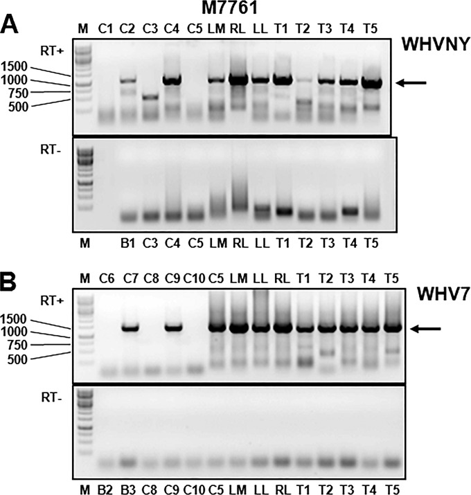
Detection of WHVNY RNA and WHV7 RNA in the samples of nonmalignant liver tissues and matching HCC tissues of woodchuck M7761. The total RNA isolation and conventional PCR assays that amplified exclusively either RNA of the strain WHVNY or RNA of the strain WHV7 are described in Materials and Methods. The tissue samples used for the analysis were collected during necropsy. The PCR products were resolved in 1% agarose gels stained with ethidium bromide. (A) Results of WHVNY-specific PCR assay. (B) Results of WHV7-specific PCR assay. At the top of each panel, the gel image shows the results of RNA analysis done in the presence of the reverse transcriptase (RT+). The bottom gel image of each panel demonstrates the results obtained in the absence of reverse transcriptase (RT−). The tissues and control samples are indicated at the top or bottom of the gel images. The HCC tissues are identified as T numbers, i.e., T1 stands for HCC1, etc. The controls and standards are the same as the ones described in the legend to Fig. 3. DNA controls were amplified in the absence of RT. For woodchuck M7761, WHVNY RNA was found in all examined tissue samples during at least two independent RNA isolations (Table 1). The WHV strain-specific PCR products of the expected sizes (see the legend to Fig. 3 for details) are indicated with arrows on the right. The DNA size markers are indicated on the left (lanes labeled M).
TABLE 1.
Frequency of WHVNY RNA detection in nonmalignant liver tissues and HCC samples
| Woodchuck and tissue typea | Tissue sampleb | Frequency of WHVNY RNA detectionc |
|---|---|---|
| F7808 | ||
| Liver | LM | 1/7 |
| RL | 2/8 | |
| LL | 1/11 | |
| HCC | T1 | 1/7 |
| T2 | 3/10 | |
| T3 | 2/5 | |
| F7806 | ||
| Liver | LM | 2/2 |
| RL | 2/3 | |
| LL | 3/6 | |
| HCC | T1 | 2/6 |
| T2 | 3/4 | |
| T3 | 3/3 | |
| M7761 | ||
| Liver | LM | 3/3 |
| RL | 4/4 | |
| LL | 2/2 | |
| HCC | T1 | 3/5 |
| T2 | 2/2 | |
| T3 | 2/2 | |
| T4 | 3/3 | |
| T5 | 3/3 |
The current study used three WHV7 carrier woodchucks, F7808, F7806, and M7761, which were superinfected with another WHV strain, WHVNY.
The analyzed tissue samples were harvested during necropsy at 6 weeks after superinfection with WHVNY. For each animal, the nonmalignant liver tissues from left medial liver lobe (LM), right lateral liver lobe (RL), and left liver lobe (LL) were collected. In addition, the tissues samples (the center cores) also were harvested from each of the WHV-induced HCCs that were identified in the livers during necropsy. T1 stands for HCC1, T2 for HCC2, etc.
For each tissue sample, two or more independent isolations of the total RNA and the subsequent analysis for the presence of WHVNY RNA were conducted. The numbers in the table reflect the number of occasions during which WHVNY RNA was detected using a conventional seminested PCR assay relative to the total number of independent RNA isolations performed.
Figures 4 and 5 represent the results of detection of WHVNY RNA and WHV7 RNA in tissue samples harvested from woodchucks F7806 and M7761, respectively. For both animals, the presence of WHVNY RNA was detected in all nonmalignant liver tissue samples (LM, RL, and LL), as judged by the appearance of the WHVNY-specific PCR product of the expected size. In addition, all HCC tissue samples collected from both woodchucks (three samples from animal F7806 and five samples from woodchuck M7761) tested positive for WHVNY RNA as well (Fig. 4A and 5A). The WHVNY-specific PCR products for the tissues collected from woodchucks F7806 and M7761 were obtained during at least two independent isolations/examinations (Table 1). Similar to woodchuck F7808, all liver/HCC tissue samples of animals F7806 and M7761 also were positive for the presence of WHV7 RNA (Fig. 4B and 5B). To further confirm the detection of WHVNY RNA, for each animal, selected PCR products derived from nonmalignant liver tissue/HCC tissue samples were analyzed further by cloning and sequencing as described in detail in Materials and Methods. The amplification of the WHVNY RNA sequences has been verified for all examined PCR products (data not shown). In addition, during the analysis of the biopsy tissue samples collected prior to the superinfection (from nonmalignant liver tissues and from HCC tissues [see Materials and Methods]), we were able to detect WHV7 RNA in each examined tissue, while no WHVNY RNA was detected in any of the tissue samples (data not shown).
FIG 4.
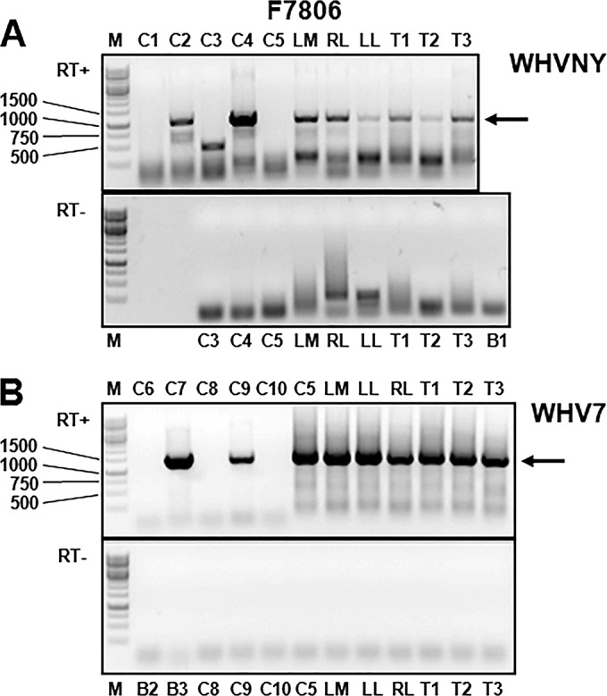
Detection of WHVNY RNA and WHV7 RNA in the samples of nonmalignant liver tissues and matching HCC tissues of woodchuck F7806. The isolation of total RNA and conventional WHV strain-specific PCR assays that amplified either RNA of WHVNY or RNA of WHV7 are described in Materials and Methods. The tissue samples were harvested during necropsy. The PCR products were resolved in 1% agarose gels stained with ethidium bromide. (A) Results of WHVNY-specific PCR assay. (B) Results of WHV7-specific PCR assay. The gel image at the top of each panel demonstrates the results of RNA analysis performed in the presence of reverse transcriptase (RT+). The gel image at the bottom of each panel shows the results obtained in the absence of reverse transcriptase (RT−). The examined tissue samples and the control samples are indicated at the top or bottom of the gel images. The HCC tissues are identified as T numbers, i.e., T1 stands for HCC1, etc. The controls and DNA and RNA standards are the same as those described for Fig. 3. The preparation of WHV strain-specific DNA and RNA control standards is described in Materials and Methods. DNA controls were amplified in the absence of RT. WHVNY RNA was detected in all assayed tissue samples during at least two independent RNA isolations/examinations (Table 1). The specific PCR products of the expected sizes (see the legend to Fig. 3 for details) are indicated with arrows on the right. The DNA size markers are shown on the left (lanes labeled M).
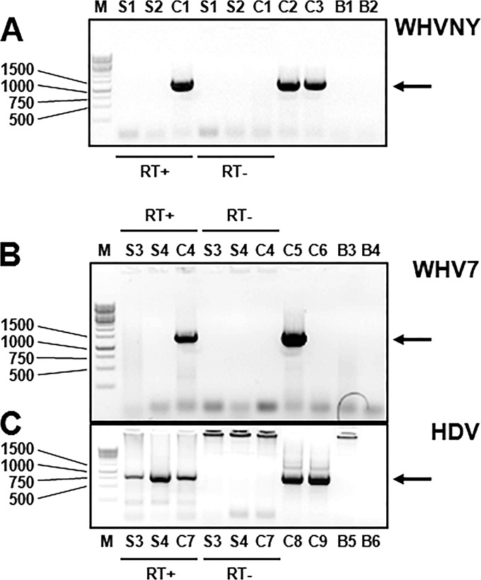
Additional control experiments were conducted in order to demonstrate that WHV RNA detected in tissue samples was produced by the replication of WHVNY and WHV7 in cells and could not be associated with virions from the sera. The results of the analysis of serum virions collected from two woodchucks that were transiently infected with WHVNY are shown in Fig. 6A. The serum titers of WHVNY were 2.37 × 10⁹ GE/ml (woodchuck F6541) and 3.15 × 10⁹ GE/ml (woodchuck F6678). The strategies that were employed for the isolation of total RNA from the serum samples, DNase treatment, and subsequent analysis for the presence of WHV RNA are described in Materials and Methods. The low copy numbers of 5.0 GE of in vitro-transcribed and gel-purified WHVNY RNA standard and 2.0 and 20 GE of WHVNY DNA standard were easily detectable by the assay. However, no WHVNY RNA was detected in either serum sample (Fig. 6A). Similar results were obtained for the serum samples harvested from two woodchucks chronically infected with WHV7 and then superinfected with wHDV. While being superinfected with wHDV, both WHV7 carrier woodchucks maintained high titers of WHV7 in sera. For woodchuck F6438, the titer was 1.74 × 10¹⁰ WHV7 GE/ml, while for animal M6593, the titer was 1.04 × 10¹⁰ WHV7 GE/ml. As shown in Fig. 6B, the WHV7-specific assay described above was able to readily detect 5.0 GE of in vitro-prepared and gel-purified WHV7 RNA standard and 20 copies of WHV7 DNA standard, while 2 copies of WHV7 DNA were not amplified. No WHV7 RNA was detected in either of the serum samples tested (Fig. 6B). The use of the material harvested from WHV7 carriers superinfected with wHDV provided us with the unique opportunity to demonstrate that the RNA extraction procedure used was adequate and did not compromise the integrity of the serum-associated virion RNA. The same RNA preparations that were examined in Fig. 6B were subjected to HDV-specific PCR in order to detect virion-associated genomic RNA of HDV. The HDV titers in the tested serum samples were determined by HDV-specific qPCR assay as described previously (31) and were 2.30 × 10⁷ HDV GE/ml (woodchuck F6438) and 2.48 × 10⁷ HDV GE/ml (woodchuck M6593). In agreement with the qPCR data, the genomic HDV RNA was detected in the presence of RT in both serum samples (Fig. 6C) by conventional reverse transcription-nested PCR assay. Therefore, based on the results presented in Fig. 3 to 6, it was concluded that the detected RNAs of WHV7 and WHVNY in liver/HCC tissue samples represented cell-associated RNA intermediates of WHV replication.
FIG 6.
Experimental proof that WHV RNA was not detected in the sera of woodchucks that were either transiently or chronically infected with WHV. The conventional PCR assays that amplified exclusively either WHVNY RNA or WHV7 RNA are the same as the ones that were used in experiments presented in Fig. 3 to 5. The HDV-specific nested PCR assay that amplifies the genomic HDV RNA, which is present in HDV virions, is detailed in Materials and Methods. In all panels, the reactions performed in the presence of the reverse transcriptase are labeled RT+, and the reactions performed in the absence of the reverse transcriptase are marked RT−. The details regarding WHV strain-specific RNA and DNA control standards are described in Materials and Methods. The PCR products were examined in 1% agarose gels stained with ethidium bromide. (A) Analysis of total RNA isolated from the serum samples of woodchucks F6678 and F6541 using WHVNY-specific PCR assay. For RNA isolation, serum collected from animal F6678 at week eight after monoinfection with the strain WHVNY and serum from animal F6541 harvested at week 11 after monoinfection with WHVNY were used. The samples that were assayed were the following: S1, total RNA from the serum of F6678; S2, total RNA from serum of F6541; C1, 5.0 GE of WHVNY RNA standard; C2, 20 GE of WHVNY DNA standard; C3, 2.0 GE of WHVNY DNA standard; B1, no template in the 1st PCR; B2, no template in the 2nd PCR. Panels B and C show the results of the analysis of total RNA isolated from the sera collected from two woodchucks, F6438 and M6593, which were chronic carriers of WHV7 that also were superinfected with WHV-enveloped hepatitis delta virus (wHDV). A serum sample from woodchuck F6438 was collected at 4 weeks after wHDV superinfection, while serum from M6593 was harvested at 5 weeks after wHDV superinfection. (B) Analysis conducted using WHV7-specific nested PCR assay. (C) Results of the analysis that used HDV-specific nested PCR assay. The samples that have been assayed were the following: S3, total RNA isolated from serum of F6438; S4, total RNA from serum of M6593; C4, 5.0 GE of WHV7 RNA standard; C5, 20 GE of WHV7 DNA standard; C6, 2.0 GE of WHV7 DNA standard; C7, 5.0 × 10³ GE of HDV genomic RNA standard (prepared as described previously [31]); C8, 3.7 × 10¹⁰ HDV DNA standard (plasmid pSVLD3, which harbors the trimer of the HDV genome [51]); C9, 3.7 × 10⁹ HDV DNA standard; B3, no template in the 1st PCR; B4, no template in 2nd PCR; B5, no template in the 1st PCR; B6, no template in 2nd PCR. The DNA control standards were amplified in the absence of RT. The specific PCR products of the expected sizes are indicated with arrows on the right. The expected sizes of WHVNY RNA-derived and WHV7 RNA-derived PCR products are described in the legend to Fig. 3. The expected size of the HDV-specific PCR product was 819 bp. The DNA size markers are shown on the left (lanes labeled M).
Overall, the data regarding RNA detection confirmed the occurrence of WHVNY superinfection of the livers of the three WHV7 carriers and demonstrated that WHVNY replication was maintained at 6 weeks after the superinfection. The results of the histopathology analysis of the tissues harvested at necropsy are presented in Table 2. As can be seen, 7/11 of the harvested HCC center core samples were classified as partial HCC tissues with the percentage of malignant HCC cells ranging between 15% and 95% of the total number of cells. For four HCC samples, T2 of woodchuck F7808 and T1, T2, and T4 of animal M7761, only HCC cells were detected during the examination by the pathologist (B. V. Kallakury). Therefore, these four HCCs were classified as total (pure) HCCs. Furthermore, since the tumors, T2 of woodchuck F7808 and T1 of animal M7761, which were classified as pure HCCs (Table 2), tested positive for the presence of WHVNY RNA during at least two independent assays (Table 1) and initially were identified and located in the livers of WHV7 carriers prior to the superinfection with WHVNY (Table 2), our results give preliminary indication that in vivo, at least fractions of the cells of WHV-induced HCCs are susceptible to reinfection with a hepadnavirus, WHV.
TABLE 2.
Histopathology analysis of collected tissue samples
| Woodchuck and tissue typea | Tissue sample | Histopathologyb | Tumor detected prior to WHVNY superinfectionc |
|---|---|---|---|
| F7808 | |||
| Liver | LM | Totally normal liver tissue | NA |
| RL | Totally normal liver tissue | NA | |
| LL | Partial normal liver; 30% scattered foci of altered hepatocytes (FAH) | NA | |
| HCC | T1 | Partial HCC; 40% scattered nodular HCC, 60% nonneoplastic liver tissue; chronic hepatitis | Yes |
| T2 | Total HCC | Yes | |
| T3 | Partial HCC; 15% scattered nodular HCC, 85% nonneoplastic liver tissue; chronic hepatitis | Yes | |
| F7806 | |||
| Liver | LM | Totally normal liver tissue | NA |
| RL | Totally normal liver tissue | NA | |
| LL | Totally normal liver tissue | NA | |
| HCC | T1 | Partial HCC; 90% HCC, 10% nonneoplastic liver tissue | Yes |
| T2 | Partial HCC; 60% scattered | No | |
| Nodular HCC; 40% nonneoplastic | |||
| Liver tissue; chronic hepatitis | |||
| T3 | Partial HCC; 50% scattered | No | |
| Nodular HCC, 50% nonneoplastic | |||
| Liver tissue; chronic hepatitis | |||
| M7761 | |||
| Liver | LM | Normal liver tissue; mild chronic hepatitis | NA |
| RL | Normal liver tissue; mild chronic hepatitis | NA | |
| LL | Normal liver tissue; mild chronic hepatitis | NA | |
| HCC | T1 | Total HCC | Yes |
| T2 | Total HCC | No | |
| T3 | Partial HCC; 95% HCC, 5% nonneoplastic liver tissue | Yes | |
| T4 | Total HCC | No | |
| T5 | Partial HCC; 75% HCC, 25% nonneoplastic liver tissue | No |
The description of the animals and tissue samples collected at necropsy that were used for the analysis is provided in the footnotes to Table 1.
The tissue samples were examined by the pathologist (B. V. Kallakury), and the findings are summarized in the table. The findings describe the results of the analysis of the morphology/histopathology of the collected tissue samples. For each HCC tissue sample analyzed, whether the tissue consisted of only malignant HCC cells is indicated. If the tissue sample represented a partial HCC, the percentage of HCC cells was indicated and the rest of the tissue was characterized. For nonmalignant liver tissues, when the tissue was concluded to be partial normal liver tissue, the rest of the tissue also was briefly characterized.
The HCCs that were identified and located in the livers prior to superinfection with WHVNY are indicated. NA, nonapplicable.
Next, the data on the frequency of WHVNY RNA detection in liver/HCC tissue samples, which are summarized in Table 1, have been considered further. Clearly, for a number of tissues (11 tissues out of 20 tested tissues from all three animals), it was observed that while several independent RNA isolations/examinations were conducted per tissue sample, not every RNA preparation (that was obtained for the same tissue sample) tested positive for WHVNY RNA. This was the case for all liver and HCC tissue samples harvested from woodchuck F7808 (6/6 tissues), for 4/6 tissues collected from animal F7806, and for 1/8 tissue samples obtained from woodchuck M7761. Furthermore, for woodchuck F7808, in three tissues, LM, LL, and T1, the RNA of WHVNY was detected only during 1/7, 1/11, and 1/7 independent RNA isolations, respectively. However, each tissue sample (20/20) tested positive for WHVNY RNA at least once (Table 1). These results suggest (i) that cells that were superinfected with WHVNY have been scattered throughout the livers, and (ii) that the superinfection with WHVNY was not uniform. Because it appears that at least in 11/20 tissue samples tested only certain fractions of the cells were infected with WHVNY, we interpreted that WHVNY occupied the replication space, which was limited to a certain extent at 6 weeks after the superinfection. In addition, it needs to be emphasized that it was necessary to modify the reverse transcription-PCR procedure for the detection of WHVNY RNA so very small amounts of the RNA of interest could be captured. The necessity of such modifications and data on the frequency of detection of WHVNY RNA together suggest that the efficiency of WHVNY superinfection was rather low, and likely a limited number of cells replicated WHVNY.
Quantitative analysis of WHVNY superinfection. (i) Measurements of serum rcDNA titers of WHVNY and WHV7.
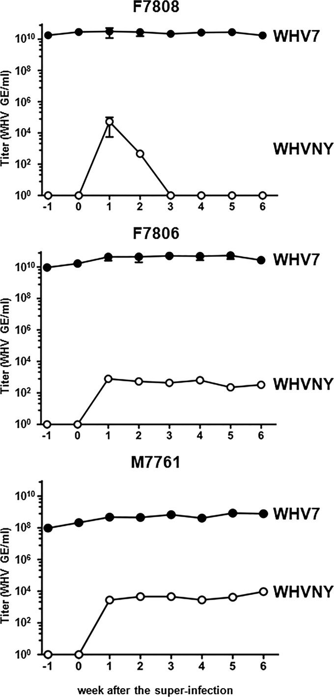
Our next focus was the quantitative characterization of WHVNY superinfection. First, we measured the WHVNY and WHV7 levels in serum samples using WHV strain-specific qPCR assays specially developed by us that were able to selectively detect either WHVNY or WHV7 sequences in the mixtures of DNAs of both WHV strains. The details regarding the isolation of rcDNAs and qPCRs used are described in Materials and Methods. The amounts of isolated rcDNA used to quantify WHVNY titers were 100-fold higher than the amounts used for measurements of the titers of WHV7, which was consistent with the low abundance of WHVNY in serum and was in agreement with the results of detection of WHVNY RNA in tissue samples described in the previous section. The results of the WHV rcDNA titers' measurements are presented in Fig. 7. It was possible to quantify WHVNY titers in the sera of all woodchucks starting one week after the superinfection. However, as expected, WHVNY rcDNA was not detected in the serum samples collected prior to the superinfection. For woodchuck F7808, WHVNY serum concentration reached the highest level of 5.29 × 10⁴ GE/ml at week one, and then it quickly declined to about 500 GE/ml by week two, and from week 3 to the remainder of the experiment it was below the qPCR detection limit, which indicates inefficient superinfection in this animal. This result is consistent with the fact that for the tissue samples collected from woodchuck F7808 at necropsy, only one nonmalignant liver tissue out of three tested and the tissue samples from two HCCs (T2 and T3) out of three examined tested positive for WHVNY RNA on more than one separate occasion (Table 1). For the same animal, at all times, the titer of WHV7 was stably high, between 1.74 × 10¹⁰ and 3.18 × 10¹⁰ GE/ml. Therefore, during the whole time of the experiment, the titers of WHV7 were higher than that of WHVNY by at least a factor of 6.0 × 10⁵ (Fig. 7). In the other two woodchucks, F7806 and M7761, the superinfection with WHVNY seemed to be more efficient. For woodchuck F7806, during weeks one through six after the superinfection, the WHVNY rcDNA serum levels persisted within a range between about 230 and 795 GE/ml. The WHV7 titers were consistently high during the entire experiment, within a range of 9.43 × 10⁹ to 5.45 × 10¹⁰ GE/ml. Therefore, the ratio of the WHV7 to WHVNY virions in the serum of woodchuck F7806 was above a factor of 5.5 × 10⁷ during all 6 weeks after the superinfection (Fig. 7). The most efficient WHVNY superinfection was observed in the liver of woodchuck M7761. The WHVNY titers were within a range between 2.77 × 10³ and 9.50 × 10³ GE/ml. Interestingly, at week six, there was an apparent increase in WHVNY titer. Similar to woodchuck F7806, no signs of the resolution of WHVNY superinfection were detected. The titers of WHV7 were between 9.71 × 10⁷ and 8.51 × 10⁸ GE/ml, which was somewhat lower than the values that were observed for the animals F7806 and F7808. The WHV7/WHVNY ratio was in the range of 8.28 × 10⁴ to 2.03 × 10⁵ (Fig. 7). The fact that relatively low titers of WHVNY persisted in both animals F7806 and M7761 without the indication of significant declining during the entire observation period of 6 weeks demonstrated that the superinfecting virus WHVNY was not progressively cleared from the superinfected livers. Therefore, the data showed that WHVNY displayed a potential for persistence after the superinfection, which was consistent with the results of conventional PCR assays used to test for (i) WHVNY rcDNA in the serum samples and (ii) WHVNY RNA in tissue samples (Fig. 2 to 5). Low WHVNY titers again indicated that WHVNY occupied a limited replication space in the superinfected livers.
FIG 7.
Quantification of rcDNA of WHVNY and WHV7 in sera of superinfected woodchucks F7808, F7806, and M7761. The isolation of WHV rcDNA from serum samples and WHV strain-specific real-time PCR assays that were used to quantify the rcDNA levels of WHVNY and WHV7 are described in Materials and Methods. The y axis represents the titer values in WHV GE/ml of serum (logarithmic scale). The x axis shows the time after the superinfection in weeks. The time of 1 week prior to WHVNY superinfection is shown as week −1, and the week of the superinfection is shown as week 0. The woodchuck number is indicated above the corresponding graph. The results for WHV7 rcDNA are shown as black circles, and the data for WHVNY rcDNA are shown as open circles. The standard deviations are indicated.
(ii) Quantification of RI-DNA and cccDNA of WHVNY and WHV7 in the livers and HCC tissue samples harvested from superinfected woodchucks.
The tissue samples harvested at necropsy were analyzed further in order to quantify the RI-DNA and cccDNA of both WHV strains. The strategies of quantification of RI-DNA and cccDNA were based on previously described protocols (38). The details of DNA preparations that were used to quantify RI-DNA and cccDNA of WHV and the respective qPCR assays are described in Materials and Methods. On a number of occasions, an additional modification was introduced in a preparation of the fraction used for the cccDNA analysis. Thus, the DNA fraction obtained using the Hirt extraction was further treated with Plasmid-Safe-DNase (PSD), which resulted in a preparation of DNA fraction that was not only enriched for cccDNA content but also was profoundly depleted of other WHV DNA intermediates. Tables 3 and 4 serve as typical illustrations of the features of the procedures used to quantify RI-DNA and cccDNA. Four sets of WHV strain-specific controls were used to evaluate (i) the efficiency and specificity of the extraction procedures, (ii) the recovery of different kinds of WHV DNA replication intermediates, and (iii) the efficiency and specificity of the WHV strain-specific qPCR assays. The uncut plasmids that bear specific WHV sequences were used as the surrogates for cccDNA species. The double-stranded, linear, greater-than-genome-size, PCR-made DNAs of WHVNY and WHV7 that have the right end at position 1945 and left end at nucleotide 1935 (38) served as the surrogates of double-stranded linear DNA (DSL) WHV genomes. The rcDNA purified from woodchuck serum was used to represent the protein-free rcDNA (pf-rcDNA). pf-rcDNA is an important intermediate of hepadnavirus replication that may represent about 10% of cytoplasmic rcDNA and about 40% of nuclear rcDNA of HBV and that was suggested to be a precursor for cccDNA. Unlike protein-bound rcDNA, pf-rcDNA partitions to the aqueous phase during phenol extraction, while polymerase-bound rcDNA goes into organic phase (in the absence of proteinase treatment) (44). Finally, the fourth kind of controls were serum rcDNAs (i.e., not extracted and, therefore, protein-bound rcDNA). Each of the control samples was added to a piece of liver tissue (approximately 50 to 100 mg) harvested from a WHV-negative (naive) woodchuck, and then the extractions of total DNA and Hirt extraction with or without subsequent PSD treatment were conducted. Table 3 summarizes the analysis of the WHVNY-specific controls, while Table 4 presents the results of analysis of WHV7-specific controls. For the quantification of RI-DNA, the total DNA extraction procedure was used in combination with a qPCR assay that would amplify all types of WHV DNA replication intermediates. For both WHVNY and WHV7, the same strain-specific qPCR assays that were used for the quantification of rcDNA (see above) were used for the quantification of RI-DNA in total DNA fractions. When the plasmid controls were used as the surrogates for the WHV strain-specific cccDNA species, it can be seen from Tables 3 and 4 that WHV strain-specific qPCR assays (that are used to measure RI-DNA) showed cccDNA recovery levels that ranged between 40% and complete recovery. The same qPCRs also demonstrated that the recovery of the DSL substitutes in total DNA fractions was in a range of between about 35% and complete recovery. The observed recovery of the pf-rcDNA was between about 30% and 54% for WHV7 and between 30% and complete recovery for WHVNY, when the amounts of the input samples were 2.0 × 10⁴ WHV GE and higher. When the starting amounts of pf-rcDNA were 2.0 × 10³ WHV GE, the recovery was between 0 and 27%, which likely represents the limitations of the procedure related to relatively low copy numbers of the input material. The serum-associated rcDNA was recovered by a total DNA extraction procedure (that employs the treatment with pronase and transforms the protein-bound serum-derived rcDNA into pf-rcDNA) within a range between 48% and complete recovery (the starting amounts of rcDNA in these experiments were 2.0 × 10⁴ GE and higher). It needs to be noted that for both strains of WHV, the controls were tested in sufficient ranges of concentrations that allowed fair evaluation of the outcomes of the procedures (Tables 3 and 4).
TABLE 3.
Analysis of control WHVNY DNA samples during the detection of RI-DNA and cccDNA using WHVNY-specific PCR assaysa
| Sample and amountb (GE) | RI-DNA (total DNA prepn) |
cccDNA (Hirt) |
cccDNA (Hirt + PSD) |
|||
|---|---|---|---|---|---|---|
| GE | Recovery (%) | GE | Recovery (%) | GE | Recovery (%) | |
| Plasmidc | ||||||
| 1.09 × 10⁷ | 1.07 × 10⁷ | 97.8 | 8.71 × 10⁶ | 80.0 | 9.82 × 10⁶ | 90.1 |
| 1.05 × 10⁴ | 1.29 × 10⁴ | 122.3g | 1.76 × 10⁴ | 167.5g | 1.23 × 10⁴ | 116.5g |
| 9.02 × 10³ | 7.44 × 10³ | 82.5 | 1.09 × 10⁴ | 120.8g | 4.75 × 10³ | 52.6 |
| 7.43 × 10² | 7.95 × 10² | 107.0g | 1.28 × 10³ | 172.2g | 3.46 × 10² | 46.6 |
| 1.42 × 10² | 1.11 × 10² | 78.0 | 2.28 × 10² | 160.5g | 1.96 × 10² | 137.9g |
| DSLd | ||||||
| 3.30 × 10⁴ | 1.90 × 10⁴ | 57.7 | 0.0 | 0.0 | 0.0 | 0.0 |
| 1.04 × 10⁴ | 1.69 × 10⁴ | 162.5g | 0.0 | 0.0 | 0.0 | 0.0 |
| 1.97 × 10³ | 1.97 × 10³ | 99.6 | 0.0 | 0.0 | 0.0 | 0.0 |
| 3.85 × 10² | 5.55 × 10² | 144.0g | 0.0 | 0.0 | 0.0 | 0.0 |
| 2.03 × 10² | 2.34 × 10² | 115.1g | 0.0 | 0.0 | 0.0 | 0.0 |
| 4.62 × 101 | 5.64 × 101 | 122.2g | 0.0 | 0.0 | 0.0 | 0.0 |
| pf-rcDNAe | ||||||
| 2.00 × 10⁶ | 1.74 × 10⁶ | 87.0 | 3.19 × 10³ | 0.16 | 2.00 × 101 | 0.001 |
| 2.00 × 10⁶ | 6.84 × 10⁵ | 34.2 | 3.47 × 10³ | 0.17 | 0.0 | 0.0 |
| 2.00 × 10⁴ | 2.38 × 10⁴ | 118.8g | 9.53 × 100 | 0.05 | 0.0 | 0.0 |
| 2.00 × 10⁴ | 6.05 × 10³ | 30.3 | 4.52 × 101 | 0.23 | 0.0 | 0.0 |
| 2.00 × 10³ | 1.62 × 10² | 8.1 | 0.0 | 0.0 | 0.0 | 0.0 |
| 2.00 × 10³ | 3.76 × 10² | 18.8 | 0.0 | 0.0 | 0.0 | 0.0 |
| Serum rcDNAf | ||||||
| 2.00 × 10⁶ | 1.88 × 10⁶ | 93.0 | 1.27 × 10² | 0.01 | 0.0 | 0.0 |
| 2.00 × 10⁶ | 3.11 × 10⁶ | 155.7g | 3.38 × 10² | 0.02 | 0.0 | 0.0 |
| 2.00 × 10⁴ | 1.72 × 10⁴ | 86.0 | 0.0 | 0.0 | 0.0 | 0.0 |
| 2.00 × 10⁴ | 2.43 × 10⁴ | 121.6g | 0.0 | 0.0 | 0.0 | 0.0 |
The WHVNY strain-specific qPCR assays that amplified either RI-DNA or cccDNA, the isolation of total DNA, Hirt extract preparation, and subsequent treatment with PSD are described in detail in Materials and Methods.
WHVNY DNA control samples were used to evaluate the isolation/treatment procedures, the specificity of the qPCR assays, and the degree of recovery of certain DNA intermediates of WHVNY replication. Each control sample (the number of WHVNY GE is indicated) was added to a piece of liver tissue (approximately 50 to 100 mg) harvested from a WHV-negative (naive) adult woodchuck, and then the preparation of DNA for analysis was conducted as indicated. The results are shown in WHVNY DNA GE and as the percentage of recovery relative to the starting amounts of the input control sample. In addition, all WHVNY-specific control DNA samples listed were tested using analogous WHV7-specific qPCR assays (for the detection of the RI-DNA in total DNA preparation and cccDNA in Hirt and Hirt + PSD preparations), which, as expected, did not detect any of the WHVNY DNA samples.
Uncut plasmid pLR-WHVNY-001 harboring WHVNY sequences (described in Materials and Methods) was used as the surrogate to evaluate the parameters of analysis of cccDNA.
A surrogate of DSL of WHVNY was prepared by PCR, using the plasmid pJSWHVNY-4C4 as the template, followed by gel purification of the obtained PCR product. The WHVNY DSL mimic is a linear, double-stranded, greater-than-genome-length DNA that, like its natural counterpart, has the left-hand position at nucleotide 1935 and the right-hand position at nucleotide 1945 (38). The details of the preparation of the WHVNY DSL surrogate are described in Materials and Methods. For the surrogate of cccDNA and surrogate of DSL, the initial concentration measurements were performed using a NanoDrop 2000. The dilutions were made based on these measurements, and then the concentrations of diluted samples were verified by qPCR that amplified the RI-DNA (the same qPCR as that used to quantify the rcDNA in sera), and the actual qPCR measurements are shown as the starting amounts of input samples.
To evaluate the parameters of analysis of protein-free rcDNA (pf-rcDNA), the rcDNA of WHVNY was isolated from the serum of woodchuck F6541, which was monoinfected with WHVNY.
To examine the parameters of analysis of serum-associated rcDNA (i.e., protein-bound rcDNA), the serum that contained rcDNA of WHVNY from woodchuck F6541, which was monoinfected with the strain WHVNY, was used. The concentrations of isolated rcDNA were measured by qPCR specific for RI-DNA, and the dilutions for input material (both kinds of rcDNA control samples) were made based on the actual qPCR measurements.
On several occasions the calculated recovery exceeded 100%, which was less than 2-fold different from the actual 100% value, reflected a variation in qPCR measurements, and was considered a complete recovery of the input sample.
TABLE 4.
Analysis of control WHV7 DNA samples during the detection of RI-DNA and cccDNA by WHV7-specific PCR assaysa
| Sample and amountb (GE) | RI-DNA (total DNA prepn) |
cccDNA (Hirt) |
cccDNA (Hirt + PSD) |
|||
|---|---|---|---|---|---|---|
| GE | Recovery (%) | GE | Recovery (%) | GE | Recovery (%) | |
| Plasmidc | ||||||
| 2.21 × 10⁹ | 2.49 × 10⁹ | 112.7g | 7.00 × 10⁸ | 31.7 | 6.23 × 10⁸ | 28.2 |
| 1.04 × 10⁹ | 4.88 × 10⁸ | 46.9 | 7.63 × 10⁸ | 73.4 | 7.42 × 10⁸ | 71.4 |
| 1.03 × 10⁷ | 4.21 × 10⁶ | 40.9 | 7.41 × 10⁶ | 72.0 | 8.99 × 10⁶ | 87.3 |
| 8.92 × 10⁶ | 3.63 × 10⁶ | 40.7 | 1.36 × 10⁷ | 152.6g | 1.26 × 10⁷ | 140.7g |
| DSLd | ||||||
| 1.82 × 10⁸ | 1.78 × 10⁸ | 97.8 | 0.0 | 0.0 | 0.0 | 0.0 |
| 7.50 × 10⁷ | 4.75 × 10⁷ | 63.3 | 1.68 × 10⁵ | 0.22 | 0.0 | 0.0 |
| 5.09 × 10⁶ | 1.78 × 10⁶ | 35.1 | 0.0 | 0.0 | 0.0 | 0.0 |
| 7.39 × 10⁵ | 3.78 × 10⁵ | 51.2 | 0.0 | 0.0 | 0.0 | 0.0 |
| 4.49 × 10³ | 6.67 × 10³ | 148.7g | 0.0 | 0.0 | 0.0 | 0.0 |
| 6.02 × 10² | 9.45 × 10² | 157.2g | 0.0 | 0.0 | 0.0 | 0.0 |
| pf- rcDNAe | ||||||
| 2.00 × 10⁶ | 1.07 × 10⁶ | 53.3 | 1.78 × 10⁴ | 0.89 | 0.0 | 0.0 |
| 2.00 × 10⁶ | 7.91 × 10⁵ | 39.5 | 8.87 × 101 | 0.004 | 0.0 | 0.0 |
| 2.00 × 10⁴ | 8.31 × 10³ | 41.5 | 0.0 | 0.0 | 0.0 | 0.0 |
| 2.00 × 10⁴ | 6.01 × 10³ | 30.1 | 0.0 | 0.0 | 0.0 | 0.0 |
| 2.00 × 10³ | 0.00 × 100 | 0.0 | 0.0 | 0.0 | 0.0 | 0.0 |
| 2.00 × 10³ | 5.29 × 10² | 26.5 | 9.96 × 101 | 0.005 | 0.0 | 0.0 |
| Serum rcDNAf | ||||||
| 6.74 × 10⁶ | 5.83 × 10⁶ | 87.0 | 0.0 | 0.0 | 0.0 | 0.0 |
| 2.00 × 10⁶ | 1.13 × 10⁶ | 56.3 | 0.0 | 0.0 | 0.0 | 0.0 |
| 6.74 × 10⁴ | 4.28 × 10⁴ | 63.8 | 0.0 | 0.0 | 0.0 | 0.0 |
| 2.00 × 10⁴ | 9.72 × 10³ | 48.6 | 0.0 | 0.0 | 0.0 | 0.0 |
The WHV7 strain-specific qPCR assays that amplified either RI-DNA or cccDNA, the isolation of total DNA, and Hirt extract preparation and subsequent treatment with PSD are described in Materials and Methods.
WHV7 DNA control samples were used to evaluate the isolation/treatment procedures, the specificity of the qPCR assays, and the degree of recovery of certain DNA intermediates of WHV7 replication. Each control sample was added to a piece of liver tissue (∼50 to 100 mg) from a WHV-negative adult woodchuck, and then DNA preparation for the analysis was done as indicated. The results are shown in WHV7 DNA GE and as the percentage of recovery relative to the starting amounts of the input DNA sample. All WHV7 control DNA samples listed also were tested using WHVNY-specific qPCR assays (for detection of the RI-DNA in total DNA preparation and cccDNA in Hirt and Hirt + PSD preparations), which, as expected, did not detect any WHV7 DNA samples.
An uncut plasmid, pUC-CMVWHV (37), was used as the surrogate for WHV7 cccDNA.
A surrogate of WHV7 DSL was made by PCR, using the plasmid pJSWHV7-2C6 as the template. The generated PCR product then was gel purified (see Materials and Methods). A WHV7 mimic of DSL is a greater-than-genome-length double-stranded linear DNA that has the right-hand position at nucleotide 1945 and the left-hand position at nucleotide 1935 (38). For the surrogates of cccDNA and DSL, the initial concentrations were quantified using a NanoDrop 2000. Using these concentrations' values, the desired dilutions were prepared. The concentrations of diluted samples then were verified using qPCR for the RI-DNA (the same qPCR as that used to measure the serum rcDNA concentrations), and the actual qPCR measurements are presented as the starting amounts of input samples.
To examine the parameters of protein-free rcDNA (pf-rcDNA) analysis, the rcDNA of WHV7 was isolated from the serum of woodchuck F6438, which was a chronic WHV7 carrier that was superinfected with HDV.
To evaluate the parameters of analysis of serum-associated rcDNA, the woodchuck serum that contained rcDNA of WHV7 from WHV7-infected woodchuck F6438 was used. The concentrations of rcDNA were quantified by qPCR specific for RI-DNA analysis. The dilutions for both kinds of rcDNA input material were prepared based on the determined qPCR values.
On several occasions, the calculated recovery exceeded 100%, which was less than a 2-fold difference from the actual 100% value, reflected a variation in qPCR measurements, and was considered a complete recovery of the input control sample.
We next analyzed the combinations of isolations/treatments and qPCRs designed to assay the cccDNA species with high specificity. For both WHVNY and WHV7, the cccDNA-specific qPCR assays were developed. It was found that in Hirt extracts, the cccDNA-specific qPCRs demonstrated a recovery of plasmids (the cccDNA substitutes) between 31% and complete recovery. As expected, the DSL surrogate of WHVNY was not recovered by the above-described procedure. Similarly, the mimic of WHV7 DSL showed a very poor recovery as well. On only one occasion, when a large amount of starting material (7.50 × 10⁷ GE) was employed, recovery of 0.22% was detected. On all other occasions (when starting amounts were either 1.82 × 10⁸ GE or lower), no WHV7 DSL surrogate was detected by this procedure (Tables 3 and 4). On the other hand, when starting amounts of pf-rcDNA were 2.0 × 10⁶ GE, the cccDNA-specific qPCR assays detected only between 0.004 and 0.89% of the input material. The WHVNY-specific assay did not detect any pf-rcDNA when the amount of the input material was 2.0 × 10³ GE, while for 2.0 × 10⁴ GE of the starting material the recovery was between 0.05% and 0.23%. Similar results were obtained for the WHV7-specific assay, which did not detect any pf-rcDNA when the initial amount of the material was 2.0 × 10⁴ GE. With a starting amount of 2.0 × 10³ GE, during one independent repeat the assay was able to detect 0.005% recovery, while during another repeat no pf-rcDNA was recovered. These results demonstrate a significant specificity of the assay for cccDNA when the combination of Hirt extraction and cccDNA-specific qPCR discriminated well against DSL and pf-rcDNA. In addition, the Hirt extraction is designed to eliminate the protein-bound rcDNA. As expected, WHV7-specific assay failed to detect any recovery of serum rcDNA, even when the initial amount of rcDNA in the starting sample was 6.74 × 10⁶ GE. For WHVNY-specific assay, no recovery was observed for the starting rcDNA amount of 2.0 × 10⁴ GE. When the starting amount was 2.0 × 10⁶ GE of serum rcDNA, the recovery was between 0.01% and 0.02% (Tables 3 and 4).
The additional treatment of Hirt DNA with PSD aimed to efficiently eliminate the residual amounts of rcDNA (the remainders of serum-associated rcDNA and pf-rcDNA) and DSL-like material. As expected, for both WHV7 and WHVNY, no serum-derived rcDNA was detected after the additional enzymatic treatment of Hirt DNA. The WHV7-specific assay also did not detect any residues of pf-rcDNA, while the WHVNY-specific assay was able to detect a residual amount of 0.001% pf-rcDNA in only one independent repeat out of two conducted when the starting amount of pf-rcDNA in the sample was 2.0 × 10⁶ GE. On all other occasions, no residual pf-rcDNA of WHVNY was found. No DSL surrogates were recovered after PSD treatment as well. The recovery of the plasmids (cccDNA substitutes) was between 28% and complete recovery (Tables 3 and 4).
The same above-described WHV7 control DNA samples were subjected to isolation of total DNA, Hirt extraction, or Hirt extraction with subsequent PSD treatment and then examined by WHVNY-specific qPCRs (for RI-DNA or for cccDNA detection). The analogous analysis by WHV7-specific qPCRs also was done for WHVNY DNA control samples that first were subjected to different DNA isolation procedures. No WHV7 DNA controls were detected by WHVNY-specific qPCR assays, and no WHVNY DNA controls were detected by WHV7-specific qPCR assays (data not shown). Overall, our assays were WHV strain specific and sufficiently sensitive. For example, the acceptable recovery was achieved even when just 142 GE of WHVNY plasmid DNA was used as the starting material (Table 3). The above-mentioned combinations of isolation procedures and qPCR assays demonstrated acceptable recovery levels of the DNAs of interest and, when needed, selectivity for a particular type(s) of DNA intermediate of WHV replication. Overall, the data validated the above-described combinations of the extraction procedures and qPCR assays as appropriate to conduct the analysis of the harvested tissue samples.
The tissue samples harvested at necropsy from the three woodchucks F7808, F7806, and M7761 were analyzed for RI-DNA and cccDNA of either strain of WHV using the combinations of DNA isolation procedures and qPCRs described above. The results are summarized in Table 5. It is important to note that template amounts used for the detection of WHVNY RI-DNA were 100-fold higher than those for WHV7. As was done for the analysis of WHV RNA, three kinds of nonmalignant liver tissues (LM, RL, and LL) and three HCCs (T1, T2, and T3) were examined for woodchuck F7808. The RI-DNA of WHVNY was found in all six kinds of the above-mentioned tissues, including HCC tissue samples. However, for HCC2 (T2), WHVNY RI-DNA was found only during one out of seven independent isolations (Table 6 summarizes the frequency of detection of RI-DNA and cccDNA of WHVNY in all examined tissue samples). The levels of WHVNY RI-DNA were very low and varied within a range of about 0.3 to 6.5 GE/μg of total DNA. In the same tissues, the levels of WHV7 RI-DNA were within a range of about 6.0 × 10⁶ to 8.5 × 10⁸ GE/μg of total DNA. The cccDNA levels were analyzed next. As detailed in Materials and Methods, for the quantification of WHVNY cccDNA, measures were taken in order to use the increased amounts of Hirt DNA for analysis compared to that of WHV7, and the preamplification step (conventional PCR) was introduced prior to qPCR. However, the cccDNA-specific qPCR was unable to detect the cccDNA of WHVNY in the Hirt extracts (without PSD treatment) in 5/6 of the tissue samples regardless of repeated independent isolations that were performed for each kind of the above-mentioned tissues (Table 6). Only for HCC3 (T3), and only on one occasion, was it possible to measure the level of WHVNY cccDNA, which was 6.4 × 10⁻⁴ GE/μg of total DNA. On the contrary, the cccDNA of WHV7 has been detected in each tissue sample, and the levels of WHV7 cccDNA (Hirt extracts with additional PSD treatment) varied from approximately 1.4 × 10⁴ to 9.1 × 10⁵ GE/μg of total DNA (Table 5). These results confirmed inefficient superinfection with WHVNY of the liver of woodchuck F7808 and were consistent with the observed very low WHVNY titers that were above the qPCR detection limit only for weeks one and two after WHVNY superinfection (Fig. 7). For woodchuck F7806, WHVNY RI-DNA also was found in all tested tissues, which include LM, RL, and LL nonmalignant liver tissues and samples harvested from HCCs T1, T2, and T3. The levels of RI-DNA of WHVNY ranged from approximately 1.3 to 9.5 GE/μg of total DNA. In the same tissues, the levels of WHV7 RI-DNA ranged from about 2.2 × 10⁷ to 4.3 × 10⁸ GE/μg of total DNA. The cccDNA of WHVNY was detected in the Hirt extract preparations (without PSD treatment) of 4/6 tissues, with the exception of the RL and T3 tissue samples. The levels of WHVNY cccDNA were very low and ranged between 4.0 × 10⁻⁵ and 0.38 GE/μg of total DNA. It needs to be emphasized that for tissue T2, the cccDNA of WHVNY was detected in only 1/6 independent isolations (Table 6). For both animals F7808 and F7806, the very low (or undetectable) levels of WHVNY cccDNA in Hirt extracts precluded measurements of WHVNY cccDNA after the additional treatment with PSD. For woodchuck F7806, the corresponding measured levels of WHV7 cccDNA (Hirt extracts with additional PSD treatment) were between approximately 3.8 × 10⁴ and 7.4 × 10⁵ GE/μg of total DNA (Table 5). Consistent with the levels of serum WHVNY rcDNA (Fig. 7), the levels of WHVNY replication in the liver of woodchuck M7761 were the highest among the three superinfected animals. As can be seen in Table 5, the RI-DNA of WHVNY was quantified in all examined tissues and ranged between about 2.0 and 535 GE/μg of total DNA. The quantified levels of WHV7 RI-DNA in the same tissues ranged from approximately 1.6 × 10⁶ to 1.6 × 10⁸ GE/μg of total DNA. The detected WHVNY cccDNA (Hirt extracts with additional PSD treatment) levels ranged from about 0.9 to 14.4 GE/μg of total DNA. In only one sample out of eight tested, HCC1 (T1), WHVNY cccDNA was not detected, even though three Hirt extracts were combined together and the concentration of DNA in input sample was therefore increased (see Materials and Methods). For all other samples, both WHVNY RI-DNA and cccDNA were detected during each independent isolation (Table 6). These data are consistent with the notion that among the three superinfected animals, the efficiency of WHVNY superinfection apparently was the highest in woodchuck M7761. The WHV7 cccDNA (Hirt extracts with additional PSD treatment) levels have been measured within a range of about 300 to 1.6 × 10⁶ GE/μg of total DNA. Overall, the levels of RI-DNA and cccDNA of WHV7 were significantly higher than that of WHVNY in all analyzed tissues (Table 5). In addition, when the biopsy samples taken prior to the superinfection from nonmalignant liver tissues from each animal were examined, WHV7 DNA was found in each sample. The levels of WHV7 RI-DNA ranged from approximately 4.1 × 10⁷ to 3.4 × 10⁸ GE/μg of total DNA, while the cccDNA levels of WHV7 (Hirt plus PSD treatment) were within a range of about 1.9 ×10⁵ to 5.3 ×10⁵ GE/μg of total DNA. However, WHVNY RI-DNA was not detected.
TABLE 5.
Detection of RI-DNA and cccDNA of WHVNY and WHV7 in collected tissue samplesa
| Animal and tissue type | Tissue sample | WHVNYb |
WHV7b |
||
|---|---|---|---|---|---|
| RI-DNA | cccDNAc | RI-DNA | cccDNA (Hirt + PSD) | ||
| F7808 | |||||
| Liver | LM | 2.08 ± 2.05 | ND | (6.04 ± 1.65) × 108 | (7.17 ± 3.19) × 105 |
| RL | 1.01 ± 0.90 | ND | (8.52 ± 7.05) × 108 | (9.14 ± 4.98) × 105 | |
| LL | 2.50 ± 1.35 | ND | (54.8 ± 5.30) × 107 | (6.43 ± 3.82) × 105 | |
| HCC | T1 | 6.46 ± 1.70 | ND | (1.54 ± 1.60) × 108 | 1.71 × 10⁴ ± NA |
| T2 | 0.32 ± NA | ND | (6.04 ± 4.26) × 106 | (14.0 ± 9.34) × 103 | |
| T3 | 0.79 ± 0.36 | 6.44 × 10⁻⁴ ± NA | (753 ± 2.15) × 105 | (19.6 ± 5.81) × 104 | |
| F7806 | |||||
| Liver | LM | 9.49 ± 10.96 | 0.38 ± 0.53 | (23.9 ± 5.99) × 107 | (2.73 ± 1.69) × 105 |
| RL | 4.65 ± 2.16 | ND | (42.6 ± 9.69) × 107 | (4.16 ± 3.08) × 105 | |
| LL | 6.53 ± 9.76 | 0.27 ± 0.38 | (2.27 ± 1.85) × 108 | (7.35 ± 5.86) × 105 | |
| HCC | T1 | 1.26 ± 1.11 | 0.01 ± 0.01 | (2.17 ± 2.60) × 107 | (3.82 ± 2.95) × 104 |
| T2 | 8.29 ± 6.24 | 4.00 × 10⁻⁵ ± NA | (26.8 ± 4.77) × 107 | (1.13 ± 1.01) × 105 | |
| T3 | 3.43 ± 0.74 | ND | (9.22 ± 3.39) × 107 | (6.96 ± 4.36) × 104 | |
| M7761 | |||||
| Liver | LM | (5.30 ± 2.23) × 102 | 14.4 ± 5.53 | (13.8 ± 5.43) × 107 | (1.21 ± 1.26) × 106 |
| RL | (4.95 ± 1.15) × 102 | 6.50 ± 1.54 | (15.6 ± 1.47) × 107 | (1.61 ± 1.81) × 106 | |
| LL | (5.35 ± 1.95) × 102 | 9.30 ± 1.25 | (11.6 ± 1.67) × 107 | (1.11 ± 1.08) × 106 | |
| HCC | T1 | 2.00 ± 2.60 | ND | (16.4 ± 3.70) × 105 | (3.24 ± 2.27) × 102 |
| T2 | (47.8 ± 1.81) × 101 | 7.71 ± 1.02 | (10.6 ± 2.43) × 107 | (9.80 ± 7.17) × 105 | |
| T3 | (2.80 ± 1.11) × 101 | 2.53 ± 0.33 | (8.79 ± 2.74) × 106 | (3.94 ± 2.89) × 104 | |
| T4 | (4.90 ± 2.70) × 101 | 0.87 ± 1.10 | (10.7 ± 5.79) × 106 | (20.1 ± 7.55) × 103 | |
| T5 | (5.08 ± 2.58) × 102 | 7.96 ± 7.80 | (15.1 ± 5.63) × 107 | (7.35 ± 7.86) × 105 | |
The description of the animals and collected tissue samples that were used for the analysis is provided in the footnotes to Table 1. For each tissue assayed, the RI-DNA and cccDNA of WHV7 were detected during each independent DNA preparation. The RI-DNA and cccDNA of WHVNY were not always detected in each independent DNA preparation conducted for the same tissue sample. The frequency of detection of RI-DNA and cccDNA for WHVNY is summarized in Table 6. The values shown for RI-DNA and cccDNA of WHV are the averages from the analysis of at least two independent DNA preparations, with exceptions for the cases when WHVNY DNA was found in only one independent isolation per tissue sample. The results are expressed as WHV GE/μg of total DNA. The amounts of total DNA used for the calculations are based on the results of the measurements in the total DNA preparations. Thus, the cccDNA numbers also were calculated per μg of DNA in the corresponding total DNA preparations. For calculating the average values, only data from experiments when WHV DNA was detected have been used. The standard deviations are provided. NA (not applicable) indicates that WHVNY DNA was detected in only one DNA preparation.
The preparation of DNA samples for the subsequent quantification of either RI-DNA or cccDNA and WHV strain-specific qPCR assays used are described in Materials and Methods.
Hirt extracts were used to quantify cccDNA of WHVNY in the samples from woodchucks F7808 and F7806; Hirt extracts treated with PSD were used to measure cccDNA of WHVNY in the samples from animal M7761. ND, not detected.
TABLE 6.
Frequency of detection of WHVNY DNA in nonmalignant liver tissues and HCC samplesa
| Woodchuck and tissue type | Tissue sample | Frequency of detectionb |
|
|---|---|---|---|
| RI-DNA | cccDNA | ||
| F7808 | |||
| Liver | LM | 3/5 | 0/5 |
| RL | 2/5 | 0/5 | |
| LL | 4/6 | 0/6 | |
| HCC | T1 | 2/2 | 0/2 |
| T2 | 1/7 | 0/7 | |
| T3 | 4/9 | 1/9 | |
| F7806 | |||
| Liver | LM | 6/7 | 2/5 |
| RL | 3/4 | 0/4 | |
| LL | 6/9 | 2/7 | |
| HCC | T1 | 4/9 | 2/9 |
| T2 | 5/6 | 1/6 | |
| T3 | 2/2 | 0/2 | |
| M7761 | |||
| Liver | LM | 3/3 | 3/3 |
| RL | 3/3 | 3/3 | |
| LL | 3/3 | 3/3 | |
| HCC | T1 | 4/5 | 0/5 |
| T2 | 3/3 | 3/3 | |
| T3 | 3/3 | 3/3 | |
| T4 | 2/2 | 2/2 | |
| T5 | 3/3 | 3/3 | |
The description of the animals and collected tissue samples that were used for the analysis is provided in the footnotes to Table 1.
For each tissue sample, several independent DNA preparations with the subsequent analysis for the presence of either RI-DNA or cccDNA of WHVNY were conducted. The data reflect the number of occasions during which WHVNY DNA (either RI-DNA or cccDNA, as indicated) was detected relative to the total number of independent DNA preparations performed.
Interestingly, for HCC1 of woodchuck M7761 and HCC2 of F7808, which (i) were identified as total (pure) HCC tissues, (ii) were found in woodchuck livers before superinfection with WHVNY (Table 2), and (iii) contained WHVNY RNA (Table 1 and Fig. 3 and 5), the RI-DNA of WHVNY was found, while the cccDNA of WHVNY was not detected regardless of the fact that several independent DNA isolations were conducted for each of these tissues (Tables 5 and 6).
It was noted that the values of standard deviations on several occasions appeared to be comparable to the average values measured for either RI-DNA or cccDNA (Table 5). Examples of relatively high standard deviations were observed for both strains of WHV. These observations are consistent with the absence of the uniformity of WHV infection in the infected livers, which basically suggests that different areas in the livers have different numbers of hepatocytes that replicate WHV to sufficiently different levels.
All of the data regarding the frequencies of the detection of RI-DNA and cccDNA of WHVNY in the examined tissue samples are summarized in Table 6. Because, for a number of tissue samples, DNA intermediates of WHVNY replication were detected only in some of the obtained DNA preparations, and not in the other preparations that were obtained during independent DNA isolations which were conducted for the same tissue sample, these data can be considered another line of evidence that suggested that a limited number of hepatocytes participated in facilitation of WHVNY superinfection. However, it is important to emphasize that areas of cells infected with WHVNY were found in different regions located throughout of the superinfected livers.
DISCUSSION
Several lines of evidence, which include the detection of (i) WHVNY rcDNA in all serum samples collected from all three superinfected woodchucks between weeks one and six after the superinfection with WHVNY (Fig. 2); (ii and iii) WHVNY RNA and WHVNY RI-DNA in the tissue samples harvested from the livers and HCCs of the three woodchucks 6 weeks after the superinfection (Fig. 3 to 5 and Tables 1, 5, and 6); and (iv) cccDNA of WHVNY in a number of tissue samples collected after the superinfection (Tables 5 and 6), demonstrated successful in vivo superinfection with the strain WHVNY of the livers chronically infected with the strain WHV7. The observations that for a number of examined tissue samples, RNA and/or DNA intermediates of WHVNY were detected only in some of the obtained RNA or DNA preparations, respectively, and not in the other preparations that were obtained during independent isolations which were performed for the same tissue sample (Tables 1 and 6), and the fact that we had to modify the procedures in order to detect small amounts of WHVNY RNA and DNA intermediates, suggested that (i) the numbers of WHVNY-infected hepatocytes were likely limited in each of the three livers, (ii) the superinfecting virus, WHVNY, likely occupied a limited replication space in the superinfected livers, and (iii) WHVNY superinfection was not uniform and WHVNY-infected hepatocytes likely were scattered and maybe also clustered throughout the superinfected livers. These interpretations are in agreement with the measurements of WHVNY serum titers and WHVNY DNA replication intermediates in the tissue samples (Fig. 7 and Table 5). Clearly, there were individual differences between the three woodchucks in terms of the extent of WHVNY superinfection. The least efficient superinfection was observed for woodchuck F7808, while the most efficient superinfection occurred in woodchuck M7761. The generated data show that for all three woodchucks, during 6 weeks after the superinfection with WHVNY, strain WHV7 maintained its dominance over WHVNY, which was evidenced by the levels of WHVNY rcDNA, RI-DNA, and cccDNA compared to those of WHV7 (Fig. 7 and Table 5). At the same time, it is important to emphasize that (i) WHVNY replication intermediates were found to be spread throughout each of the superinfected livers, which indicates that the cells susceptible to the superinfection were present in different parts of each liver, and (ii) based on the patterns of WHVNY titers in sera of woodchucks F7806 and M7761, no signs of clearance of WHVNY were observed (Fig. 7). It seemed that strain WHVNY could persist further in superinfected livers. Therefore, follow-up studies are warranted to address the persistence in the livers of the superinfecting virus. While the evidence of WHVNY superinfection of the woodchuck livers chronically infected with WHV7 clearly has been obtained, our study did not provide the demonstration that two strains, WHV7 and WHVNY, replicated in the same hepatocyte/nucleus. This also would be an interesting subject for follow-up research. In addition, the results obtained do not provide an estimation of what fraction of the cells in the livers can be susceptible to reinfection with a hepadnavirus. In other words, based on our data, which demonstrated the occurrence of limited superinfection, we cannot say whether only a limited number of cells can be superinfected while the rest of the cells would not support the superinfection with hepadnavirus at any given time. To answer this question and to investigate possible persistence of the superinfecting virus, future studies may consider attempting different multiplicities of superinfection and also increasing the duration of the monitoring period after superinfection.
The observed levels of WHVNY superinfection were very limited. For comparison, the spread of WHV throughout the liver of a naive adult woodchuck could occur within the first several weeks after the infection, at which point about 95% of all hepatocytes would test positive for the presence of WHV core antigen (immunostaining) and RI-DNA (in situ hybridization) (38). It appears that the superinfection in our experimental settings was efficiently downregulated. Our findings seem to be in agreement with the previous suggestion that if the virus cell-to-cell spread occurs during chronic hepadnavirus infection, it is extremely inefficient (16, 27). The reason for such inefficiency could be the selection that was driven by the immune system, as previously suggested. In other words, the liver that is chronically infected with a hepadnavirus is significantly different from the naive noninfected liver. During the course of hepadnavirus infection, the hepatocytes that were able to replicate the virus efficiently have been eliminated by immune-mediated cell killing, which led to the selection/survival of hepatocytes that did not replicate hepadnavirus to considerable levels. Such hepatocytes were not targeted by the immune system and divided to compensate the elimination of infected cells that efficiently replicated the virus. The clonal outgrowth of the hepatocytes during chronic hepadnaviral infection has been demonstrated. It also was suggested that only a limited number of cccDNA molecules/cell can be present in the infected hepatocytes, so they would not become the subjects of the attack by cytotoxic T lymphocytes. Therefore, it is likely that in the chronically infected liver, the available replication space for the superinfecting virus is very limited, and thus, virus spread likely does not occur or will be very inefficient (16, 17, 21, 22, 27, 45, 46). Overall, our data are consistent with continuous but inefficient virus spread and superinfection during chronic WHV infection.
An additional point has to be taken into consideration. In a separate study, we have compared the strains WHVNY and WHV7 in terms of their abilities to induce productive transient WHV monoinfection in naive adult woodchucks. In a nutshell, we found that WHVNY was able to induce efficient transient infection in 3/3 naive adult woodchucks and did not demonstrate any significant deficiency compared to the strain WHV7 (data not shown). Therefore, the limitations of the superinfection observed in the current study did not occur on the level of the superinfecting virus but likely were mediated by the properties of hepatocytes and the influence of the immune system.
Previously, superinfection exclusion was demonstrated for infection with HBV-related duck hepatitis B virus (28). Our results, however, support the interpretation that WHV cell-to-cell spread and superinfection likely occur during the chronic stage of WHV infection. Our findings further advance the understanding of the mechanism of chronic hepadnavirus infection. It was suggested previously that chronic hepadnavirus infection can be maintained solely by division of infected hepatocytes in the absence of virus spread (15–17, 21, 22, 27–30). Given our new findings, it became conceivable that, regardless of the overall efficiency, continuous hepadnavirus spread and superinfection may play important roles in the maintenance of the chronic infection state along with the division of hepadnavirus-infected hepatocytes. There are several categories of cells, the infection or reinfection of which with a hepadnavirus may be needed in order to maintain the state of chronic infection. Among those cells are (i) uninfected hepatocytes that arise by the differentiation of uninfected progenitor cells (47), (ii) uninfected hepatocytes that are originated from the initially infected hepatocytes via cell division that causes dilution/loss of cccDNA during mitosis (48), and (iii) cells that either completely lost cccDNA or experienced a significant reduction in cccDNA copy number as a result of a noncytopathic action(s) of cytokines (49). However, further functional studies, such as follow-up experiments that will investigate if the blocking/inhibiting of hepadnavirus spread and superinfection in chronically infected livers leads to suppression of chronic infection, are needed in order to evaluate whether limited superinfection and cell-to-cell spread of hepadnavirus can be considered critical determinants of maintenance of chronic hepadnavirus infection.
Two tumors, T2 of woodchuck 7808 and T1 of animal M7761, were located in the livers prior to superinfection with WHVNY and were considered by the pathologist (B. V. Kallakury) as representatives of total (pure) HCCs (Table 2). The RNA of WHVNY was found during 3/10 independent isolations for T2 tissue sample of F7808 and in 3/5 attempts for T1 of M7761 (Table 1), which was indicative of the infection of HCC cells with WHVNY. In terms of WHVNY DNA replication intermediates, the RI-DNA of WHVNY was found on 1/7 occasions for T2 of F7808 and on 4/5 occasions for T1 of M7761. However, the cccDNA of WHVNY was not detected in these tissue samples during seven independent DNA isolations for T2 of F7808 and five independent attempts for T1 of M7761 (Table 6). Therefore, it became apparent that in the future, more HCC samples from the superinfected woodchuck livers need to be acquired and analyzed before a conclusion regarding the susceptibility of hepadnavirus-induced HCCs to reinfection with a hepadnavirus can be made. A recently published study stated that still much is unknown about the relationship between HBV infection and liver carcinogenesis (50); therefore, further efforts directed to understanding how ongoing hepadnavirus infection can affect the development of HCC are warranted.
ACKNOWLEDGMENTS
S.O.G. and S.M. were supported by NIH grant NCI R01CA166213. S.O.G. was also supported by the University of Kansas Endowment Association.
We acknowledge the help of Jessica Salisse. We thank Betty Baldwin, Lou Ann Graham, and Christine Bellezza of Cornell University for their expert assistance with the animal work. We also thank Bud Tennant for support and for providing the serum of woodchuck 2761 that contained strain WHVNY.
REFERENCES
- 1.Seeger C, Mason WS. 2000. Hepatitis B virus biology. Microbiol Mol Biol Rev 64:51–68. doi: 10.1128/MMBR.64.1.51-68.2000. [DOI] [PMC free article] [PubMed] [Google Scholar]
- 2.Seeger C, Zoulim F, Mason WS. 2007. Hepadnaviruses, p 2977–3029. In Knipe DM, Howley PM, Griffin DE, Lamb RA, Martin MA, Roizman B, Straus SE (ed), Fields virology, 5th ed. Lippincott Williams & Wilkins, Philadelphia, PA. [Google Scholar]
- 3.Schaefer S. 2005. Hepatitis B virus: significance of genotypes. J Viral Hepat 12:111–124. doi: 10.1111/j.1365-2893.2005.00584.x. [DOI] [PubMed] [Google Scholar]
- 4.Schaefer S. 2007. Hepatitis B virus taxonomy and hepatitis B virus genotypes. World J Gastroenterol 13:14–21. doi: 10.3748/wjg.v13.i1.14. [DOI] [PMC free article] [PubMed] [Google Scholar]
- 5.Lupberger J, Hildt E. 2007. Hepatitis B virus-induced oncogenesis. World J Gastroenterol 13:74–81. doi: 10.3748/wjg.v13.i1.74. [DOI] [PMC free article] [PubMed] [Google Scholar]
- 6.London WT, Evans AA. 1996. Epidemiology of hepatitis viruses B, C and D. Clin Lab Med 16:251–271. [PubMed] [Google Scholar]
- 7.Kremsdorf D, Soussan P, Paterlini-Brechot P, Brechot C. 2006. Hepatitis B virus-related hepatocellular carcinoma: paradigms for viral-related human carcinogenesis. Oncogene 25:3823–3833. doi: 10.1038/sj.onc.1209559. [DOI] [PubMed] [Google Scholar]
- 8.Block TM, Mehta A, Fimmel CJ, Jordan R. 2003. Molecular viral oncology of hepatocellular carcinoma. Oncogene 22:5093–5107. doi: 10.1038/sj.onc.1206557. [DOI] [PubMed] [Google Scholar]
- 9.Altekruse SF, McGlynn KA, Reichman ME. 2009. Hepatocellular carcinoma incidence, mortality, and survival trends in the United States from 1975 to 2005. J Clin Oncol 27:1485–1491. doi: 10.1200/JCO.2008.20.7753. [DOI] [PMC free article] [PubMed] [Google Scholar]
- 10.Beasley RP, Hwang LY, Lin CC, Chien CS. 1981. Hepatocellular carcinoma and hepatitis B virus. A prospective study of 22,707 men in Taiwan. Lancet ii:1129–1133. [DOI] [PubMed] [Google Scholar]
- 11.Parkin DM, Pisani P, Ferlay J. 1999. Estimates of the worldwide incidence of 25 major cancers in 1990. Int J Cancer 80:827–841. doi:. [DOI] [PubMed] [Google Scholar]
- 12.Parkin DM, Bray FI, Devesa SS. 2001. Cancer burden in the year 2000. The global picture. Eur J Cancer 37(Suppl 8):S4–S66. doi: 10.1016/S0959-8049(01)00267-2. [DOI] [PubMed] [Google Scholar]
- 13.Shi YH, Shi CH. 2009. Molecular characteristics and stages of chronic hepatitis B virus infection. World J Gastroenterol 15:3099–3105. doi: 10.3748/wjg.15.3099. [DOI] [PMC free article] [PubMed] [Google Scholar]
- 14.Schutte K, Bornschein J, Malfertheiner P. 2009. Hepatocellular carcinoma-epidemiological trends and risk factors. Dig Dis 27:80–92. doi: 10.1159/000218339. [DOI] [PubMed] [Google Scholar]
- 15.Mason WS, Litwin S, Xu C, Jilbert AR. 2007. Hepatocyte turnover in transient and chronic hepadnavirus infection. J Viral Hepat 14(Suppl 1):S22–S28. doi: 10.1111/j.1365-2893.2007.00911.x. [DOI] [PubMed] [Google Scholar]
- 16.Mason WS, Litwin S, Jilbert AR. 2008. Immune selection during chronic hepadnavirus infection. Hepatol Intl 2:3–16. doi: 10.1007/s12072-007-9024-3. [DOI] [PMC free article] [PubMed] [Google Scholar]
- 17.Xu C, Yamamoto T, Zhou T, Aldrich CE, Frank K, Cullen JM, Jilbert AR, Mason WS. 2007. The liver of woodchucks chronically infected with the woodchuck hepatitis virus contains foci of virus core antigen-negative hepatocytes with both altered and normal morphology. Virology 359:283–294. doi: 10.1016/j.virol.2006.09.034. [DOI] [PMC free article] [PubMed] [Google Scholar]
- 18.Yang D, Alt E, Rogler CE. 1993. Coordinate expression of N-myc 2 and insulin-like growth factor II in precancerous altered hepatic foci in woodchuck hepatitis virus carriers. Cancer Res 53:2020–2027. [PubMed] [Google Scholar]
- 19.Radaeva S, Li Y, Hacker HJ, Burger V, Kopp-Schneider A, Bannasch P. 2000. Hepadnaviral hepatocarcinogenesis: in situ visualization of viral antigens, cytoplasmic compartmentation, enzymic patterns, and cellular proliferation in preneoplastic hepatocellular lineages in woodchucks. J Hepatol 33:580–600. doi: 10.1016/S0168-8278(00)80010-0. [DOI] [PubMed] [Google Scholar]
- 20.Li Y, Hacker H, Kopp-Schneider A, Protzer U, Bannasch P. 2002. Woodchuck hepatitis virus replication and antigen expression gradually decrease in preneoplastic hepatocellular lineages. J Hepatol 37:478–485. doi: 10.1016/S0168-8278(02)00233-7. [DOI] [PubMed] [Google Scholar]
- 21.Mason WS, Liu C, Aldrich CE, Litwin S, Yeh MM. 2010. Clonal expansion of normal-appearing human hepatocytes during chronic hepatitis B virus infection. J Virol 84:8308–8315. doi: 10.1128/JVI.00833-10. [DOI] [PMC free article] [PubMed] [Google Scholar]
- 22.Mason WS, Low HC, Xu C, Aldrich CE, Scougall CA, Grosse A, Clouston A, Chavez D, Litwin S, Peri S, Jilbert AR, Lanford RE. 2009. Detection of clonally expanded hepatocytes in chimpanzees with chronic hepatitis B virus infection. J Virol 83:8396–8408. doi: 10.1128/JVI.00700-09. [DOI] [PMC free article] [PubMed] [Google Scholar]
- 23.Robinson WS. 1994. Molecular events in the pathogenesis of hepadnavirus-associated hepatocellular carcinoma. Annu Rev Microbiol 45:297–323. [DOI] [PubMed] [Google Scholar]
- 24.Negro F, Korba BE, Forzani B, Baroudy BM, Brown TL, Gerin JL, Ponzetto A. 1989. Hepatitis delta virus (HDV) and woodchuck hepatitis virus (WHV) nucleic acids in tissues of HDV-infected chronic WHV carrier woodchucks. J Virol 63:1612–1618. [DOI] [PMC free article] [PubMed] [Google Scholar]
- 25.Fu XX, Su CY, Lee Y, Hintz R, Biempica L, Snyder R, Rogler CE. 1988. Insulinlike growth factor II expression and oval cell proliferation associated with hepatocarcinogenesis in woodchuck hepatitis virus carriers. J Virol 62:3422–3430. [DOI] [PMC free article] [PubMed] [Google Scholar]
- 26.Kairat A, Beerheide W, Zhou G, Tang ZY, Edler L, Schroder CH. 1999. Truncated hepatitis B virus RNA in human hepatocellular carcinoma: its representation in patients with advancing age. Intervirology 42:228–237. doi: 10.1159/000024982. [DOI] [PubMed] [Google Scholar]
- 27.Litwin S, Toll E, Jilbert AR, Mason WS. 2005. The competing roles of virus replication and hepatocyte death rates in the emergence of drug-resistant mutants: theoretical consideration. J Clin Virol 34(Suppl 1):S96–S107. doi: 10.1016/S1386-6532(05)80018-6. [DOI] [PubMed] [Google Scholar]
- 28.Walters KA, Joyce MA, Addison WR, Fischer KP, Tyrrell DL. 2004. Superinfection exclusion in duck hepatitis virus infection is mediated by large surface antigen. J Virol 78:7925–7937. doi: 10.1128/JVI.78.15.7925-7937.2004. [DOI] [PMC free article] [PubMed] [Google Scholar]
- 29.Zhou T, Saputelli J, Aldrich CE, Deslauriers M, Mason WS. 1999. Emergence of drug-resistant populations of woodchuck hepatitis virus in woodchucks treated with the antiviral nucleoside lamivudine. Antimicrob Agents Chemother 43:1947–1954. [DOI] [PMC free article] [PubMed] [Google Scholar]
- 30.Zoulim F, Locarnini S. 2009. Hepatitis B virus resistance to nucleos(t)ide analogues. Gastroenterology 137:1593–1608. doi: 10.1053/j.gastro.2009.08.063. [DOI] [PubMed] [Google Scholar]
- 31.Freitas N, Salisse J, Cunha C, Toshkov I, Menne S, Gudima SO. 2012. Hepatitis delta virus infects the cells of hepadnavirus-induced hepatocellular carcinoma in woodchucks. Hepatology 56:76–85. doi: 10.1002/hep.25663. [DOI] [PMC free article] [PubMed] [Google Scholar]
- 32.Cote PJ, Korba BE, Miller RH, Jacob JR, Baldwin BH, Hornbuckle WE, Purcell RH, Tennant BC, Gerin JL. 2000. Effect of age and viral determinants on chronicity as an outcome of experimental woodchuck hepatitis virus infection. Hepatology 31:190–200. doi: 10.1002/hep.510310128. [DOI] [PubMed] [Google Scholar]
- 33.Kew MC, Chestnut T, Baldwin BH, Hornbuckle WE, Tennant BC, Purcell RH, Miller RH. 1993. Heterogeneity of the woodchuck hepatitis virus genome in a chronically infected woodchuck. Virus Res 27:229–237. doi: 10.1016/0168-1702(93)90035-L. [DOI] [PubMed] [Google Scholar]
- 34.Gunther S, Li BC, Miska S, Kruger DH, Meisel H, Will H. 1995. A novel method for efficient amplification of whole hepatitis B virus genomes permits rapid functional analysis and reveals deletion mutants in immunosuppressed patients. J Virol 69:5437–5444. [DOI] [PMC free article] [PubMed] [Google Scholar]
- 35.Gunther S, Sommer G, Von Breung F, Iwanska A, Kalinina T, Sterneck M, Will H. 1998. Amplification of full-length hepatitis B virus genomes from samples from patients with low levels of viremia: frequency and functional consequences of PCR-introduced mutations. J Clin Microbiol 36:531–538. [DOI] [PMC free article] [PubMed] [Google Scholar]
- 36.Wang J, Michalak T. 2006. Inhibition by woodchuck hepatitis virus of class I major histocompatibility complex presentation on hepatocytes is mediated by virus pre-S2 protein and can be reversed by treatment with gamma interferon. J Virol 80:8541–8553. doi: 10.1128/JVI.00830-06. [DOI] [PMC free article] [PubMed] [Google Scholar]
- 37.Yu M, Miller RH, Emerson S, Purcell RH. 1996. A hydrophobic heptad repeat of the core protein of woodchuck hepatitis virus is required for capsid assembly. J Virol 70:7085–7091. [DOI] [PMC free article] [PubMed] [Google Scholar]
- 38.Mason WS, Xu C, Low HC, Saputelli J, Aldrich CE, Scougall C, Grosse A, Colonno R, Litwin S, Jilbert AR. 2009. The amount of hepatocyte turnover that occurred during resolution of transient hepadnavirus infection was lower when virus replication was inhibited with entecavir. J Virol 83:1778–1789. doi: 10.1128/JVI.01587-08. [DOI] [PMC free article] [PubMed] [Google Scholar]
- 39.Gudima SO, Kazantseva EG, Kostyuk DA, Shchaveleva IL, Grishchenko OI, Memelova LV, Kochetkov SN. 1997. Deoxynucleotide-containing RNAs: a novel class of templates for HIV-1 reverse transcriptase. Nucleic Acids Res 25:4614–4618. doi: 10.1093/nar/25.22.4614. [DOI] [PMC free article] [PubMed] [Google Scholar]
- 40.Gudima SO, Chang J, Taylor J. 2004. Features affecting the ability of hepatitis delta virus RNAs to initiate RNA-directed RNA synthesis. J Virol 78:5737–5744. doi: 10.1128/JVI.78.11.5737-5744.2004. [DOI] [PMC free article] [PubMed] [Google Scholar]
- 41.Bui M, Liu Z. 2009. Simple allele-discriminating PCR for cost-effective and rapid genotyping and mapping. Plant Methods 5:1. doi: 10.1186/1746-4811-5-1. [DOI] [PMC free article] [PubMed] [Google Scholar]
- 42.Kuo M, Goldberg J, Coates L, Mason W, Taylor J. 1988. Molecular cloning of hepatitis delta virus RNA from an infected woodchuck liver: sequence, structure, and applications. J Virol 62:1855–1861. [DOI] [PMC free article] [PubMed] [Google Scholar]
- 43.Chang J, Sigal L, Lerro A, Taylor J. 2001. Replication of the human hepatitis delta virus genome is initiated in mouse hepatocytes following intravenous injection of naked DNA or RNA. J Virol 75:3469–3473. doi: 10.1128/JVI.75.7.3469-3473.2001. [DOI] [PMC free article] [PubMed] [Google Scholar]
- 44.Kock J, Rosler C, Zhang JJ, Blum HE, Nassal M, Thoma C. 2010. Generation of covalently closed circular DNA of hepatitis B viruses via intracellular recycling is regulated in a virus specific manner. PLoS Pathog 6:e1001082. doi: 10.1371/journal.ppat.1001082. [DOI] [PMC free article] [PubMed] [Google Scholar]
- 45.Mason WS, Jilbert AR, Summers J. 2005. Clonal expansion of hepatocytes during chronic woodchuck hepatitis virus infection. Proc Natl Acad Sci U S A 102:1139–1144. doi: 10.1073/pnas.0409332102. [DOI] [PMC free article] [PubMed] [Google Scholar]
- 46.Zhang YY, Zhang BH, Theele D, Litwin S, Toll E, Summers J. 2003. Single-cell analysis of covalently closed circular DNA copy numbers in hepadnavirus infected liver. Proc Natl Acad Sci U S A 100:12372–12377. doi: 10.1073/pnas.2033898100. [DOI] [PMC free article] [PubMed] [Google Scholar]
- 47.Petersen J, Lutgehetmann M, Volz T, Dandri M. 2007. What is the role of cccDNA in chronic HBV infection? Impact on HBV therapy. Hepatol Rev 4:9–13. [Google Scholar]
- 48.Levrero M, Pollicino T, Petersen J, Belloni L, Raimondo G, Dandri M. 2009. Control of cccDNA function in hepatitis B virus infection. J Hepatol 51:581–592. doi: 10.1016/j.jhep.2009.05.022. [DOI] [PubMed] [Google Scholar]
- 49.Yang HC, Kao JH. 2014. Persistence of hepatitis B virus covalently closed circular DNA in hepatocytes: molecular mechanisms and clinical significance. Emerg Microbes Infect 3:e64. doi: 10.1038/emi.2014.64. [DOI] [PMC free article] [PubMed] [Google Scholar]
- 50.Shlomai A, de Jong YP, Rice CM. 2014. Virus associated malignancies: the role of viral hepatitis in hepatocellular carcinoma. Semin Cancer Biol 26:77–88. doi: 10.1016/j.semcancer.2014.01.004. [DOI] [PMC free article] [PubMed] [Google Scholar]
- 51.Kuo M, Chao M, Taylor J. 1989. Initiation of replication of human hepatitis delta virus genome from cloned DNA: role of delta antigen. J Virol 63:1945–1950. [DOI] [PMC free article] [PubMed] [Google Scholar]


