Abstract
The α1A-AR is thought to couple predominantly to the Gαq/PLC pathway and lead to phosphoinositide hydrolysis and calcium mobilization, although certain agonists acting at this receptor have been reported to trigger activation of arachidonic acid formation and MAPK pathways. For several G protein-coupled receptors (GPCRs) agonists can manifest a bias for activation of particular effector signaling output, i.e. not all agonists of a given GPCR generate responses through utilization of the same signaling cascade(s). Previous work with Gαq coupling-defective variants of α1A-AR, as well as a combination of Ca2+ channel blockers, uncovered cross-talk between α1A-AR and β2-AR that leads to potentiation of a Gαq-independent signaling cascade in response to α1A-AR activation. We hypothesized that molecules exist that act as biased agonists to selectively activate this pathway. In this report, isoproterenol (Iso), typically viewed as β-AR-selective agonist, was examined with respect to activation of α1A-AR. α1A-AR selective antagonists were used to specifically block Iso evoked signaling in different cellular backgrounds and confirm its action at α1A-AR. Iso induced signaling at α1A-AR was further interrogated by probing steps along the Gαq /PLC, Gαs and MAPK/ERK pathways. In HEK-293/EBNA cells transiently transduced with α1A-AR, and CHO_α1A-AR stable cells, Iso evoked low potency ERK activity as well as Ca2+ mobilization that could be blocked by α1A-AR selective antagonists. The kinetics of Iso induced Ca2+ transients differed from typical Gαq- mediated Ca2+ mobilization, lacking both the fast IP3R mediated response and the sustained phase of Ca2+ re-entry. Moreover, no inositol phosphate (IP) accumulation could be detected in either cell line after stimulation with Iso, but activation was accompanied by receptor internalization. Data are presented that indicate that Iso represents a novel type of α1A-AR partial agonist with signaling bias toward MAPK/ERK signaling cascade that is likely independent of coupling to Gαq.
Introduction
Adrenoceptors (AR) belong to the large family of G protein-coupled receptors (GPCRs), also known as seven-transmembrane receptors (7-TMRs), which transduce extracellular stimuli into cellular responses. Adrenoceptors respond to the endogenous catecholamines norepinephrine and epinephrine, and mediate critical functions of the central and peripheral nervous systems. They were initially subdivided into two main types, α- and β-, based on the rank orders of potency of norepinephrine, epinephrine and Iso as well as the physiological outcome of the response (contraction vs. relaxation) [1,2]. With the discovery of new synthetic and more selective ligands, new receptor subtypes have been identified within each of the two groups. β- AR now includes β1, β2, and β3- subtypes while α- is subdivided into α1- and α1- [3–6]. Introduction of molecular cloning confirmed the existence of these genetically and pharmacologically distinct subtypes of β- AR and allowed a final classification of the α1- subgroup into α1A-, α1B- and α1D- [7] and α1- into α2A-, α2B- and α2C- [8] ARs.
Iso has been one of the most commonly used agonists for differentiation of α- and β-ARs. At low concentrations (1–100 nM) Iso causes smooth muscle relaxation through its action at β-ARs, a property that prompted its introduction for the treatment of asthma, chronic bronchitis and emphysema. Even though very selective for the β-AR class, several groups reported that Iso, at high doses (4 μM and higher) also evoked α- mediated responses leading to the contraction of smooth muscles of rabbit aorta and posterior vena cava as well as of rat vas deferens [9–14]. High doses of Iso were also shown to increase blood pressure in rabbits [1], and cause arterial hypertension in anesthetized cats and dogs [15,16]. The involvement of α-AR in mediating the physiological effects of Iso was implicated in these and other studies by the ability of antagonists dibenamine, phenoxybenzamine or phentolamine to block responses [11,14,15].
More recently, observations of Ca2+ mobilization responses in rat parotid acinar cells in response to high concentrations of Iso (1–200 μM) led to a long running debate of how Ca2+ is involved in cAMP-mediated amylase release, and whether this response is mediated solely by β-AR [17,18]. Subsequent studies in rat parotid acinar cell preparations revealed prazosin sensitivity for the Iso-mediated Ca2+ mobilization response, indicating Iso activation of α-AR [19,20] although the subtype involved was not identified. Thus, although compelling historical precedents exist for Iso agonism at α1-ARs, no studies focused on the signaling mechanisms or α1-AR receptor sub-types involved. The use of Iso in basic and clinical studies would clearly benefit from greater mechanistic understanding of Iso- mediated signaling via α-ARs.
Iso binds with relatively high affinity to all three β-AR subtypes (K i: 0.22 μM at β1-, 0.46 μM at β2- and 1.6 μM at β3-AR in presence of GTP; 0.02 μM at β2-AR in its absence [21,22]), acting as a high intrinsic efficacy (full) agonist. Thus, Iso-bound β-AR couples to GαS leading to stimulation of adenylyl cyclase, cAMP production, and phosphorylation of protein kinase A (PKA). In addition to the activation of this “canonical” pathway, Iso is highly efficacious at inducing β2-AR mediated signaling via G-protein coupled receptor kinases-(GRK) and β-arrestin. This leads to receptor phosphorylation, recruitment of c-Src and activation of MAPK signaling pathways among others (reviewed in [23].
It has also been shown that Iso at higher doses (above 100 nM) induces G protein-independent signaling at β2-ARs [24]. In mouse embryonic fibroblasts (MEFs), stimulation of β2-AR by Iso results in a biphasic concentration–dependent increase in extracellular signal-regulated protein kinase (ERK) activity. The high potency phase was found to be dependent on GαS while the low potency phase was not. Sun et al. combined the use of MEF cells from various knock-out mice with biophysical studies testing the interaction of the purified components (β-AR and Src) to show that the low potency phase reflects direct interaction and activation of Src by the Iso-activated β2-AR. The high concentrations of Iso used in this and several other studies [18,25,26] may also lead to activation of α-ARs with signaling consequences that are not well-characterized at the molecular level. Thus, there is a great need to explore the mechanism of Iso signaling through α-ARs.
We investigated the Iso initiated signaling in transiently transduced HEK-293/EBNA cells expressing quasi-physiological levels of α1A-AR. Since HEK-293 cells endogenously express β2-AR, this system gave us the ability to monitor Iso activation of several cellular events in the presence and absence of α1A-AR [27]. In untransduced cells, we observed Iso- induced monophasic cAMP and ERK activation as well as Ca2+ mobilization with efficacy within the range expected for this agonist acting at β2-AR. In contrast, in cells transiently transduced with α1A-AR, Iso evoked biphasic concentration-dependent activation of ERK activity as well as Ca2+ mobilization. The high potency phase of the concentration-effect relation was sensitive to a β2- selective antagonist, while the low potency phase was blocked by application of α1A-AR-selective antagonists. Iso was found to be an agonist at recombinantly expressed α1A-AR subtype in a manner which recruits signaling mechanisms distinct from those seen with NE or selective α1A-AR agonists. Our data suggest that Iso induces a α1A-AR-mediated signaling mode biased toward MAPK/ERK and likely independent Gαq. We also show evidence indicating that this signaling mode involves receptor internalization.
Materials and Methods
Materials and reagents
Reagents were purchased from the following commercial suppliers: A-61603, xamoterol, procaterol and fenoterol from Tocris (Ellisville, MO); norepinephrine, prazosin, phentolamine, ICI 118551, atenolol, salbutamol, propranolol, probenecid, BSA and glucose from Sigma-Aldrich (St. Louis, MO); isoproterenol and oxymetazoline from MP Biomedicals (Irvine, CA), sodium butyrate from Alfa Aesar (Ward Hill, MA); HEPES, HBSS, 3-isobutyl-1-methylxanthine (IBMX) from Axxora (San Diego, CA) and Fluo3-AM from Invitrogen (Carlsbad, CA). [3H]-Prazosin and [125I]-CYP were purchased from Perkin Elmer (Boston, MA). Crude membranes were prepared from transduced or transfected HEK-293/EBNA cells as published [28] with one modification. Cells were resuspended in the lysis buffer without sucrose and broken using a Polytron homogenizer (3 × 30 s pulses).
Cell culture and transient transductions
Cell-based experiments were performed using suspension-adapted HEK-293/EBNA cells [27], HEK293 or CHO cells. HEK-293/EBNA cells were grown in Free Style 293 serum free medium (Invitrogen) and maintained in an incubator at 37°C, 7% CO2 atmosphere with constant shaking at 150 rpm. Prior to experiments, cells were transduced with baculovirus strains designed for transient expression of α1A-AR or aldehyde oxidase (negative control). This was performed by incubating cells with virus (MOI 100–150) for 3 to 4 h, followed by exchange into fresh serum free growth medium supplemented with 4 mM sodium butyrate (NaBu). Cells were grown for another 14 to 18 h and then examined for agonist-evoked responses in transient Ca2+ release, IP accumulation or pERK activation assays. The surface receptor expression density was determined by flow cytometry via immunofluorescence labeling of an N-terminal HA epitope tag on the receptor and radioligand binding in partially purified membranes.
HEK293 cells were grown in Eagle’s minimum essential medium (MEM) supplemented with 10% (v/v) heat-inactivated fetal bovine serum (Invitrogen Gibco, Carlsbad, CA) at 37°C in a humidified atmosphere of 95% air and 5% CO2.
Ca2+ transient response assay
Cells were resuspended in Hank’s balanced salt solution (Invitrogen) supplemented with 2 mM CaCl2, 10 mM HEPES pH 7.4, 2.5 mM probenecid, plus 1g/L each of glucose and bovine serum albumin (BSA), and seeded in poly-D-lysine coated 96-well black plates with transparent bottom (Costar) at a density of 50,000 cells/0.1 mL per well. Cells were then incubated for 1 h at 37°C with an additional 0.1 mL of buffer containing 4 μM fluo3-AM that was diluted from a stock solution containing 1 mM fluo3-AM dye in DMSO with 10% pluronic acid. After dye loading incubation, plates were washed twice with 100 μL of buffer and refilled with 100 μL of assay buffer. In experiments testing the effect of antagonists, 25 μL of buffer from wells of washed cells were replaced with 25 μL of vehicle or 4× antagonist solution. Pre-incubation of ligand with dye-loaded cells proceeded for 5–30 minutes immediately prior to measurements of agonist-evoked responses. Agonist-evoked Ca2+ mobilization responses of cell populations were monitored at room temperature simultaneously in all wells of the assay plate using the plate-imaging fluorometric reader FLIPR (MDS Analytical Technologies, Sunnyvale, CA). Measurements consisted of recording baseline fluorescence signal for 10 seconds followed by addition of the test substance and 1–2 minute readings of Ca2+ transient responses as reported by changes in Ca2+ dye fluorescence (excitation 488 nm, emission 510–570 nm). The amplitudes of Ca2+ transient responses are reported as ΔF/F0, or fold-change in Ca2+ dye fluorescence relative to the baseline signal ΔF/F0 = (F-F0)/F0 +1 (where ΔF is maximal fluorescence intensity observed following agonist application, F0 = baseline fluorescence, or average intensity measured over the 10 s interval prior to agonist application). Image acquisition rates were varied from 1 Hz during the first 100 s of measurements to 0.5 Hz for the remaining time of the recording.
IP and cAMP Accumulation Assays
Virally transduced HEK-293/EBNA cells were washed by centrifugation at 150 × g for 8 min, before resuspension in assay buffer (20 mM HEPES, 10 mM glucose, 1.8 mM CaCl2, 0.5 mM MgSO4, in HBSS 1X buffer, 50 mM LiCl) at 108 cells/mL. Cells were placed in 384-well black polystyrene plates (Costar 3912) at 10 μL/well, and incubated with 10 μL of antagonist or vehicle at room temperature. After 10–20 minutes, 10 μL of agonist was added followed by incubation for 5 to 30 minutes. Stimulation was stopped by addition of lysis buffer. Second messenger levels were determined using a homogeneous immunoassay method with time-resolved FRET detection (IP-One HTRF, Cisbio International) according to protocols provided by the supplier. Sample fluorescence was measured with a Nanoscan plate reader (IOM, Berlin). Each data point represents the average of quadruplicate determinations and each experiment was performed at least two times independently.
Detection of IP1, IP2 and IP3 inositol phosphates
Virally transduced HEK-293/EBNA cells were resuspended in growth medium and seeded in poly-D-lysine coated 96-well black plates at a density of 100,000 cells/0.1 mL per well, Cells were allowed to attach for 2 hours at 37°C in a humidified atmosphere of 95% air and 5% CO2. After that, the medium was replaced with agonist solution in assay buffer (20 mM HEPES, 10 mM glucose, 1.8 mM CaCl2, 0.5 mM MgSO4, in HBSS 1X buffer, 50 mM LiCl) and cells were incubated at 37°C for 30 min. Stimulation was stopped by exchange of assay buffer with lysis buffer (100 μL 0.1N HCl/MeOH), and lysis proceeded for 30 min at 4°C. Cellular levels of inositol phosphates were determined using a Thermo LC-MS detection system following a published protocol [29]. Chromatography was performed employing a BioBasic column AX (50 × 2.1 mm, Thermo). The mobile phase consisted of solvent A: 10 mM ammonium acetate, pH 6 in 30/70 acetonitrile/water, and solvent B: 1 mM ammonium acetate, pH 11 in 30/70 acetonitrile/water. A gradient solvent system was used starting with 0% solvent B, and analytes were eluted by increasing solvent B to 100% over 3 min. Each data point represents the average of triplicate determinations and each experiment was performed at least three times independently.
MAPK activation assays
MAP kinase activation was monitored using the bead proximity-based AlphaScreen assay to detect phosphorylated ERK (SureFire p-ERK, PerkinElmer, Boston, MA). Virally transduced HEK-293/EBNA cells were seeded in 96 well plates with serum-free growth medium at 50,000 cells/well, followed by incubation for six hours at 37°C in 7% CO2. Cells were then stimulated with vehicle or agonist for 5 min at 37°C in 7% CO2. Agonist response was terminated following rapid removal of the medium by adding 50 μL per well of SureFire lysis buffer, followed by incubation of cells for 10 min at room temperature. Plates were stored at −80 C prior to analysis for p-ERK levels. Processing of cells for phospho-ERK detection was performed using an AlphaScreen SureFire p-ERK assay kit (PerkinElmer, Boston, MA) according to specifications from the manufacturer. Briefly, 10 μL of lysate were transferred to 384 well plates (OptiPlate) and combined with 17 μL of SureFire buffer containing AlphaScreen beads. Plates were incubated for 2 h at room temperature and the fluorescence signal was recorded using a Fusion plate reader (PerkinElmer), adjusted to standard AlphaScreen settings.
Radioligand binding studies
Ligand binding was monitored using membranes prepared from NaBu-treated untransduced HEK-293/EBNA cells or cells virally transduced with the α1A-AR construct. [3H]-prazosin and [125I]-CYP were used as radioligands and 100 μM phentolamine or 10 μM propranolol as respective non-radioactive competitors to determine non-specific binding. For competition binding assays, unlabeled ligands (A-61603, NE, and Iso) were used to compete [3H]-prazosin or [125I]-CYP binding. Reactions were set up as described previously [27]. Affinity (pK i) values were calculated from IC50 values using the Cheng-Prusoff correction [30].
Immunofluorescence staining and confocal microscopy
HEK293 cells were transiently co-transfected with FLAG-tagged α1A – AR and one of the following GFP-tagged proteins: Rab5 (Rab5 Q79L) variant, Rab11 (Rab11 S25N) variant, β-arrestin-1 or β-arrestin-2. Rab5 and Rab11 were kindly provided by Dr. Marino Zerial (Max Planck Institute of Molecular Cell Biology and Genetics, Dresden, Germany). Following serum-deprivation for 24h, cells were stimulated with 1 μM A-61603 or 1 mM ISO for 2h. After treatment, cells were fixed with freshly prepared 3.7% paraformaldehyde in PBS for 15 min at room temperature. Subsequently, cells were permeabilized with 0.1% Triton X-100 (Sigma-Aldrich) in PBS for 5 min, followed by nonspecific binding site blocking with 3% normal serum (Santa Cruz Biotechnology) in PBS for 1 h. Incubation with Alexa Fluor-568 conjugated anti-FLAG antibodies in blocking solution was done as directed by the manufacturer. Confocal microscopy was performed using a Zeiss LSM 510 META laser scanning microscope (Carl Zeiss, Inc., Thornwood, NY) equipped with a 60X objective, using the following laser wavelengths: excitation at 488 nm and emission at 505–530 nm; excitation at 543 nm and emission at 560–615 nm.
Reversible biotinylation of Cell Surface Proteins
HEK293 cells transiently transfected with α1A – AR were serum deprived for 24h prior to treatments. Cells were washed with ice-cold PBS and incubated with 0.5 mg/mL of cell-impermeable sulfo-NHS-biotin (Pierce) for 30 min at 4°C to label surface proteins, followed by washing with 15 mM glycine to quench excess, unreacted biotin. Cells were further washed with PBS and incubated in serum-free medium at 37°C for 1 h, then treated with vehicle, 1 μM A-61603 or 1 mM ISO for 5, 30, or 60 min. After treatment, cells were rinsed briefly with ice-cold PBS, and either collected following stripping of cell surface biotin (intracellular receptors), or collected without biotin stripping (total cell surface and intracellular receptors). Stripping of cell surface biotinylated receptors was performed at 4°C by washing cells three times for 5 min each with ice-cold GSH cleavage buffer (50 mM GSH, 75 mM NaCl, 1mM EDTA, 1% BSA, 0.075 N NaOH). Cellular proteins were extracted with Triton lysis buffer [50 mM Tris-HCl (pH 7.4), 150 mM NaCl, 1% Triton X-100, and 5 mM EDTA] supplemented with protease inhibitor cocktail III (EMD Calbiochem, San Diego, CA), 1 mM PMSF, and phosphatase inhibitors (HALT phosphatase inhibitor cocktail, Pierce). Equal amounts of proteins (0.5 mg) were precleared by incubation for 30 min at 4°C with 30 μL of protein A/G Agarose beads (Santa Cruz Biotechnology). After brief centrifugation, supernatants were removed and incubated overnight at 4°C with 50 μL of streptavidin-agarose beads (Novagen, Madison, WI). Samples were then centrifuged and washed three times with 1 mL of Triton lysis buffer. Proteins were eluted from the beads using Laemmli sample buffer, followed by analysis via SDS-PAGE and Western blotting.
Data Analysis
Experiments were carried out in independent replicates (n indicated in figure legends). Graphs shown reflect either pooled or representative data (see figure legends). For concentration-response analysis, results from Ca2+ mobilization experiments were plotted as ΔF/F0 vs. agonist concentration and fit to a sigmoidal concentration-response equation using the GraphPad Prism software package. In cases where biphasic concentration-response curves were observed data were fit to a two-site concentration-response model:
| (1) |
, where X is the logarithm of the agonist concentration, R0 and Rmax are respectively the response minimum and maximum; Fr1 is the response fraction attributable to receptor characterized by half effective concentration of the first response phase (EC50,1), with the reminder response attributable to the second half effective concentration (EC50,2). For radioligand binding experiments, B max values were calculated from maximum binding using the equation B max = (SB × (IC50 + [L])/[L]), where SB is the specific binding expressed as fmol per mg of membrane protein, and [L] is the ligand concentration.
Results
Iso induces biphasic concentration-response behavior in Ca2+ mobilization in α1A- AR transduced HEK-293/EBNA cells
In baculovirus-transduced HEK-293/EBNA cells homogenously expressing low levels of α1A-AR close to the ones observed in primary cells (∼400 fmol/mg protein, [27]), the β-AR selective agonist Iso evoked transient responses in Ca2+ mobilization. A plot of the peak amplitude of the Ca2+ transient, i.e. maximal ΔF/F0, as a function of agonist concentration yielded a biphasic concentration-response curve (Fig. 1A). This suggested the presence of two distinct mechanisms mediating the observed Ca2+ response. The half-maximal amplitude of the high potency phase was observed at 4.0 nM Iso, whereas the half-maximal amplitude of the lower potency phase occurred at 2.6 μM Iso (Table 1). Since HEK-293/EBNA cells are known to express β2-AR endogenously (∼100–300 fmol/mg protein [27,31]), we examined if the parental cells responded to Iso in a similar fashion. Application of Iso to HEK-293/EBNA cells transduced with negative control vector (aldehyde oxidase) resulted in monophasic concentration-dependent Ca2+ mobilization with EC50 = 4 ± 1 nM (Fig. 1A, Table 1), equivalent to the EC50 determined for the high potency phase of the biphasic dose-response curve in α1A-AR_HEK-293/EBNA cells. Furthermore, the maximal response (ΔF/F0 = 1.3) matched that of the first, high potency phase observed in α1A-AR-expressing cells, derived from fitting data to a biphasic concentration-response equation (ΔF/F0 = 1.3). This finding indicates that Iso occupancy of endogenous β2-AR leads to a small magnitude Ca2+ transient response in untransduced HEK-293/EBNA cells and potentially in the α1A-AR-transduced ones. Since the Iso EC50 in non-transduced cells corresponds to the higher potency phase of the biphasic Iso response observed in α1A-AR_HEK-293/EBNA cells, we used receptor-selective antagonists to further characterize the observed Ca2+ response. The lower potency phase of response to Iso was sensitive to the highly selective α1A-AR antagonist, RS100329 (pK B = 9.6 for α1A-AR) (10 nM, Fig. 1B) [32]. The magnitude of the Iso response in presence of RS100329 (ΔF/F0 = 1.2) matched the magnitude of the high potency response phase observed in vehicle pretreated cells, as determined using the two-site model (ΔF/F0 = 1.2). Consistent with this, the potency of Iso in the presence of RS100329 (EC50 = 4.6 nM) corresponded well to the potency calculated for the high potency phase of biphasic dose-response curves in cells pretreated with vehicle (EC50 = 4.0 nM, Table 1). This finding indicates virtually complete blocking of the low potency phase by RS100329 with no effect on the high potency phase. On the other hand, pretreatment of α1A-AR_HEK-293/EBNA cells with the β2-AR selective antagonist ICI 118,551 (10 nM) resulted in monophasic concentration-response to Iso (Fig. 1B). The calculated potency of Iso in the presence of this β2-AR selective antagonist (EC50 = 2.4 μM) matched the EC50 of 2.6 μM determined for the low potency phase of the bipasic response to Iso in vehicle-treated cells. Interestingly, the maximal response amplitude for Iso in the presence of ICI 118,551 (ΔF/F0 = 1.3, estimated using a monophasic concentration-response model) was lower than the amplitude of the lower potency phase observed in vehicle treated cells (ΔF/F0 ∼ 1.5, estimated using a biphasic concentration-response model). This suggests some level of synergy when Iso simultaneously occupies α1A-AR and β2-AR. The β1-AR selective antagonist atenolol (1 μM) had no effect on the Iso concentration-response relationship (Fig. 1B). In untransduced HEK-293/EBNA cells, response to Iso was blocked by 10 nM ICI118551, a β2-selective antagonist, but not by the α1A-AR-selective antagonist RS100329 (Fig. 1C). Thus, the biphasic Ca2+ transient concentration-response relationship observed with Iso seems to require co-expression of both the α1A- and β2-adrenoceptors.
Figure 1. Biphasic concentration-response relationship for Iso-mediated Ca2+ responses in α1A-AR transduced HEK-293/EBNA cells.
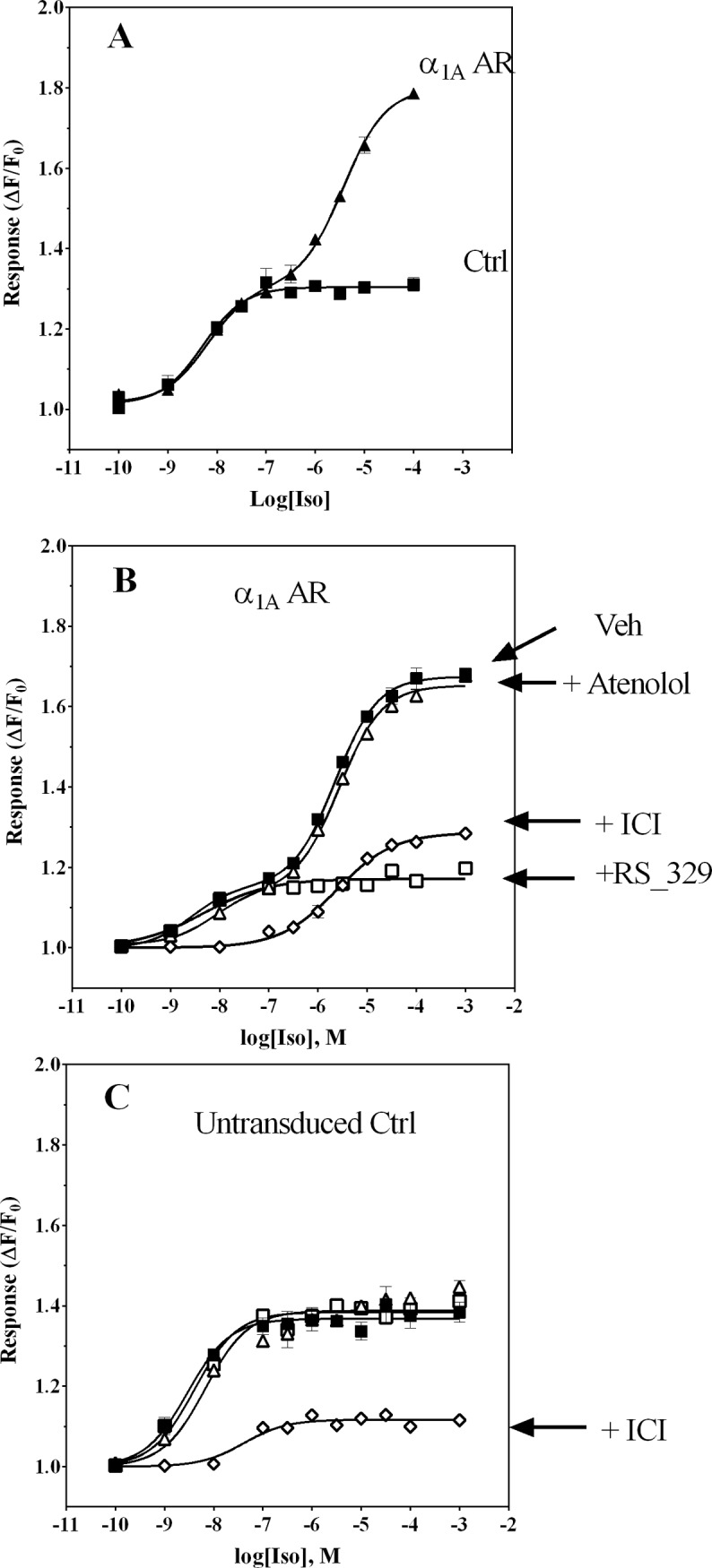
Ca2+ transient responses (expressed as ΔF/F0) were measured as a function of Iso concentration in fluo3-loaded cells by fluorometric plate imaging (FLIPR). HEK-293/EBNA cells were first exposed for 3–4 h to baculovirus encoding α1A-AR or an irrelevant negative control protein (aldehyde oxidase), then cultured in fresh medium containing 4 mM NaBu for 18 h prior to use in experiments as described in Materials and Methods. Panel A: Control cell (■) Ca2+ transient responses exhibited a monophasic increase in amplitude, whereas responses in cells transiently expressing α1A-AR (▲) revealed a biphasic concentration-response relationship, with large response amplitude at high Iso concentrations. Panel B: Blockade of Iso responses by pre-treatment of cells with antagonists selective for β-ARs or α1A-ARs reveals roles for both receptors in mediating Ca2+ transient responses in HEK-293/EBNA cells transiently expressing α1A-AR. Although the β2-AR-selective antagonist ICI 118551 at 10 nM (◇) abolished responses to low Iso concentrations, responses to high Iso concentrations were attenuated by only ∼ 50%. The α1A-AR-selective antagonist RS100329 (□) blocked only responses to high Iso concentrations. Neither phase of the concentration-response profile was affected by the β1-AR-selective antagonist atenolol (1 μM, Δ). Vehicle-treated cell responses represented by ■. Panel C: In non-transduced control cells, only the β2-AR-selective antagonist ICI 118551 at 10 nM (◇) was effective at blocking responses to Iso: both α1A-AR-selective RS100329 (10 nM, □) and β1-AR-selective atenolol (1 μM, Δ) were ineffective. Each data point represents an average of duplicate determinations; results shown are representative of experiments repeated at least 3 times.
Table 1. Pharmacological parameters determined from IP and cAMP accumulation, Ca2+ mobilization and p-ERK1/2 assays performed with transiently transduced HEK-293/EBNA cells.
| Cell line | Agonist | Ki (μM) | EC50 IP (μM) | EC50 Ca2+ (μM) | EC50 cAMP (μM) | EC50 p-ERK (μM) | |
|---|---|---|---|---|---|---|---|
| α1A-AR HEK293/EBNA | NE | 13.1 | 10.90 ± 0.20 | 10.070 ± 0.010 | 20.67 ± 0.04 | 0.09 ± 0.04 | |
| A-61603 | 10.13 | 10.040 ± 0.020 | 10.003 ± 0.001 | 20.019 ± 0.005 | 0.0008 ± 0.0003 | ||
| Iso | 1 | 50.44 | 3BQL | 40.004 ± 0.002 | 0.021 ± 0.020 | 40.005 ± 0.004 | |
| 2 | 93 | BQL | 42.6 ± 2.0 | BQL | 46.0 ± 2.0 | ||
| HEK293/EBNA | NE | 1a34 | BQL | 10.50 ± 0.10 | 4.9 ± 0.04 | 6ND | |
| A-61603 | 1a24 | BQL | 10.003 ± 0.001 | BQL | BQL | ||
| Iso | 50.45 | BQL | 0.004 ± 0.002 | 0.026 ± 0.020 | 0.001 ± 0.001 |
Data were fitted to a sigmoidal dose-response or two-site concentration-response model using the GraphPad Prism software package to determine EC50 values, expressed as mean ± standard error.
1[27]
1a [125I-CYP] used as radioligand
2Values determined in assays performed with HEK-293/EBNA cells not treated with NaBu and transduced with baculovirus construct yielding high pmol level expression of α1A-AR
3BQL: Below quantifiable levels
4 EC 50 values for the Ca2+ mobilization and p-ERK assays were determined from fitting data to a biphasic concentration-effect model (Eq 1, Materials and methods section).
5 K i of Iso at β2-AR and α1A-AR were determined as described in Materials and Methods by radioligand competition binding methods using [125I]CYP and [3H]-Prazosin, respectively, as the radioligands.
6ND –not determined
Iso induces Ca2+-mobilization in α1A-AR transduced HEK-293/EBNA cells with atypical, slower kinetics
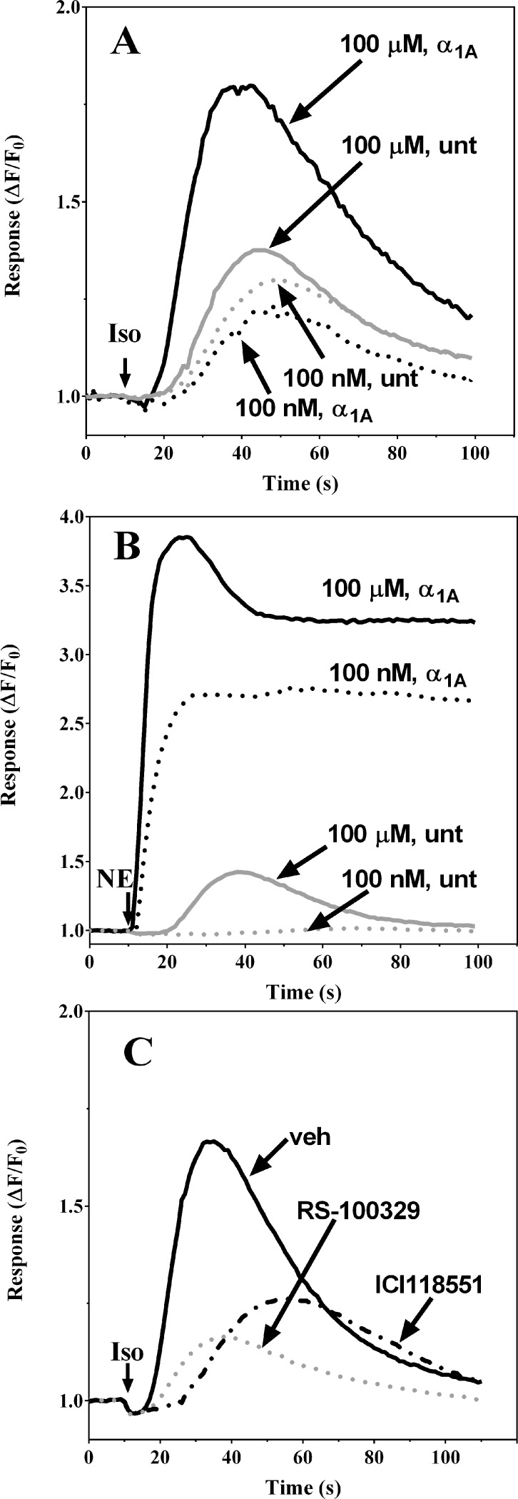
It has been shown previously that the kinetics of intracellular Ca2+ accumulation mediated by α vs. β2-AR are very different, due to the distinct sources of Ca2+ involved with each pathway downstream from the receptor. The Gαq-mediated α1-AR transient is very fast in onset relative to the Gαs-mediated β2-AR transient [31,33–35]. To further dissect Iso-mediated contributions to observed transients, we analyzed the kinetics of Ca2+ responses to Iso and norepinephrine (NE) in α1A-AR_HEK-293/EBNA transduced and untransduced cells (Fig. 2). Stimulation of α1A-AR_HEK-293/EBNA cells with 100 μM Iso (a concentration that produces high agonist occupancy at both α1A-AR and β2-ARs) elicited a slow onset Ca2+ transient response (5–10 second delay from addition of agonist to the rise in Ca2+) that return towards baseline (i.e. decline from peak amplitude >75%) within 100 sec and lacked the sustained phase (Fig. 2A, solid black line). A smaller magnitude response with similar slow onset and no sustained elevated phase was observed in response to 100 nM Iso, a concentration that would achieve high fractional occupancy of β2-AR, yet insignificant at α1A-AR (Fig. 2A, dashed black line). Responses to application of 100 nM or 100 μM Iso in untransduced negative control cells are shown by the gray dashed and solid traces, respectively. By contrast, treatment of α1A-AR_HEK-293/EBNA transduced cells with 100 nM NE resulted in an almost immediate Ca2+ transient onset, with peak response amplitude for the population average attained within 10 seconds post-agonist addition (Fig. 2B, dashed black line). This response was characterized by a sustained plateau phase of elevated cytosolic Ca2+, not found in untransduced control cells (Fig. 2B, black lines vs. solid gray trace). Because NE possesses ∼ 10-fold higher affinity at α1A-AR relative to β2-AR (K i = 3.1 μM at α1A-AR vs. 34 μM at β2-AR, Table 1), the observed response to 100 nM NE is likely driven mostly through occupancy at α1A-AR (Fig. 2B, dashed black line). Thus, the rapid and sustained Ca2+ elevation in response to 100 nM NE reveals an ability of this agonist to elicit Ca2+ transient responses through a mechanism distinct from the slow onset and transient mobilization response induced by Iso. Responses to 100 μM Iso in cells that were preincubated with the α1A-AR selective antagonist RS100329 or the β2-AR-selective antagonist ICI 118551 revealed the functional contributions of both receptors (Fig. 2C; gray dashed and black dashed lines, respectively): both antagonists significantly diminished responses and delayed their onset when compared to the vehicle control (Fig. 2C; solid black line). Interestingly, the biggest delay was observed in cells pretreated with the β2-AR-selective antagonist ICI 118551, a condition that isolates the α1A-AR component of Iso-mediated Ca2+ transients (Fig. 2C; black, dashed line).
Figure 2. The Iso-induced Ca2+ mobilization response in α1A-AR transduced HEK-293/EBNA cells is slower in onset and shorter in duration than the response to NE.
Ca2+ transient response kinetics (expressed as ΔFt/F0) are shown for fluo3-loaded cells monitored fluorometrically following addition of Iso or NE. HEK-293/EBNA cells were first exposed to recombinant baculoviral strains (3–4 h) encoding either α1A-AR or an irrelevant negative control protein (aldehyde oxidase), then cultured in fresh culture medium containing 4 mM NaBu for 18 h prior to use in experiments as described in Materials and Methods. Panel A: Slow onset of the Iso agonist response which returns to baseline within a 2 min interval is evident in representative traces from α1A-AR transduced HEK-293/EBNA cells (black lines) and negative control cells (gray lines) during responses elicited by addition of Iso (↓) at 100 nM (dashed lines) or 100 μM (solid lines). Panel B: Representative Ca2+ transients in α1A-AR transduced HEK-293/EBNA cells showing rapid onset and sustained NE response following application of NE (↓) at 100 nM (dashed lines) or 100 μM (solid lines) to α1A-AR transduced (black lines) or negative control-transduced cells (gray lines). Panel C: α1A-AR transduced HEK-293/EBNA cells exhibit distinct kinetics for Iso-mediated responses via occupancy of β-AR vs α1A-AR transduced, as revealed by monitoring of responses following pre-treatment of cells for 20 min with vehicle (solid black line), 10 nM RS-100329 (dashed gray line) or 10 nM ICI118551 (dashed black line). Representative traces are shown for responses to 100 μM Iso (↓).
Effect of extracellular Ca2+ removal on the β2- and α1A-AR-dependent Ca2+ mobilization response to Iso stimulation
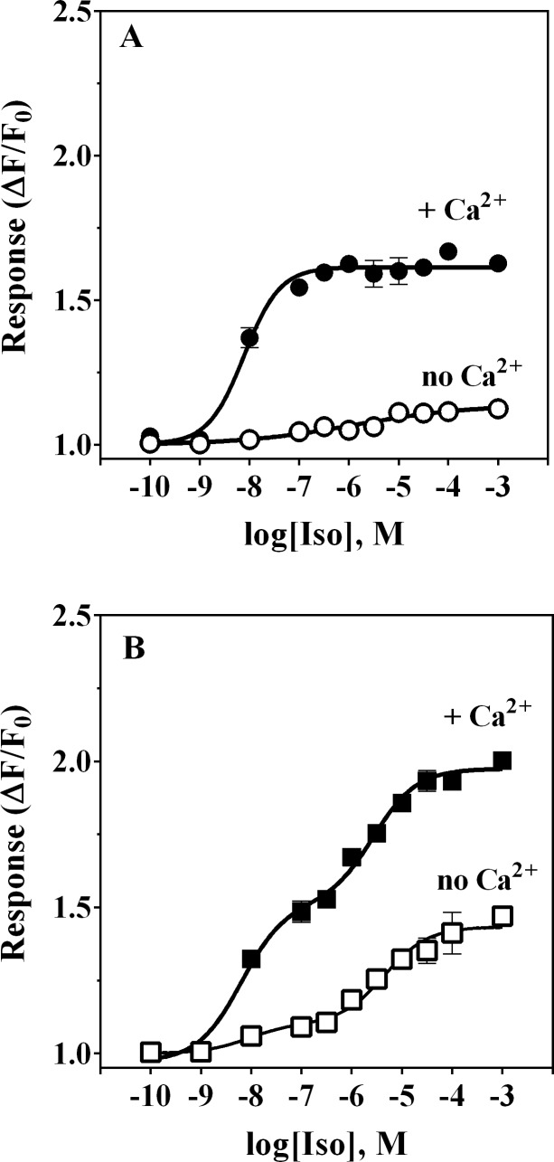
We next addressed whether observed rises in cytosolic Ca2+ were due to influx of extracellular Ca2+ by measuring Ca2+ mobilization in HEK-293/EBNA cells upon exposure to Iso in the presence or absence of external Ca2+. Omitting Ca2+ from the extracellular buffer eliminated Iso-evoked Ca2+ responses in untransduced HEK-293/EBNA cells (Fig. 3A). In α1A-AR_ HEK-293/EBNA cells, Iso-mediated responses were greatly reduced in amplitude in the absence of extracellular Ca2+ (Fig. 3B). However, the low potency phase of the concentration-response relationship was only partially affected, unlike responses to Iso < 100 nM, which were almost fully inhibited. Fitting of those data to a biphasic concentration-response model yielded EC50 values of 8.6 nM and 4.5 μM for the high and low potency phases, respectively, close to the EC50 values of 6.3 nM and 2.5 μM measured in the same cells responding to Iso in the presence of extracellular Ca2+. Moreover, the amplitude of the Iso-induced maximal responses under nominally Ca2+-free conditions (ΔF/F0 = 1.1 and 1.3 for the high and low potency phases, respectively) was lower than that determined in the same cells in the presence of extacellular Ca2+ (ΔF/F0 = 1.5 and 1.4). Thus, under near-zero Ca2+ concentration both the Iso response of untransduced cells and the high potency phase of the response in α1A-AR transduced cells were largely absent. This finding suggests that the observed β2-AR dependent Ca2+ mobilization requires an influx of Ca2+ from the extracellular compartment, likely via activation of cAMP nucleotide gated-channels. As described in the previous section, the time course of intracellular Ca2+ mobilization in response to 100 μM Iso (a concentration that produces high occupancy of α1A-ARs) was very similar to that observed for β2-AR response and dramatically different from typical Gαq-initiated signaling canonical for α1A-AR. Remarkably, Ca2+ mobilization in response to Iso occupancy of both β2-AR and α1A-AR was found to be less sensitive to removal of extracellular Ca2+ (Fig. 3B), indicating intracellular stores as the source of Ca2+.
Figure 3. Iso-induced Ca2+ mobilization in α1A-AR transduced HEK-293/EBNA cells is partially dependent on the presence of extracellular Ca2+.
Ca2+ transient responses (expressed as ΔF/F0) were measured as a function of Iso concentration in untransduced control cells (Panel A) or α1A-AR transduced HEK-293/EBNA cells (Panel B), either in the presence (filled symbols) or absence (open symbols) of 2 mM Ca2+ in the assay buffer. Responses in untransduced cells were virtually abolished in the absence of extracellular Ca2+ (Panel A). In α1A-AR transduced cells, responses to low concentrations of Iso in the absence of extracellular Ca2+ were also essentially abolished whereas the low potency phase of response was diminished by approximately 50% (Panel B). Each data point is an average of duplicate determinations; this experiment was repeated twice.
Iso induced Ca2+ mobilization in α1A-AR transduced HEK-293/EBNA cells does not reflect coupling to Gαi
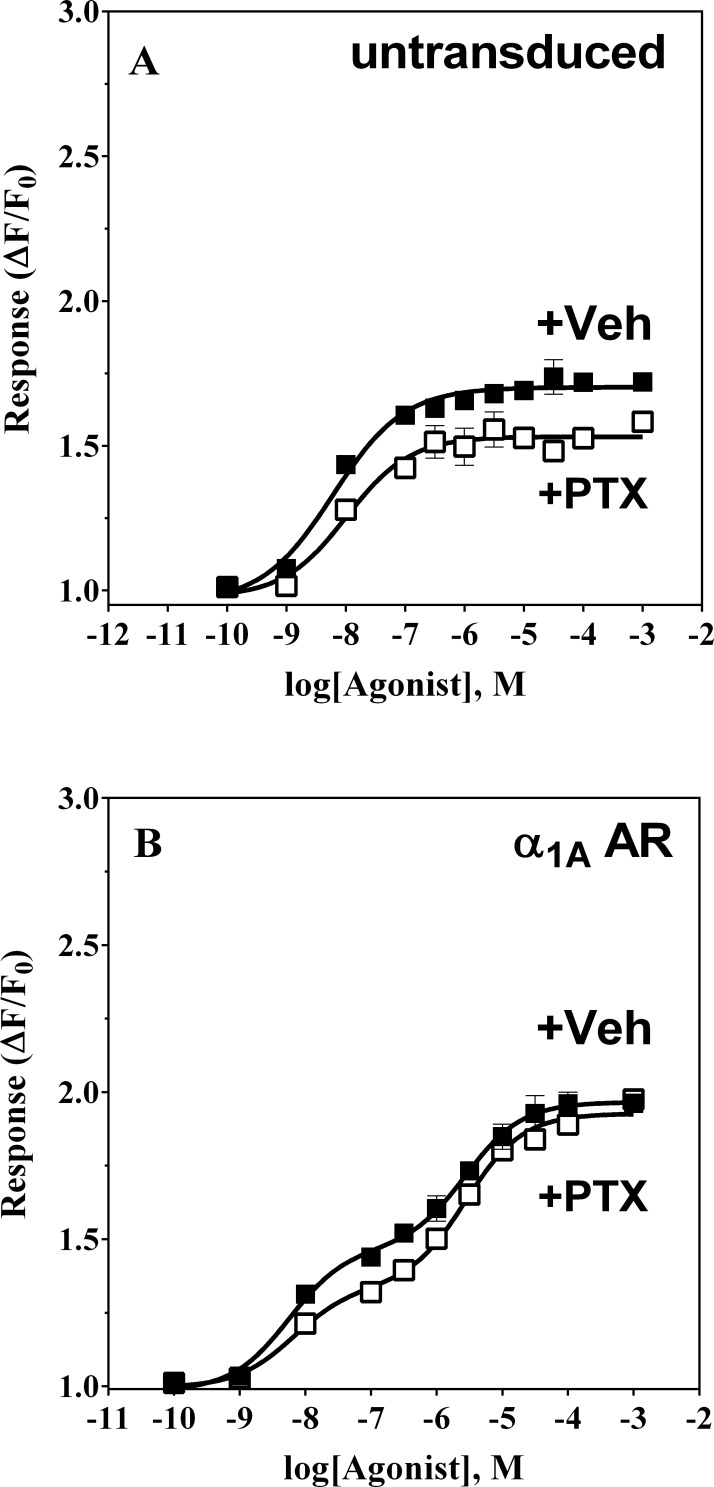
Receptor coupling to Gαi, the most abundant G protein α subunit, has been shown to lead to activation of PLC activity via the Gβ/γ subunits and thus, can lead to IP accumulation and release of intracellular Ca2+ (Reviewed in [36]. Since this pathway is different from Gαq-mediated signaling, the kinetics of Ca2+ mobilization may also differ. We next searched for a role for Gαi activation in response to Iso-treatment in untransduced and α1A-AR transduced cells, pretreated for 18 hours with pertussis toxin or vehicle. Pretreatment of untransduced cells with pertussis toxin (100 ng/mL) resulted in a slight increase in the maximum amplitude of the Ca2+ transient response at saturating concentrations of Iso (ΔF/F0 = 1.5 vs. 1.7), with a negligible increase in EC50 from 6 to 10 nM (Fig. 4A). A similar increase was observed in the β2-AR–dependent phase of the Iso concentration-response curve (from ΔF/F0 = 1.3 to 1.5) in α1A-AR expressing cells, also with no change in observed potency (EC50 = 6.6 vs. 5.6 nM) (Fig. 4B). Since in these cells induction of cAMP leads to Ca2+ mobilization [27], increase in Ca2+ mobilization following PTX pretreatment may be a result of an increase in intracellular cAMP. This observation would be consistent with previous findings indicating that β2-AR can couple to both Gαs and Gαi in HEK-293 [37]. On the other hand, in α1A-AR transduced HEK-293/EBNA cells, pertussis toxin treatment did not significantly affect Ca2+ mobilization responses to Iso (EC50 = 2.8 vs. 2.7 μM with PTX and ΔF/F0 = 1.6 vs. 1.5 with PTX), suggesting that coupling to Gαi is not necessary for this Iso-mediated function of α1A-AR (Fig. 4B).
Figure 4. Pertussis toxin pretreatment does not impair Ca2+ responses to Iso.
HEK-293/EBNA cells were exposed for 3 h to a baculoviral strain carrying α1A-AR or to viral medium. Cells were treated with 100 ng/mL of pertussis toxin (PTX) or vehicle for 18 h prior to experiments. Panel A: untransduced HEK-293/EBNA cells exposed to PTX (filled symbols) or vehicle (open symbols) were treated with Iso (■,□) during fluorometric imaging of the Ca2+-tracking dye. Panel B: untreated (open symbols) or PTX-pretreated (filled symbols) α1A-AR HEK-293/EBNA cells were stimulated with Iso (■,□) during fluorometric imaging of the Ca2+-tracking dye. Each experiment was performed in duplicate two independent times.
Iso does not stimulate formation of inositol phosphates (IP1, IP2 or IP3) nor IP accumulation in HEK-293/EBNA_α1A AR cells
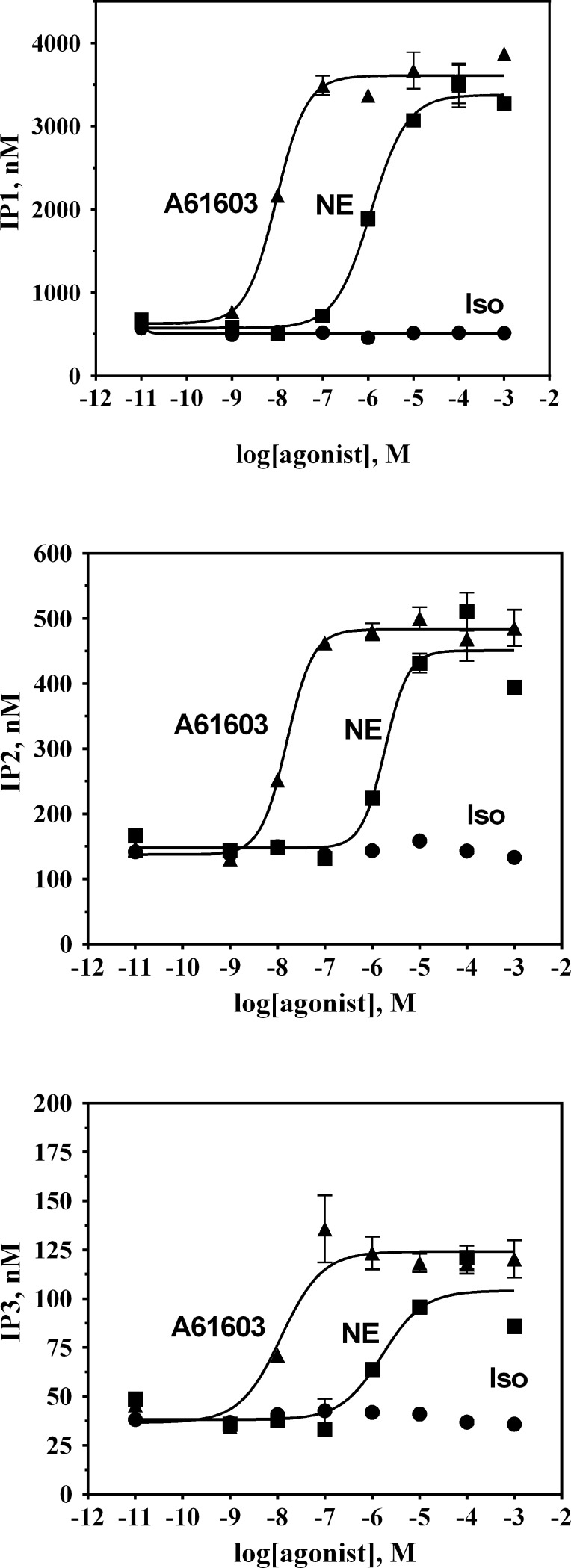
Given that the α1A-AR-dependent phase of the Iso concentration-response relationship was found to be only partially dependent on the extracellular Ca2+ and insensitive to pertussis toxin treatment, we asked whether Iso-occupancy at α1A-AR results in Gαq activation. In α1A-AR transduced HEK-293/EBNA cells, the selective α1A-AR agonist A-61603 elicited concentration-dependent IP accumulation (S1 Fig.). A-61603-stimulated IP accumulation was best described by a single phase sigmoidal equation yielding an EC50 of 40 nM (Table 1). Furthermore, preincubation of those cells for 5 minutes with 10 nM of the α1A-AR selective antagonist RS100329 significantly attenuated IP accumulation and produced a large, rightward shift in A-61603 potency. In contrast, when replicate cells were treated with Iso (up to 10 mM) no significant accumulation of IP was detected. Furthermore, IP formation in untransduced cells upon treatment with either A-61603 or Iso was not detectable (data not shown). We employed an LC-MS method to increase our detection sensitivity and to measure individual inositol phosphates rather than total IPx accumulation (Fig. 5). In α1A-AR transduced HEK-293/EBNA both NE and the selective α1A-AR agonist A-61603, produced concentration-dependent increases in cellular levels of inositol phosphates IP1, IP2 and IP3 that were best described by a monophasic sigmoidal equation (Fig. 5). On the other hand, when replicate cells were treated with Iso (up to 10 mM), no significant changes in intracellular inositol phosphates were detected. Thus, it appears that Iso occupancy at α1A-AR biases receptor signaling toward a Gαq-independent pathway.
Figure 5. Inositol phosphates production in α1A-AR transduced HEK-293/EBNA cells occurs in response to A-61603 and NE, but not in response to Iso.
HEK-293/EBNA cells were exposed to baculovirus encoding α1A-AR for 3–4 h, then cultured in fresh medium containing 4 mM NaBu for 18 h prior to use in experiments as described in Materials and Methods. IP1 (top), IP2 (middle) and IP3 (bottom) formation was measured in α1A-AR transduced HEK-293/EBNA cells stimulated with increasing concentrations of A-61603 (▲), NE (■) or Iso(●). IP1, IP2, and IP3 levels were determined via LC-MS. Plots are representative of three independent experiments with each data point being the average of triplicates.
Iso induces biphasic concentration-response behavior for p-ERK formation in α1A- AR transduced HEK-293/EBNA cells
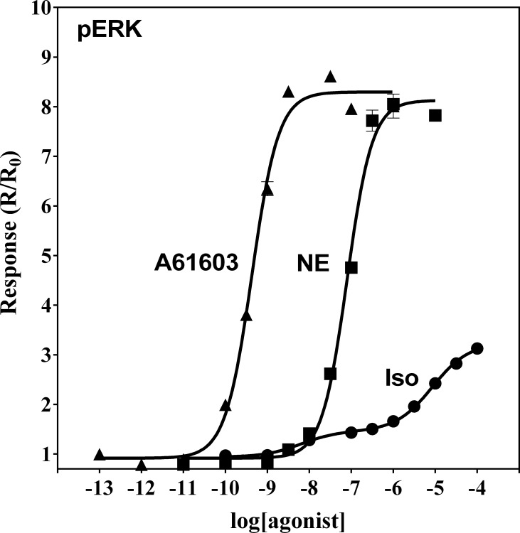
Significant efforts have been placed in understanding the role of the mitogen-activated protein kinase (MAPK) cascade in adrenoceptor signaling (e.g. in cardiomyocyte physiology, see [38]). We decided to test whether the Iso-mediated signaling that we observed involved this signaling pathway. For this, we examined whether Iso induces ERK1/2 activation. Treatment of α1A-AR transduced HEK-293/EBNA cells with Iso induced a dose-dependent increase of p-ERK formation (Fig. 6, filled circles). The resulting concentration-response relationship was best fit by a biphasic model. The calculated EC50 values were 5 nM and 6 μM for the high and low potency phases, respectively. In untransduced cells a monophasic relationship was observed (EC50 = 1 nM, S2 Fig.). Interestingly, in α1A-AR transduced HEK-293/EBNA cells, A-61603 or NE yielded concentration-dependent activation of ERK that was best fit by a monophasic sigmoidal model. The measured EC50 values were 0.8 and 90 nM for A-61603 and NE, respectively (Fig. 6, filled triangles or squares, and Table 1). In those cells, A-61603 appeared to be almost 4 times more potent at inducing p-ERK formation than in effecting Ca2+ mobilization (EC50 = 3 nM, Table 1), while NE had about the same potency for both response modes.
Figure 6. MAPK activation in α1A-AR transduced HEK-293/EBNA cells treated with A-61603, NE or Iso.
α1A-AR transduced HEK-293/EBNA cells were pre-treated with NaBu for 18 h to induce receptor expression. Cells were stimulated with A-61603 (▲), NE (■) or Iso(●) for 5 min. Agonist treatment was terminated by addition of of SureFire lysis solution. Samples were analyzed for levels of phospho-ERK using an AlphaScreen SureFire p-ERK assay kit. Plots are representative of four independent experiments, with each data point being the average of triplicates.
Iso stimulation of α1A-AR _ HEK-293/EBNA cells resulted in a biphasic concentration-response relationship for both p-ERK formation and Ca2+ mobilization transients. The high potency phase of response to Iso (via occupancy of β2-AR) and the low potency phase (via occupancy of both β2-AR and α1A-AR) were quite similar for the two different readouts (Table 1).
Stimulation of α1A-AR transduced HEK-293/EBNA with A-61603 and Iso leads to an increase in the level of intracellular α1A – AR without recruitment of arrestins
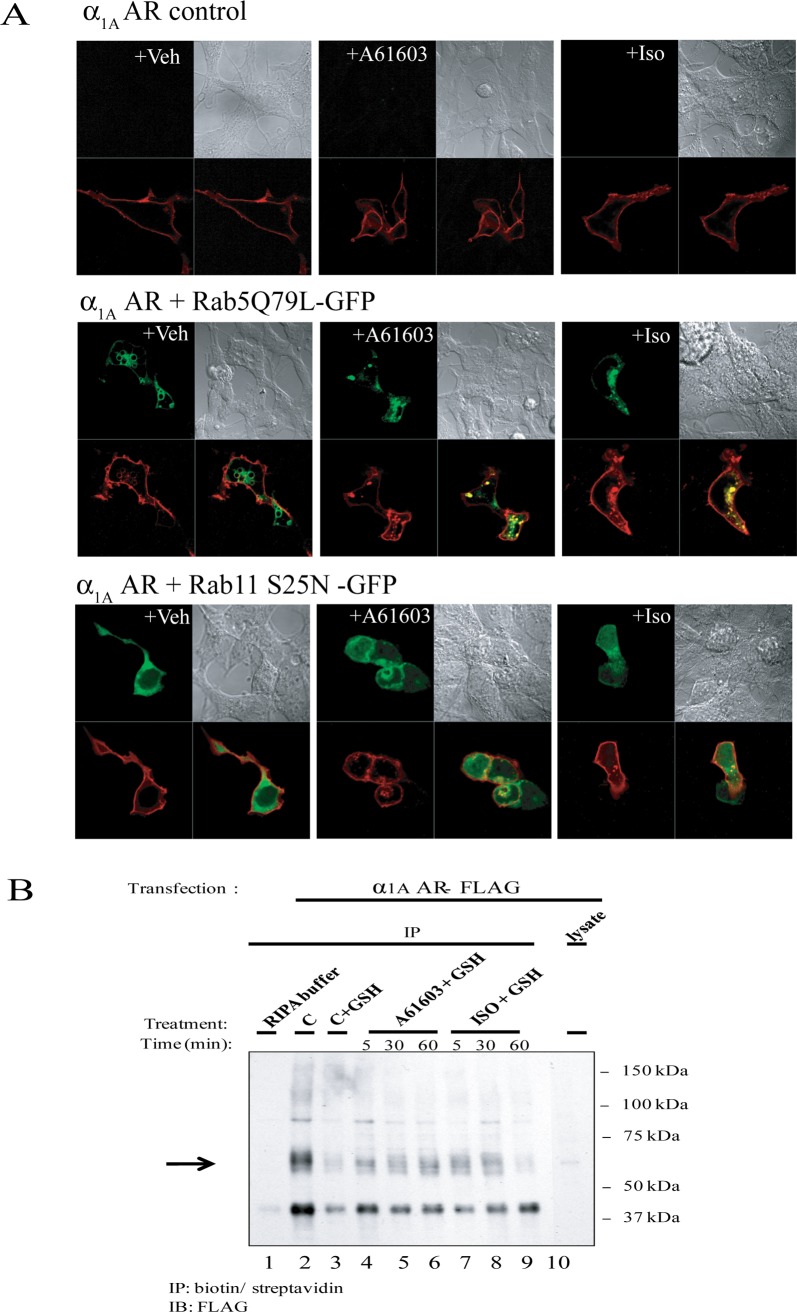
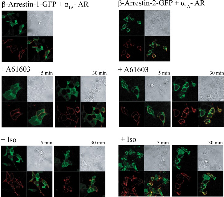
We next investigated whether or not α1A– AR internalizes upon stimulation with A-61603 or ISO. In several prior studies α1A– AR has been found to undergo constitutive, ligand independent trafficking and to internalize rather modestly (∼20%) upon agonist stimulation [39–41]. Furthermore this receptor cycling involves clathrin-coated vesicles and entry to early endosomes [39]. To eliminate some of the experimental challenges and uncertainty related to detection of the modest and transient internalization of α1A– AR, we employed constitutively active and dominant-negative forms of Rab5 and Rab11, respectively, to the analysis of α1A– AR endocytosis using confocal microscopy. Constitutively active Rab5 Q79L enhances endocytosis and early endosome fusion, causing the formation of enlarged early endosomes, and its overexpression has been shown to inhibit transferrin recycling. Rab11 S25N is defective in GTP binding and impairs recycling by inhibiting exit from sorting endosomes (early endosomes) to the recycling endosomes and/or plasma membrane. In HEK-293/EBNA cells transfected with α1A – AR only and stimulated with A-61603 or ISO, α1A – AR was mostly localized at the plasma membrane, as was the case for the vehicle control (Fig. 7A). Overexpression of Rab5Q79L caused some intracellular α1A – AR accumulation in vehicle control cells, indicative of constitutive trafficking, as well as significant α1A – AR accumulation upon A-61603 or ISO treatment. Overexpression of Rab11 S25N led to some accumulation in A-61603 or ISO-treated cells, not as pronounced as the one observed upon Rab5Q79L overexpression. In vehicle-treated cells, α1A – AR appeared predominantly at the plasma membrane. To confirm the observation that stimulation with Iso led to internalization of α1A – AR, we took advantage of a cleavable biotin labeling reagent to discern levels of surface versus internalized α1A – AR by means of immunoprecipitation (Fig. 7B and S3 Fig.). Intracellular α1A – AR could be detected at low levels in control cells (lane 3, “C+GSH”) indicative of constitutive trafficking. Treatment with A-61603 caused an increase in intracellular α1A – AR levels after 5, 30, and 60 min of treatment (lanes 4–6, “A-61603+GSH”) relative to control cells (lane 3,“C+GSH”). Similarly, treatment with ISO caused an increase in the intracellular α1A – AR (lanes 7–8, “ISO+GSH 5 or 30”), as compared to control cells (lane 3, “C+GSH”). We next examined whether Iso-induced internalization of α1A – AR in HEK-293 cells involved the recruitment of β-arrestins. The ability of the N-terminally FLAG-tagged α1A-ARs to trigger the translocation of GFP-tagged β-arrestins in those cells was investigated by confocal microscopy. As shown in Fig. 8, in vehicle treated cells, the α1A-AR was localized mainly at the plasma membrane while both β-arrestins 1 (left panels) and 2 (right panels) were homogenously distributed throughout the cytoplasmic compartment with no visible co-localization with the surface receptor. In cells expressing the α1A-AR, stimulation with A-61603 for 30 min did not induce significant translocation of either β-arrestin-1 nor β-arrestin-2, to the plasma membrane. Similarly exposure of the same cells to Iso did not cause visible β-arrestin rearrangement. Internalization of the α1A-AR was not detected with either agonist by this method.
Figure 7. Stimulation of α1A-AR transduced HEK-293/EBNA with A-61603 and Iso leads to an increase in intracellular α1A – AR.
A. HEK293 cells were transiently transfected with α1A – AR only (top panels), or co-transfected with α1A – AR and Rab5 variant Q79L (middle panels) or Rab11 variant S25N (bottom panels). Following serum-deprivation, cells were stimulated with vehicle, 1μM A-61603 or 1mM ISO for 2h. Cells were then fixed and analyzed by confocal microscopy. B. HEK293 cells were transiently transfected with α1A – AR. After serum deprivation for 24h, cells were pre-treated with a membrane impermeable, disulfide-cleavable biotin reagent to label plasma membrane α1A – AR. Cells were then left untreated, or stimulated 1 μM A-61603 or 1mM ISO for 5, 30, or 60 min. After treatment, one dish of control cells was harvested without any further manipulations (C: total α1A – AR). The remaining seven dishes were divided into one control (C+GSH), three treated with A-61603 (A-61603+GSH) and three treated with ISO (ISO+GSH). They were stripped of surface biotin label using a reducing agent, in order to reveal internalized, labeled α1A – AR. Samples were then analyzed by immunoprecipitation (IP) with streptavidin followed by immunoblotting (IB) with an anti-FLAG antibody.
Figure 8. Treatment of α1A-AR transduced HEK-293/EBNA with A-61603 and Iso does not trigger intracellular redistribution of arrestins.
HEK293 cells were co-transfected with FLAG-tagged α1A – AR and GFP- tagged β-arrestin-1 (left panels) or β-arrestin-2 (right panels). Following serum-deprivation for 24h, cells were left untreated (top panels), or stimulated with 1μM A-61603 (middle panels) or 1mM Iso (bottom panels) for the indicated amount of time. Cells were then fixed, permeabilized, stained with Alexa Fluor-568 conjugated anti-FLAG antibodies, and analyzed employing confocal microscopy.
Isoproterenol-induced Ca2+ mobilization in α1A-AR_CHO cells is not accompanied by detectable increases in intracellular inositol phosphates, yet correlates with activation of the MAPK kinase pathway
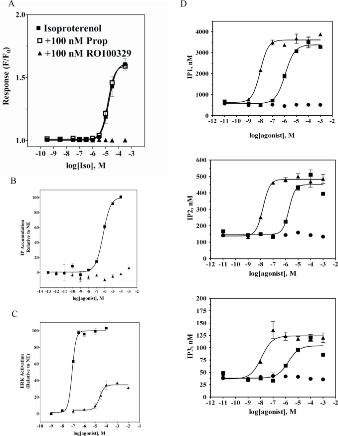
Since in HEK-293 cells Iso can stimulate both endogenous β2-AR as well as transduced α1A-AR, we next tested the activity of this β–agonist at α1A-AR in CHO cells stably expressing moderate levels of the receptor (∼1 pmol/mg of membrane proteins) [42]. The β-AR selective agonist Iso evoked a Ca2+ transient response in α1A-AR_CHO cells (Fig. 9A) that was not observed in CCR5_CHO cells used as a negative control (S4 Fig.). The peak amplitude of Ca2+ transient response, ΔF/F0, as a function of agonist concentration yielded a monophasic concentration-response curve with the half-maximal amplitude occurring at 20 μM Iso (Fig. 9A). That response to Iso was blocked by preatreatment with 100 nM RO100329, but was insensitive to propranolol at 100 nM. We next examined whether Iso-mediated Ca2+ mobilization involved formation of inositol phosphates. Stimulation of α1A-AR_CHO cells with NE led to concentration-dependent IP accumulation with an EC50 of 0.6 μM (Fig. 9B). On the other hand, no IP accumulation could be detected in the same cells stimulated with Iso at concentrations up to 1 mM. Similarly, in α1A-AR CHO cells both NE, and the selective α1A-AR agonist A-61603, produced concentration-dependent increases in cellular concentration of inositol phosphates; IP1, IP2 and IP3 (Fig. 9D) that were best described by a single-site sigmoidal equation. In contrast, when replicate cells were treated with Iso (up to 1 mM) no significant changes in intracellular levels of inositol phosphates could be detected. Thus, it appears that Iso occupancy at α1A-AR biases receptor signaling to a Gαq-independent pathway. Yet, similarly to the observations made with α1A-AR_HEK-293/EBNA cells, stimulation of α1A-AR_CHO cells with Iso resulted in a concentration dependent increase in phospho-ERK formation (Fig. 9C). The amount of p-ERK formed at the maximal concentration of Iso represented 35% of the level observed in these cells when stimulated with saturating concentrations of NE or A-61603. Iso was less potent than NE and A61603 at inducing ERK activation in α1A-AR_CHO cells (Iso: EC50 = 17±5 μM; NE: EC50 = 0.13±0.02 μM; A61603: EC50 = 0.8 ± 0.2 nM). These results indicate that Iso is ineffective at inducing α1A-AR-mediated Gαq coupling and PLC activation, yet shows partial agonist activity at mediating α1A-AR induced activation of the MAPK signaling cascade.
Figure 9. Concentration-response behavior to Iso in Chinese hamster ovary cells stably expressing recombinant α1A-AR.
A. Ca2+ transients (expressed as ΔF/F0) were measured as a function of Iso concentration in fluo3-loaded cells by fluorometric plate imaging (FLIPR). Responses to Iso were monitored following pre-treatment with either vehicle (■), 100 nM propranolol (□) or 100 nM RO100329 (▲). Inositol phosphate accumulation (B) and levels of phospho-ERK (C) were measured as a function of increasing concentrations of A-61603 (■) or Iso (▲) and are represented relative to NE. D. IP1 (top), IP2 (middle) and IP3 (bottom) formation was measured in CHO cells stably expressing α1A-AR, stimulated with increasing concentrations of A-61603 (▲), NE (■) or Iso(●). IP1, IP2, and IP3 levels were determined via LC-MS. Plots are representative of three independent experiments with each data point being the average of triplicates.
Discussion
A search on “isoproterenol” in the PubMed database retrieves more than thirty thousand citations. This potent, β-AR-selective agonist has been extensively studied with respect to many aspects of its action at β-ARs. It has been applied frequently as a tool to verify the presence of β-ARs in cell-based systems or to elucidate the mechanism of β-AR signaling in non-recombinant, native cells such as cardiomyocytes, adipocytes and many others, as well as in organ tissue preparations. In several of these studies the concentrations used were in the micromolar range, which may have triggered off-target effects. Dating back to the 1950s, several reports have indicated that Iso at higher concentrations can evoke α-AR-mediated responses. For instance, Furchgott [9] observed that Iso at concentrations 1–500 μM caused contraction of rabbit thoracic aorta, a tissue in which all three α1- AR subtypes are expressed (reviewed in [43]. These researchers also noted that the rate of shortening on addition of Iso was much slower relative to epinephrine or norepinephrine, indicating a distinct mechanism of action for Iso. The maximal contractile response to Iso was about 75% of that obtained for norepinephrine indicating that Iso acted as a partial agonist.
Our studies of HEK-293/EBNA cells with transient low expression levels of α1A-ARs cells, to mimic tissues that possess both α1A and β-ARs, provide for the first time a mechanism to account for Furchgott’s original observations. In those cells, a biphasic dose-response relationship was found for Iso-evoked calcium transients. The high potency phase of the response was blocked by non-selective as well as β2-selective antagonists, while the low potency phase was sensitive only to α1A-AR-selective antagonists. Remarkably, Ca2+ transient responses to Iso mediated by α1A- ARs exhibited a time course distinctly slower than those seen with prototypical α1A AR agonists (NE, A-61603) that are highly efficacious for IPx formation responses and considered typical for Gαq-initiated signaling. Responses to Iso showed delayed onset by 5 to 10 seconds and were much slower to rise in amplitude. Moreover, Iso was a partial agonist at α1A- ARs as recorded by this readout. Finally, the observed Ca2+ transients exhibited almost complete desensitization, unlike responses to NE. These data taken together indicate a mechanism of action distinct from classical Gαq coupling for Iso at the α1A-AR, and suggest functional selectivity (effector signaling bias) for the activity of this agonist.
Agonist occupancy of any given GPCR may trigger signaling events through activation of more than one physiological response cascade. For instance, α1A-AR is widely known to couple to phospholipase C. However, this receptor also reportedly mediates activation of several other effectors such as phospholipase D, phospholipase A2, adenyl cyclase and several members of the MAPK family (reviewed in [44]). Certain synthetic agonists have been found to be capable of biasing GPCR signaling toward a particular proximal effector. This ability of a ligand to direct an activated receptor state to a particular effector output has been termed “functional selectivity” or “effector bias” (for reviews see [45–48]. Within the adrenoceptor family, there is both pharmacological and biophysical evidence for agonist dependent effector bias at β-ARs. Signaling bias has been shown for several ligands at both β1- and β2-AR toward adenyl cyclase and MAPK [49,50].
We observed that Iso occupancy of α1A-AR caused Ca2+ transient responses in HEK-293/EBNA cells without observable PLC activation: no formation of individual inositol phosphates or IP accumulation was detected in these cells under the same conditions. Unexpectedly, Iso occupancy of α1A-AR also mediated ERK1/2 activation, suggesting that Gαq coupling is not required for ERK activation by Iso. Adding to the surprise, A-61603 appeared to be significantly more potent at activating ERK as compared to activation of Gαq, whereas NE displayed equivalent potencies for both responses. Taken together, the signaling patterns observed with these agonists appear to reveal a spectrum of partially overlapping effector coupling mechanisms. Iso, and the imidazoline A-61603, appear to have a bias toward MAPK activation. Moreover, for A-61603 there seems to be greater receptor reserve or response amplification for the pERK formation response.
The α1A-AR has also been found to regulate mitogenic responses, but it is not clear if this activity is downstream of Gαq engagement [38,51]. Minneman and co-workers have in fact reported NE activation of α1A-AR-mediated MAPK phosphorylation in rat PC12 cells [51,52], independently of both Gαq-induced Ca2+ mobilization and PKC activity [53]. The same group later showed that deletion of the first three amino acids from the third intracellular loop not only uncouples the receptor from Gαq, but also abolishes activation of the MAPK pathway [54]. It was suggested that these two pathways operate independently from one another, yet may depend on similar structural elements of agonist-occupied receptor. Several lines of evidence seem to indicate that in mouse cardiomyocytes, the α1A-AR-mediated MAPK activation may not be associated with activation of Gαq pathway [38,55]. α1A-AR /α1B-AR double-knockout mice also developed heart failure after transverse aortic constriction, and reconstitution of α1A-AR signaling in cardiomyocytes from those animals rescued them from NE-induced apoptosis [56]. The expected IP generation was not detectable in those cardiomyocytes with reconstituted α1A-AR function or in wild-type mouse cardiomyocytes, pointing at a non-Gq signaling mode for this receptor in myocardial function. More importantly, Huang et al. (2007) reported that α1A-AR stimulation of an ERK-mediated pathway is critical for cardiomyocyte survival. Our data support those findings as well as provide further evidence indicating that Iso occupancy of α1A-AR induces an active receptor “state” that leads to MAPK pathway activation without activating the canonical Gαq pathway.
Our previous studies of Gαq coupling defective variants of α1A-AR combined with examination of the effects of Ca(II) channel blockers uncovered cross-talk between α1A-AR and β2-AR that leads to potentiation of a Gαq-independent signaling cascade in response to α1A-AR activation [27]. Moreover, this signaling event was accompanied by Ca(II) mobilization with unusual kinetics mirroring the kinetics observed for Iso induced Ca(II) mobilization in α1A-AR_HEK293/EBNA cells. Interestingly, in the presence of a β-AR-selective antagonist, the amplitude of the α1A-AR-mediated Ca2+ transient response to Iso (i.e. the lower potency phase of the Iso concentration-effect relationship) was not only lower than expected, but also occurred with a significant delay in onset. This may indicate a synergy occurs between these receptors in generating the lower potency phase of response. Depletion of extracellular Ca(II) did not affect Iso induced activation of α1A-AR indicating that intracellular stores were the source of Ca(II) released. Finally, this signaling cascade did not involve coupling to Gαq, Gαs or Gαi but led to receptor internalization and activation of ERK1/2. An alternate mechanism for the control of Ca2+ release from the ER involves ryanodine receptors (RyRs). They have been shown to exist in several non-excitable cell lines, although their functional expression in HEK-293 cells is controversial. RT-PCR analysis of HEK-293/EBNA cells found that significant levels of RyR2 mRNA are present in these cells.
Recent reports provide an independent line of evidence for α1A-AR-mediated activation of endocytic pathway that leads to phosphorylation of ERK1/2, independent of Gαq /PLC/PKC signaling [57,58]. In one study, dynamin mutants were employed to induce trafficking defects, along with various molecules disrupting actin and tubulin organization. Liu and coworkers show that α1A-AR induced activation of ERK1/2, but not p38 MAPK, was dependent on cytoskeleton and actin organization. On the other hand, α1A-AR-induced activation of PKC and C-Raf was not affected by endocytosis disruption. Neither PKC nor PLC inhibition blocked α1A-AR induced activation of ERK1/2 [57]. More recently, the spatial-temporal characteristics of receptor internalization and ERK1/2 activation in response to stimulation of α1A-AR in HEK293 were investigated in combination with various inhibitors of PKC, receptor internalization as well as β-arrestin 2 silencing and differential patterns were discovered for Gq/PKC dependent as compared to the Gαq-independent signaling cascade. The rapid phosphorylation of ERK1/2 was dependent on activation of PKC downstream of Gαq and resulted in pERK1/2 translocation into the nucleus while sustained activation of ERK1/2 that was limited to cytoplasmic compartment, was independent of PKC but dependent on receptor internalization into acidified endosomes and was mediated by β-arrestin 2[58]. Together, those findings and our own data provide strong evidence that α1A-AR can induce activation of ERK1/2 in a manner that is independent of the canonical Gαq /PLC/PKC signaling cascade but involves receptor internalization. We also found that Iso is a biased agonist that can preferentially stimulate α1A-AR mediated activation of MAPK signaling pathway through receptor internalization and without induction of Gαq coupling. Although, our study did not conclusively detect Iso induced α1A-AR association with β-arrestins after agonist application, involvement of β-arrestin 2 in mediating this response cannot be ruled out. Several groups have demonstrated that agonist-mediated GPCR association with β-arrestin leads to G protein-independent activation of the MAPK pathway [23]. In fact, it has been shown recently that NE occupied α1A-AR associates although weakly with both β-arrestin-1 and 2, but coupling to the MAPK pathway was not investigated in this study [59]. Further studies including β-arrestin knock-downs will be needed to fully determine whether or not this signaling mechanism is actually dependent on β-arrestin 2.
On the other hand, it has been recently shown that a genetic variant of the α1A-AR transactivates EGFR via a β-arrestin1-dependent mechanism similarly to the β1-AR mediated EGFR transactivation and leads to ERK1/2 activation downstream of EGFR [60–62]. Transactivation of EGFR by β-AR confers cardioprotection [61,62] suggesting that transactivation could be involved in the antiapoptotic and cardioprotective activity of the α1A-subtype. We have examined this potential mechanism in mediating the Iso induced α1A-AR stimulation of ERK1/2 using EGFR inhibitor AG1478 and found that activation of ERK was not affected by the inhibition of EGFR (data not shown). Further studies may be needed to fully explore this mechanism.
Our observations differ from those of Sun et al., who reported that Iso evoked biphasic dose-effect relations in MAPK activation [24]. The observed biphasic behavior was attributed to distinct processes by which MAPK activity is modulated, either through regulation of adenylyl cyclase by β2-AR/Gαs, or by formation of a macromolecular complex containing β2-AR and c-Src which directly activates c-Src. The conclusion that β2-AR was the sole mediator of both phases of the Iso response was based on their complete blockade by 1 μM ICI118551, as well as lack of detectable MAPK activation in MEF cells derived from β1-AR−/− and β2-AR−/− mice. Although these findings offer strong evidence for β2-AR involvement in mediating that response, they do not necessarily rule out a synergy between β2-AR and α1-ARs, if present in those cells.
Interestingly, we observed that in the presence of a β-AR-selective antagonist, the kinetics of Iso-evoked α1A-AR-mediated Ca(II) transient response was much slower and with the greatest delay (Fig. 2C), and the the amplitude of the α1A-AR-mediated Ca(II) transient (i.e. the lower potency phase of the Iso concentration-effect relationship- Fig. 1B) was lower than expected, indicating synergy between these receptors in generating the low potency response phase. This is consistent with previously published results with wild-type α1A AR and Gαq signaling deficient variants expressed in HEK EBNA cells with endogenous expression of β2 AR that showed that β2 AR signaling actually potentiates α1A AR- mediated Ca(II) response and the potentiation was most striking in cells expressing Gαq defective mutants [27]. Furthermore, this potentiation of Ca(II) transient was most evident for the signal not blocked by Xestospongin C/2-APB application, and consistent with the non-Gq /ERK signaling cascade.
Nonetheless, Iso can also induce intracellular Ca2+ mobilization (as well as ERK activation) in CHO cells lacking β2-AR expression, upon stable expression of α1A-ARs (Fig. 9) and in α1A-AR_ HEK-293/EBNA cells in presence of β-AR antagonists indicating that this signaling is not dependent on β2-AR. The potency of Iso at mediating the Ca(II) response in α1A-AR_ CHO cells (EC50 = 18 ± 8 μM) was somewhat lower than the potency of the second phase observed in HEK-293/EBNA cells (EC50 = 2.6 μM). This response was blocked by an α1A-AR-selective antagonist, but was not sensitive to the β-AR selective antagonist propranolol. On the other hand, in untransduced HEK-293/EBNA cells expressing β2-ARs which lack α1A-AR expression, we observed only a high potency, monophasic response to Iso for both intracellular Ca2+ mobilization and p-ERK formation.
Although, this study utilizes a model system where α1A-AR was transiently expressed at low, physiological levels together with endogenous β2-AR in HEK293(EBNA), evidence already exist suggesting that similar mechanism may be utilized in physiologically relevant settings such as e.g. cardiomyocytes where both receptors are co-expressed. Sabri et al observed that in rat cardiomyocytes ERK activation in response to NE was mostly mediated by α1-AR receptor and only a minor component of ERK was activated by β-AR[63]. In the same cardiomyocytes Isoproterenol induced p38-MAPK, cAMP and enhanced contractile function at concentrations that retained β2-AR selectivity while ERK1/2 activation required 100 fold higher dose (of 10 μM). On the other hand Zinterol induced ERK activation at β2-AR selective concentrations indicating that receptor reserve was not the problem. Role of other adrenoceptors in mediating ERK1/2 activation in response to ISO was not addressed. In a more recent publication cardioprotective and cardiotoxic effects of Isoproterenol on feline cardiomyocytes in vitro and in vivo have been reported. The cardiotoxic effect was found to be mediated by PKA and sarcoplasmic Ca(II) overload downstream of β-AR since the effect could be blocked by PKA inhibitory peptide and β-AR antagonist. On the other hand the cardioprotective effect was attributed to ERK activation mediated by EPAC downstream of β-AR [64]. Although strong evidence implicating EPAC in the Iso mediated cardioprotective signaling was presented, the response was induced by high non-selective Iso concentrations (10 μM) and could not be fully blocked by β-AR antagonist metoprolol (at 20 mg/kg body weight) implicating potentially involvement of other non-β-AR receptor. Interestingly, earlier studies performed with α1A/B-AR KO mice reported that α1A-AR induced ERK signaling pathway is cardioprotective and required for cardiomycocyte’s survival[38]. The hearts of AB KO mice had worse fibrosis and increased cardiac cell death as compared to those from wild-type littermates when pressure loaded, while NE or Iso treatment of cultured cardiomyocytes from the KO mice resulted in more necrosis and apoptosis. The enhanced susceptibility to cell death could be rescued by reintroduction of α1A –AR but not α1B-AR and required activation of ERK1/2. Similarly, in neonatal rat myocytes, α1-AR stimulation inhibited apoptosis caused by cAMP, and was abolished by a MEK inhibitor suggesting a role for ERK1/2[65]. Recent evidence points also at potential protective effects of α1A –AR signaling in the CNS (reviewed in [66]). Chronic α1A-AR stimulation was shown to increase neurogenesis, enhance learning and memory while also protecting the brain from anoxia and traumatic injury, seizures, and age-dependent neurodegeneration.
In conclusion, we have found that isoproterenol is a low potency agonist at α1A-ARs, and appears to manifest effector bias when bound at α1A-AR, inducing activation of a Gαq-independent signaling cascade. This may explain decades-old observations of unusual properties reported for this ligand. As discussed earlier, in cardiomyocytes, induction of the MAPK pathway via α1-ARs has been shown to protect those cells from apoptosis [38]. In contrast, increased Gαq signaling induced cardiac hypertrophy and loss of β-AR inotropic responsiveness [67], ultimately leading to heart failure [68]. Thus, our discovery of the apparently biased coupling at α1A-AR of the agonist ISO, adds pharmacological and chemical support for the potential to identify a novel class of therapeutics for treatment of heart failure [56,69]. Although, isoproterenol itself will not be a drug candidate in this respect, it can serve as benchmark for the search of molecules with such pharmacological profile. Meanwhile, caution should be taken while using isoproterenol in non-recombinant, undefined systems, with proper controls and/or selective antagonists included in those experiments to minimize confounds attributable to the involvement of α-ARs in monitored outcomes.
Supporting Information
Inositol phosphate accumulation responses are shown for A-61603 (squares) or Iso (circles) in α1A-AR transduced HEK-293/EBNA cells after pre-treatment with vehicle (filled symbols) or antagonist (empty symbols). HEK-293/EBNA cells were exposed to baculovirus encoding α1A-AR for 3–4 hours and then cultured in fresh media containing 4 mM NaBu for 18 hours prior to use in experiments as described in Materials and Methods. Cells were preincubated for 20 min with vehicle or 10 nM of α1A-AR antagonist RS-100329 and then exposed to agonist for 10 minutes. IP concentration was determined by homogenous time-resolved immunofluorescence detection method. The plot is representative of two independent experiments with each data point being an average of quadruplicate.
(TIF)
HEK-293/EBNA cells that were transduced with baculovirus bearing aldehyde oxidase(as negative control vector) (■) or α1A-AR (●), were pre-treated with NaBu for 18 h to induce receptor expression. Cells were stimulated for 5 min with increasing concentration of Iso. Agonist treatment was terminated by addition of of SureFire lysis solution. Samples were analyzed for levels of phospho-ERK using an AlphaScreen SureFire p-ERK assay kit. Plots are representative of two independent experiments, with each data point being the average of triplicates.
(EPS)
HEK293 cells were transiently transfected with α1A – AR. After serum deprivation for 24h, cells were pre-treated with a membrane impermeable, disulfide-cleavable biotin reagent to label plasma membrane α1A – AR. Cells were then left untreated, or stimulated 1 μM A-61603(A) or 1mM ISO(I) for 5, 30, or 60 min. After treatment, one dish of control cells was harvested without any further manipulations (C: total α1A – AR). The remaining seven dishes were divided into one control (C+GSH), three treated with A-61603 (A-61603+GSH) and three treated with ISO (ISO+GSH). They were stripped of surface biotin label using a reducing agent, in order to reveal internalized, labeled α1A – AR. Samples were then analyzed by immunoprecipitation (IP) with streptavidin followed by immunoblotting (IB) with an anti-FLAG antibody. Bands were quantified by densiometry, normalized to control. Plots are representative of three independent experiments.
(EPS)
Ca2+ transients (expressed as ΔF/F0) were measured as a function of Iso concentration (■) in fluo3-loaded cells by fluorometric plate imaging (FLIPR). Cells were stimulated with CCR5 agonist MIP1α (∇, positive control) at 1 μM concentration to confirm their responsiveness. Plots are representative of two independent experiments with each data point being the average of triplicates.
(EPS)
Acknowledgments
We are very grateful to leaders of the Discovery Technologies Group of Roche Palo Alto, Michelle Browner and Tom Novak for their critical support to enable this work, as well as to Mausumi Debnath, Nixy Zutsi, Mariola Ilnicka, Simon Lee and Juan José Fung, for help and expert advice. We thank the MUSC Hollings Cancer Center Molecular Imaging Facility for the use of Zeiss confocal microscope. We also thank members of the now disbanded Roche Palo Alto Biochemical Pharmacology Group, for their camaraderie, collegiality and passion. The GFP-tagged Rab11 was kindly provided by Dr. Marino Zerial (Max Planck Institute of Molecular Cell Biology and Genetics, Dresden, Germany).
Data Availability
All relevant data are within the paper.
Funding Statement
Most of this work was performed at Roche Palo Alto LLC. Immunofluorescence microscopy studies were performed with support from the Department of Veterans Affairs Merit and Research Enhancement Award Program grants, by National Institutes of Health Grants DK052448 and GM063909, by laboratory endowment jointly supported by the Medical University of South Carolina Division of Nephrology, and Dialysis Clinics, Inc. (to JRR). Roche Palo Alto LLC, the funder, provided support in the form of salaries for authors AJC, KN, SS, HH, AK, PJD, BF, APDWF, DB and MEM, but did not have any additional role in the study design, data collection and analysis, decision to publish, or preparation of the manuscript. The specific roles of these authors are articulated in the ‘author contributions’ section.
References
- 1. Ahlquist R (1948) A study of the adrenotropic receptors. American Journal of Physiology 153: 586–600. [DOI] [PubMed] [Google Scholar]
- 2. Furchgott RF (1959) The receptors for epinephrine and norepinephrine (adrenergic receptors). Pharmacol Rev 11: 429–441; discussion 441–422 [PubMed] [Google Scholar]
- 3. Lands AM, Arnold A, McAuliff JP, Luduena FP, Brown TG Jr. (1967) Differentiation of receptor systems activated by sympathomimetic amines. Nature 214: 597–598. 10.1038/214597a0 [DOI] [PubMed] [Google Scholar]
- 4. Arch JR, Ainsworth AT, Cawthorne MA, Piercy V, Sennitt MV, et al. (1984) Atypical beta-adrenoceptor on brown adipocytes as target for anti-obesity drugs. Nature 309: 163–165. 10.1038/309163a0 [DOI] [PubMed] [Google Scholar]
- 5. Berthelsen S, Pettinger WA (1977) A functional basis for classification of alpha-adrenergic receptors. Life Sci 21: 595–606. 10.1016/0024-3205(77)90066-2 [DOI] [PubMed] [Google Scholar]
- 6. Langer SZ (1974) Presynaptic regulation of catecholamine release. Biochem Pharmacol 23: 1793–1800. 10.1016/0006-2952(74)90187-7 [DOI] [PubMed] [Google Scholar]
- 7. Ford AP, Williams TJ, Blue DR, Clarke DE (1994) Alpha 1-adrenoceptor classification: sharpening Occam’s razor. Trends Pharmacol Sci 15: 167–170. 10.1016/0165-6147(94)90136-8 [DOI] [PubMed] [Google Scholar]
- 8. Bylund DB, Blaxall HS, Iversen LJ, Caron MG, Lefkowitz RJ, et al. (1992) Pharmacological characteristics of alpha 2-adrenergic receptors: comparison of pharmacologically defined subtypes with subtypes identified by molecular cloning. Mol Pharmacol 42: 1–5. [PubMed] [Google Scholar]
- 9. Furchgott RF, Bhadrakom S (1953) Reactions of strips of rabbit aorta to epinephrine, isopropylarterenol, sodium nitrite and other drugs. J Pharmacol Exp Ther 108: 129–143. [PubMed] [Google Scholar]
- 10. Sutter MC (1965) The Pharmacology of Isolated Veins. Br J Pharmacol Chemother 24: 742–751. 10.1111/j.1476-5381.1965.tb01630.x [DOI] [PMC free article] [PubMed] [Google Scholar]
- 11. Dorevitch N (1968) Effect of isoproterenol on adrenergic receptors in rabbit thoracic aorta. Arch Int Pharmacodyn Ther 174: 98–107. [PubMed] [Google Scholar]
- 12. Fleisch JH, Maling HM, Brodie BB (1970) Beta-receptor activity in aorta; variations with age and species. Circ Res 26: 151–162. 10.1161/01.RES.26.2.151 [DOI] [PubMed] [Google Scholar]
- 13. Turpapaty P, Carrier O Jr., jurevics H (1975) Effect of magnesium on isoproterenol-induced alpha and beta receptor responses of vascular smooth muscle. J Pharmacol Exp Ther 192: 372–380. [PubMed] [Google Scholar]
- 14. Borda ES, Agostini MC, Gimeno MF, Gimeno AL (1981) Alpha and beta sympathetic responses to isoproterenol by the isolated rat vas deferens. Pharmacol Res Commun 13: 487–499. 10.1016/S0031-6989(81)80075-6 [DOI] [PubMed] [Google Scholar]
- 15. Giudicelli R, Lefevre F (1967) [On the alpha-adrenergic effects of large doses of isoprenaline]. C R Seances Soc Biol Fil 161: 284–288. [PubMed] [Google Scholar]
- 16. Walz DT, Koppanyi T, Maengwyn-Davies GD (1960) Isoproterenol vasomotor reversal by sympathomimetic amines. J Pharmacol Exp Ther 129: 200–207. [PubMed] [Google Scholar]
- 17. Takemura H (1985) Changes in cytosolic free calcium concentration in isolated rat parotid cells by cholinergic and beta-adrenergic agonists. Biochem Biophys Res Commun 131: 1048–1055. 10.1016/0006-291X(85)90196-2 [DOI] [PubMed] [Google Scholar]
- 18. Horn VJ, Baum BJ, Ambudkar IS (1988) Beta-adrenergic receptor stimulation induces inositol trisphosphate production and Ca2+ mobilization in rat parotid acinar cells. J Biol Chem 263: 12454–12460. [PubMed] [Google Scholar]
- 19. Tanimura A, Matsumoto Y, Tojyo Y (1990) Evidence that isoproterenol-induced Ca2(+)-mobilization in rat parotid acinar cells is not mediated by activation of beta-adrenoceptors. Biochim Biophys Acta 1055: 273–277. 10.1016/0167-4889(90)90043-D [DOI] [PubMed] [Google Scholar]
- 20. Tojyo Y, Tanimura A, Nezu A, Matsumoto Y (1998) Activation of beta-adrenoceptors does not cause any change in cytosolic Ca2+ distribution in rat parotid acinar cells. Eur J Pharmacol 360: 73–79. 10.1016/S0014-2999(98)00651-7 [DOI] [PubMed] [Google Scholar]
- 21. Hoffmann C, Leitz MR, Oberdorf-Maass S, Lohse MJ, Klotz KN (2004) Comparative pharmacology of human beta-adrenergic receptor subtypes—characterization of stably transfected receptors in CHO cells. Naunyn Schmiedebergs Arch Pharmacol 369: 151–159. 10.1007/s00210-003-0860-y [DOI] [PubMed] [Google Scholar]
- 22. Del Carmine R, Ambrosio C, Sbraccia M, Cotecchia S, Ijzerman AP, et al. (2002) Mutations inducing divergent shifts of constitutive activity reveal different modes of binding among catecholamine analogues to the beta(2)-adrenergic receptor. Br J Pharmacol 135: 1715–1722. 10.1038/sj.bjp.0704622 [DOI] [PMC free article] [PubMed] [Google Scholar]
- 23. Lefkowitz RJ, Shenoy SK (2005) Transduction of receptor signals by beta-arrestins. Science 308: 512–517. 10.1126/science.1109237 [DOI] [PubMed] [Google Scholar]
- 24. Sun Y, Huang J, Xiang Y, Bastepe M, Juppner H, et al. (2007) Dosage-dependent switch from G protein-coupled to G protein-independent signaling by a GPCR. Embo J 26: 53–64. 10.1038/sj.emboj.7601502 [DOI] [PMC free article] [PubMed] [Google Scholar]
- 25. Zou Y, Komuro I, Yamazaki T, Kudoh S, Uozumi H, et al. (1999) Both Gs and Gi proteins are critically involved in isoproterenol-induced cardiomyocyte hypertrophy. J Biol Chem 274: 9760–9770. 10.1074/jbc.274.14.9760 [DOI] [PubMed] [Google Scholar]
- 26.De Arcangelis V, Soto D, Xiang Y (2008) Phosphodieasterase 4 and phosphatase 2A differentially regulate cAMP/PKA signaling for cardiac myocyte contraction under stimulation of {beta}1 adrenergic receptor. Mol Pharmacol. [DOI] [PMC free article] [PubMed]
- 27. Copik AJ, Ma C, Kosaka A, Sahdeo S, Trane A, et al. (2009) Facilitatory interplay in alpha 1a and beta 2 adrenoceptor function reveals a non-Gq signaling mode: implications for diversification of intracellular signal transduction. Mol Pharmacol 75: 713–728. 10.1124/mol.108.050765 [DOI] [PubMed] [Google Scholar]
- 28. Waugh DJ, Gaivin RJ, Damron DS, Murray PA, Perez DM (1999) Binding, partial agonism, and potentiation of alpha(1)-adrenergic receptor function by benzodiazepines: A potential site of allosteric modulation. J Pharmacol Exp Ther 291: 1164–1171. [PubMed] [Google Scholar]
- 29. Liu X, Villalta PW, Sturla SJ (2009) Simultaneous determination of inositol and inositol phosphates in complex biological matrices: quantitative ion-exchange chromatography/tandem mass spectrometry. Rapid Commun Mass Spectrom 23: 705–712. 10.1002/rcm.3923 [DOI] [PubMed] [Google Scholar]
- 30. Cheng Y, Prusoff WH (1973) Relationship between the inhibition constant (K1) and the concentration of inhibitor which causes 50 per cent inhibition (I50) of an enzymatic reaction. Biochem Pharmacol 22: 3099–3108. 10.1016/0006-2952(73)90196-2 [DOI] [PubMed] [Google Scholar]
- 31. Violin JD, DiPilato LM, Yildirim N, Elston TC, Zhang J, et al. (2008) beta2-adrenergic receptor signaling and desensitization elucidated by quantitative modeling of real time cAMP dynamics. J Biol Chem 283: 2949–2961. 10.1074/jbc.M707009200 [DOI] [PubMed] [Google Scholar]
- 32. Williams TJ, Blue DR, Daniels DV, Davis B, Elworthy T, et al. (1999) In vitro alpha1-adrenoceptor pharmacology of Ro 70-0004 and RS-100329, novel alpha1A-adrenoceptor selective antagonists. Br J Pharmacol 127: 252–258. 10.1038/sj.bjp.0702541 [DOI] [PMC free article] [PubMed] [Google Scholar]
- 33. Lohse MJ, Hein P, Hoffmann C, Nikolaev VO, Vilardaga JP, et al. (2008) Kinetics of G-protein-coupled receptor signals in intact cells. Br J Pharmacol 153 Suppl 1: S125–132. 10.1038/sj.bjp.0707656 [DOI] [PMC free article] [PubMed] [Google Scholar]
- 34. Clapham DE (2007) Calcium signaling. Cell 131: 1047–1058. 10.1016/j.cell.2007.11.028 [DOI] [PubMed] [Google Scholar]
- 35. Minneman KP (1988) Alpha 1-adrenergic receptor subtypes, inositol phosphates, and sources of cell Ca2+. Pharmacol Rev 40: 87–119. [PubMed] [Google Scholar]
- 36. Bristol JA, Rhee SG (1994) Regulation of phospholipase C-beta isozymes by G-proteins. Trends Endocrinol Metab 5: 402–406. 10.1016/1043-2760(95)92522-K [DOI] [PubMed] [Google Scholar]
- 37. Daaka Y, Luttrell LM, Lefkowitz RJ (1997) Switching of the coupling of the beta2-adrenergic receptor to different G proteins by protein kinase A. Nature 390: 88–91. 10.1038/36362 [DOI] [PubMed] [Google Scholar]
- 38. Huang Y, Wright CD, Merkwan CL, Baye NL, Liang Q, et al. (2007) An alpha1A-adrenergic-extracellular signal-regulated kinase survival signaling pathway in cardiac myocytes. Circulation 115: 763–772. 10.1161/CIRCULATIONAHA.106.664862 [DOI] [PubMed] [Google Scholar]
- 39. Morris DP, Price RR, Smith MP, Lei B, Schwinn DA (2004) Cellular trafficking of human alpha1a-adrenergic receptors is continuous and primarily agonist-independent. Mol Pharmacol 66: 843–854. 10.1124/mol.104.000430 [DOI] [PubMed] [Google Scholar]
- 40. Chalothorn D, McCune DF, Edelmann SE, Garcia-Cazarin ML, Tsujimoto G, et al. (2002) Differences in the cellular localization and agonist-mediated internalization properties of the alpha(1)-adrenoceptor subtypes. Mol Pharmacol 61: 1008–1016. 10.1124/mol.61.5.1008 [DOI] [PubMed] [Google Scholar]
- 41. Morris DP, Lei B, Wu YX, Michelotti GA, Schwinn DA (2008) The alpha1a-adrenergic receptor occupies membrane rafts with its G protein effectors but internalizes via clathrin-coated pits. J Biol Chem 283: 2973–2985. 10.1074/jbc.M705795200 [DOI] [PMC free article] [PubMed] [Google Scholar]
- 42. Ford AP, Daniels DV, Chang DJ, Gever JR, Jasper JR, et al. (1997) Pharmacological pleiotropism of the human recombinant alpha1A-adrenoceptor: implications for alpha1-adrenoceptor classification. Br J Pharmacol 121: 1127–1135. 10.1038/sj.bjp.0701207 [DOI] [PMC free article] [PubMed] [Google Scholar]
- 43. Guimaraes S, Moura D (2001) Vascular adrenoceptors: an update. Pharmacol Rev 53: 319–356. [PubMed] [Google Scholar]
- 44. Perez DM (2005) The adrenergic receptors: in the 21st century. Totowa, N.J.: Humana Press. xi, 404 p. p. [Google Scholar]
- 45. Kenakin T (1995) Agonist-receptor efficacy. II. Agonist trafficking of receptor signals. Trends Pharmacol Sci 16: 232–238. 10.1016/S0165-6147(00)89032-X [DOI] [PubMed] [Google Scholar]
- 46. Mailman RB (2007) GPCR functional selectivity has therapeutic impact. Trends Pharmacol Sci 28: 390–396. 10.1016/j.tips.2007.06.002 [DOI] [PMC free article] [PubMed] [Google Scholar]
- 47. Simmons MA (2005) Functional selectivity, ligand-directed trafficking, conformation-specific agonism: what’s in a name? Mol Interv 5: 154–157. 10.1124/mi.5.3.4 [DOI] [PubMed] [Google Scholar]
- 48. Hoffmann C, Zurn A, Bunemann M, Lohse MJ (2008) Conformational changes in G-protein-coupled receptors-the quest for functionally selective conformations is open. Br J Pharmacol 153 Suppl 1: S358–366. 10.1038/sj.bjp.0707615 [DOI] [PMC free article] [PubMed] [Google Scholar]
- 49. Galandrin S, Bouvier M (2006) Distinct signaling profiles of beta1 and beta2 adrenergic receptor ligands toward adenylyl cyclase and mitogen-activated protein kinase reveals the pluridimensionality of efficacy. Mol Pharmacol 70: 1575–1584. 10.1124/mol.106.026716 [DOI] [PubMed] [Google Scholar]
- 50. Galandrin S, Oligny-Longpre G, Bonin H, Ogawa K, Gales C, et al. (2008) Conformational rearrangements and signaling cascades involved in ligand-biased mitogen-activated protein kinase signaling through the beta1-adrenergic receptor. Mol Pharmacol 74: 162–172. 10.1124/mol.107.043893 [DOI] [PubMed] [Google Scholar]
- 51. Williams NG, Zhong H, Minneman KP (1998) Differential coupling of alpha1-, alpha2-, and beta-adrenergic receptors to mitogen-activated protein kinase pathways and differentiation in transfected PC12 cells. J Biol Chem 273: 24624–24632. 10.1074/jbc.273.38.24624 [DOI] [PubMed] [Google Scholar]
- 52. Zhong H, Minneman KP (1999) Differential activation of mitogen-activated protein kinase pathways in PC12 cells by closely related alpha1-adrenergic receptor subtypes. J Neurochem 72: 2388–2396. 10.1046/j.1471-4159.1999.0722388.x [DOI] [PubMed] [Google Scholar]
- 53. Berts A, Zhong H, Minneman KP (1999) No role for Ca++ or protein kinase C in alpha-1A adrenergic receptor activation of mitogen-activated protein kinase pathways in transfected PC12 cells. Mol Pharmacol 55: 296–303. [DOI] [PubMed] [Google Scholar]
- 54. Lee D, Robeva A, Chen Z, Minneman KP (2003) Mutational uncoupling of alpha1A-adrenergic receptors from G proteins also uncouples mitogenic and transcriptional responses in PC12 cells. J Pharmacol Exp Ther 306: 471–477. 10.1124/jpet.103.050500 [DOI] [PubMed] [Google Scholar]
- 55. Hilal-Dandan R, Kanter JR, Brunton LL (2000) Characterization of G-protein signaling in ventricular myocytes from the adult mouse heart: differences from the rat. J Mol Cell Cardiol 32: 1211–1221. 10.1006/jmcc.2000.1156 [DOI] [PubMed] [Google Scholar]
- 56. O’Connell TD, Swigart PM, Rodrigo MC, Ishizaka S, Joho S, et al. (2006) Alpha1-adrenergic receptors prevent a maladaptive cardiac response to pressure overload. J Clin Invest 116: 1005–1015. 10.1172/JCI22811 [DOI] [PMC free article] [PubMed] [Google Scholar]
- 57. Liu F, He K, Yang X, Xu N, Liang Z, et al. (2011) alpha1A-adrenergic receptor induces activation of extracellular signal-regulated kinase 1/2 through endocytic pathway. PLoS One 6: e21520 10.1371/journal.pone.0021520 [DOI] [PMC free article] [PubMed] [Google Scholar]
- 58. Perez-Aso M, Segura V, Monto F, Barettino D, Noguera MA, et al. (2013) The three alpha1-adrenoceptor subtypes show different spatio-temporal mechanisms of internalization and ERK1/2 phosphorylation. Biochim Biophys Acta 1833: 2322–2333. 10.1016/j.bbamcr.2013.06.013 [DOI] [PubMed] [Google Scholar]
- 59. Stanasila L, Abuin L, Dey J, Cotecchia S (2008) Different internalization properties of the alpha1a- and alpha1b-adrenergic receptor subtypes: the potential role of receptor interaction with beta-arrestins and AP50. Mol Pharmacol 74: 562–573. 10.1124/mol.107.043422 [DOI] [PubMed] [Google Scholar]
- 60. Oganesian A, Yarov-Yarovoy V, Parks WC, Schwinn DA (2011) Constitutive coupling of a naturally occurring human alpha1a-adrenergic receptor genetic variant to EGFR transactivation pathway. Proc Natl Acad Sci U S A 108: 19796–19801. 10.1073/pnas.1116271108 [DOI] [PMC free article] [PubMed] [Google Scholar]
- 61. Noma T, Lemaire A, Naga Prasad SV, Barki-Harrington L, Tilley DG, et al. (2007) Beta-arrestin-mediated beta1-adrenergic receptor transactivation of the EGFR confers cardioprotection. J Clin Invest 117: 2445–2458. 10.1172/JCI31901 [DOI] [PMC free article] [PubMed] [Google Scholar]
- 62. Grisanti LA, Talarico JA, Carter RL, Yu JE, Repas AA, et al. (2014) beta-Adrenergic receptor-mediated transactivation of epidermal growth factor receptor decreases cardiomyocyte apoptosis through differential subcellular activation of ERK1/2 and Akt. J Mol Cell Cardiol 72: 39–51. 10.1016/j.yjmcc.2014.02.009 [DOI] [PMC free article] [PubMed] [Google Scholar]
- 63. Sabri A, Pak E, Alcott SA, Wilson BA, Steinberg SF (2000) Coupling function of endogenous alpha(1)- and beta-adrenergic receptors in mouse cardiomyocytes. Circ Res 86: 1047–1053. 10.1161/01.RES.86.10.1047 [DOI] [PubMed] [Google Scholar]
- 64. Zhang X, Szeto C, Gao E, Tang M, Jin J, et al. (2013) Cardiotoxic and cardioprotective features of chronic beta-adrenergic signaling. Circ Res 112: 498–509. 10.1161/CIRCRESAHA.112.273896 [DOI] [PMC free article] [PubMed] [Google Scholar]
- 65. Iwai-Kanai E, Hasegawa K, Araki M, Kakita T, Morimoto T, et al. (1999) alpha- and beta-adrenergic pathways differentially regulate cell type-specific apoptosis in rat cardiac myocytes. Circulation 100: 305–311. 10.1161/01.CIR.100.3.305 [DOI] [PubMed] [Google Scholar]
- 66. Perez DM, Doze VA (2011) Cardiac and neuroprotection regulated by alpha(1)-adrenergic receptor subtypes. J Recept Signal Transduct Res 31: 98–110. 10.3109/10799893.2010.550008 [DOI] [PMC free article] [PubMed] [Google Scholar]
- 67. D’Angelo DD, Sakata Y, Lorenz JN, Boivin GP, Walsh RA, et al. (1997) Transgenic Galphaq overexpression induces cardiac contractile failure in mice. Proc Natl Acad Sci U S A 94: 8121–8126. 10.1073/pnas.94.15.8121 [DOI] [PMC free article] [PubMed] [Google Scholar]
- 68. Sakata Y, Hoit BD, Liggett SB, Walsh RA, Dorn GW 2nd (1998) Decompensation of pressure-overload hypertrophy in G alpha q-overexpressing mice. Circulation 97: 1488–1495. 10.1161/01.CIR.97.15.1488 [DOI] [PubMed] [Google Scholar]
- 69. Simpson PC (2011) Where are the new drugs to treat heart failure? Introduction to the special issue on “Key signaling molecules in hypertrophy and heart failure”. J Mol Cell Cardiol 51: 435–437. 10.1016/j.yjmcc.2011.08.005 [DOI] [PubMed] [Google Scholar]
Associated Data
This section collects any data citations, data availability statements, or supplementary materials included in this article.
Supplementary Materials
Inositol phosphate accumulation responses are shown for A-61603 (squares) or Iso (circles) in α1A-AR transduced HEK-293/EBNA cells after pre-treatment with vehicle (filled symbols) or antagonist (empty symbols). HEK-293/EBNA cells were exposed to baculovirus encoding α1A-AR for 3–4 hours and then cultured in fresh media containing 4 mM NaBu for 18 hours prior to use in experiments as described in Materials and Methods. Cells were preincubated for 20 min with vehicle or 10 nM of α1A-AR antagonist RS-100329 and then exposed to agonist for 10 minutes. IP concentration was determined by homogenous time-resolved immunofluorescence detection method. The plot is representative of two independent experiments with each data point being an average of quadruplicate.
(TIF)
HEK-293/EBNA cells that were transduced with baculovirus bearing aldehyde oxidase(as negative control vector) (■) or α1A-AR (●), were pre-treated with NaBu for 18 h to induce receptor expression. Cells were stimulated for 5 min with increasing concentration of Iso. Agonist treatment was terminated by addition of of SureFire lysis solution. Samples were analyzed for levels of phospho-ERK using an AlphaScreen SureFire p-ERK assay kit. Plots are representative of two independent experiments, with each data point being the average of triplicates.
(EPS)
HEK293 cells were transiently transfected with α1A – AR. After serum deprivation for 24h, cells were pre-treated with a membrane impermeable, disulfide-cleavable biotin reagent to label plasma membrane α1A – AR. Cells were then left untreated, or stimulated 1 μM A-61603(A) or 1mM ISO(I) for 5, 30, or 60 min. After treatment, one dish of control cells was harvested without any further manipulations (C: total α1A – AR). The remaining seven dishes were divided into one control (C+GSH), three treated with A-61603 (A-61603+GSH) and three treated with ISO (ISO+GSH). They were stripped of surface biotin label using a reducing agent, in order to reveal internalized, labeled α1A – AR. Samples were then analyzed by immunoprecipitation (IP) with streptavidin followed by immunoblotting (IB) with an anti-FLAG antibody. Bands were quantified by densiometry, normalized to control. Plots are representative of three independent experiments.
(EPS)
Ca2+ transients (expressed as ΔF/F0) were measured as a function of Iso concentration (■) in fluo3-loaded cells by fluorometric plate imaging (FLIPR). Cells were stimulated with CCR5 agonist MIP1α (∇, positive control) at 1 μM concentration to confirm their responsiveness. Plots are representative of two independent experiments with each data point being the average of triplicates.
(EPS)
Data Availability Statement
All relevant data are within the paper.


