
Figure 6.
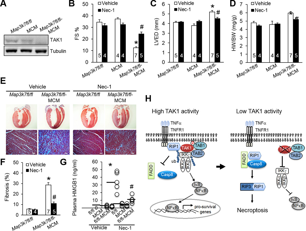
Inhibition of necroptosis with necrostatin-1 rescues pathological cardiac remodeling and dysfunction in TAK1-deficient mice. (A) Immunoblots for TAK1 and α-tubulin in cardiac extracts from Map3k7fl/fl, αMHC-MerCreMer (MCM), and Map3k7fl/fl-MCM mice 2 weeks after treatment with tamoxifen as described in Methods. (B, C, D) FS, LVED, and HW/BW in mice of the indicated genotypes 2 weeks after treatment with tamoxifen in the presence of Nec-1 or vehicle control. *P < 0.01 versus Vehicle Map3k7fl/fl; #P < 0.05 versus Vehicle Map3k7fl/fl-MCM. (E) Masson’s trichrome-stained cardiac sections from mice as described in in B, C, and D. Scale bars, 50 µm. (F) Myocardial fibrosis quantified by MetaMorph software. *P < 0.01 versus Vehicle Map3k7fl/fl; #P < 0.05 versus Vehicle Map3k7fl/fl-MCM. (G) Plasma HMGB1 levels from mice as described in B, C, and D. n=4–5 per group. *P < 0.01 versus Vehicle Map3k7fl/fl; #P < 0.05 versus Vehicle Map3k7fl/fl-MCM. (H) Proposed model: TAK1 functions as a “molecular switch” in TNFR1-mediated cell survival/death signaling.