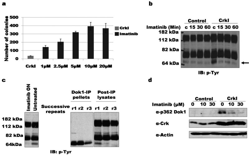
Figure 1. Decreased phosphorylation of Dok1 in CrkI-transformed cells treated with imatinib.

a) Soft agar colonies formed by Crk1-tranformed NIH3T3 cells treated continuously with the indicated concentrations of imatinib. b) Serum-starved CrkI-transformed NIH3T3 cells treated with 20 μM imatinib for indicated times were lysed and blotted with anti-pTyr. Phosphorylation of ∼64 kDa band is decreased upon imatinib treatment of CrkI-transformed cells (indicated by arrow). IB, immuno-blot; pTyr, anti-phosphotyrosine. c) Lysates of CrkI-transformed NIH3T3 cells were serially immunoprecipitated using anti-Dok1 antibody. Left panel, whole cell lystates treated with or without 2.5 μM imatinib; center and right panel, immunoprecipitate (IP) and supernatant (post-IP) fractions. ON: overnight incubation; successive rounds of immunoprecipitation indicated by r1, r2, and r3. d) CrkI-expressing or control NIH3T3 cells treated with indicated concentrations of imatinib were lysed and immunoblotted with phosphospecific Dok1 antibody (α-p362 Dok1). Immunoblotting with anti-Crk and anti-actin shown as controls.
