Fig. 1.

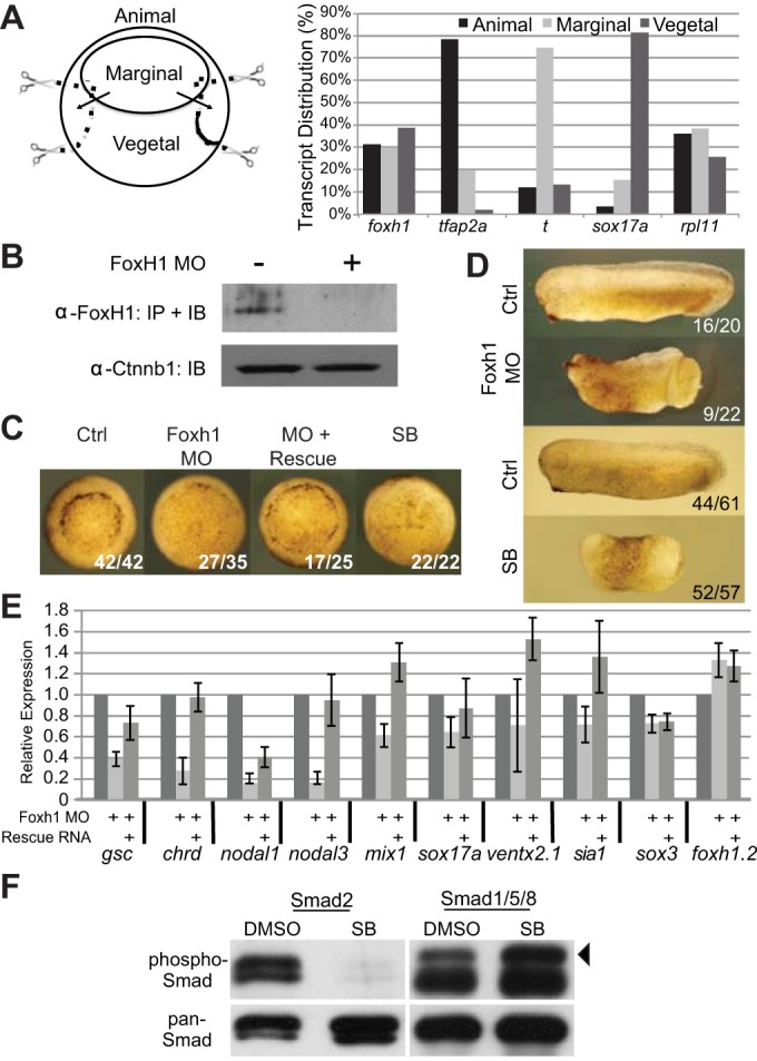
Foxh1 is crucial for mesendoderm formation. (A) Distribution of foxh1 transcripts in X. tropicalis early gastrula. Total RNAs from animal, marginal and vegetal fragments were subjected to RT-qPCR analyses. tfap2a, ectodermal marker; t/brachyury, mesodermal marker; sox17a, endodermal marker; rpl11, expressed throughout the embryo. (B) Embryonic lysates from control or foxh1-MO injected embryos were subjected to immunoprecipitation followed by western blot using anti-Foxh1 antibody. Ctnnb1 protein levels in crude embryo lysates are unaffected by the MO. (C) Both Foxh1 morphant and SB431542-treated embryos exhibit gastrulation delay (vegetal views). (D) Early tailbud stage Foxh1 morphants displaying anterior defects and incomplete blastopore closure; SB431542-treated embryos lack distinctive A-P or D-V features. (E) Examination of foxh1 MO effects on different germ-layer markers by RT-qPCR. gsc, chrd, nodal1, nodal3 and mix1 are mesoendodermal markers; sox17a is an endodermal marker; ventx2.1 is a BMP target gene; sia1 is a Wnt target gene. The ectodermally enriched markers sox3 and foxh1.2 are included as non-Foxh1 targets for comparison. (F) Early gastrula cleared lysates were immunoprecipitated using either pan anti-Smad2 or anti-Smad1 polyclonal antibodies covalently coupled to beads. Bound proteins were subjected to western immunoblotting using anti-P-Smad2 or anti-P-Smad1, respectively. After detection, membranes were re-probed with anti-Smad2 or anti-Smad1 to show efficiency of the immunoprecipitations. Arrowhead indicates the P-Smad1 band; the lower band in the P-Smad1 lanes is low level primary antibody release from the beads.
