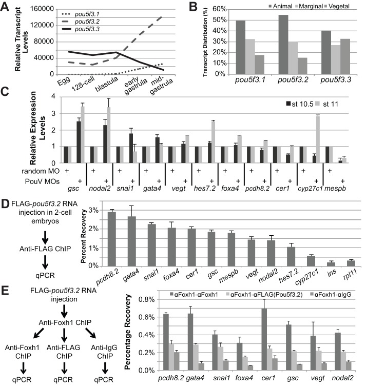
Fig. 5.
Functional analysis of PouV genes in regulating Nodal targets. (A) RT-qPCR analysis of pou5f3.1, pou5f3.2 and pou5f3.3 transcript levels at egg, 128-cell, blastula (stage 9), early (stage 10) and mid-gastrula (stage 10.5) stages. Transcript levels were normalized to the pou5f3.1 level in egg RNA. (B) RT-qPCR analysis of pou5f3.1, pou5f3.2 and pou5f3.3 in animal, marginal and vegetal fragments of the gastrula (stage 10-10.5) stage embryo. (C) RT-qPCR of mesendodermal targets in PouV-depleted embryos at early gastrula (stage 10.5) and mid-gastrula (stage 11). (D) ChIP-qPCR strategy to show FLAG-Pou5f3.2 binding to Pou motif-containing regions within Foxh1 peaks. (E) Sequential ChIP-qPCR analyses for Foxh1 and PouV co-binding on Nodal targets. Chromatin from embryos expressing FLAG-Pou5f3.2 was immunoprecipitated using anti-Foxh1 antibody, followed by a second immunoprecipitation using anti-FLAG antibody or anti-IgG antibody (negative control).

