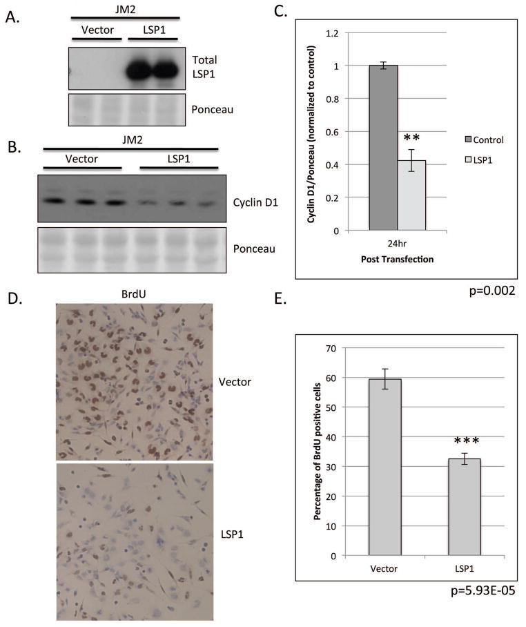
Figure 5. Functional analysis of LSP1 expression in JM2 rat hepatoma cell line.
JM2 cells were transiently transfected with LSP1 cDNA and pExpress-1 plasmid (control). A. Western blot analysis of total LSP1 expression (top panel) at 24 hours post transfection in transiently transfected JM2 cell line. Ponceau S (bottom panel) loading control. B. Western blot analysis of cyclin D1 (top panel) expression at 24 hours post transfection. (Ponceau S (bottom panel), loading control). C. Quantification of cyclin D1 protein expression from B. n=3, p=0.002. D. Representative images of BrDU staining in transiently transfected JM2, 100x magnification. E. Quantification of BrDU labeling in D. n=5 per condition, p=5.93E-05.

