

Abstract
Aim
Direct pulp capping is the treatment of an exposed vital pulp with a dental material to facilitate the formation of reparative dentin and maintenance of vital pulp. A bioengineered drug delivery vehicle has the potential to increase the success rate of pulp capping. The aim of this study was to develop an injectable and light curing drug delivery vehicle for endodontic treatment including direct pulp capping.
Methods
Polyethylene glycol-maleate-citrate (PEGMC) hydrogel was synthesized as a drug delivery vehicle that is composed of PEGMC (45% w/v), acrylic acid (AA) (5% w/v), 2,2′-Azobis(2-methylpropionamidine) dihydrochloride (AAPH) (0.1% w/v), and deionized water. The association between pre-hydrogel solution volume and visible light-curing was examined. The cytotoxicity of the hydrogel was tested using L929 cells in a cell culture system. Ca2+ release from the hydrogel was determined using calcium hydroxide as the incorporated medicine.
Results
The results showed that the light-curing time for hydrogel is comparable to composite resin. The hydrogel had cell toxicity similar to adhesive systems. Moreover, controlled Ca2+ release was obtained from the calcium hydroxide incorporated hydrogel.
Conclusion
The data suggest that hydrogel should be explored further as a promising drug delivery vehicle for vital pulp therapy and regenerative endodontics.
Keywords: hydrogel, pulp capping, drug delivery
Introduction
Direct pulp capping is a treatment for exposed vital pulp using dental materials such as calcium hydroxide,1, 2 bonded composite resins3 or mineral trioxide aggregate.4 Pulp capping has been shown to facilitate both the formation of reparative dentin from odontoblasts5-7 and the maintenance of vital pulp.8, 9 Nevertheless, the 5-year success rates for pulp capping caries-exposed permanent teeth with a mature apex are very low (33%-50%),10-12 due to the lack of methods to simultaneously improve the inflamed pulp condition.13, 14 Steroids,15-17 osteogenic protein-118 and TGF-beta19, 20 have been studied as anti-inflammatory agents towards treating inflamed pulp using drug delivery vehicles. However insufficient degradation of the drug delivery vehicle has been noted in animal studies.19, 20
Thus, a clinically relevant vehicle that possesses realistic handling and drug delivery properties is needed in direct pulp capping and regenerative endodontic treatment. A vehicle having the above mentioned properties can be found in bioengineered hydrogel, which is a colloidal gel suspended in a water medium. Biodegradable hydrogel may be a promising controlled drug delivery system. Specifically, polyethylene glycol-maleate-citrate (PEGMC) hydrogel is a synthetic polyester drug delivery vehicle that is elastomeric, biocompatible, biodegradable and has light-curing crosslinking properties.21
In this study we investigated PEGMC hydrogel as an injectable drug delivery vehicle for regenerative endodontic treatment, including direct pulp capping. The aims of this study were to examine 1) the association between PEGMC pre-hydrogel solution volume and visible light-curing time, 2) the cytotoxicity of the hydrogel drug delivery vehicle and its components in a cell culture system in vitro, and 3) the Ca2+ release from the hydrogel drug delivery vehicle using calcium hydroxide as a model drug.
Materials and Methods
Synthesis of PEGMC
PEGMC was synthesized by using the three main components: 1) polyethylene glycol (PEG) (MW 200-1000 Da), 2) maleic acid (MA) and 3) citric acid (CA) as described elsewhere.21 Briefly, the components were stirred in a three-necked round-bottom flask (250 mL) fitted with an inlet/outlet adapter for 20 minutes at 160°C under nitrogen gas flow. This was followed by 4 hours stirring at 130°C. The reaction was carefully observed until the desired viscosity was achieved, which was confirmed by the twitching (disturbance in stirring motion) of the stirring bar. Synthesized PEGMC was then purified for three-days by dialysis, followed by freeze-drying. The ratio of MA and CA in the initial composition was 6:4, and the overall ratio of the acids over the alcohol was 1:1. The material properties including structure, mechanics, swelling ability, sol-gel contents, and degradation rate were studied previously.22-24
Preparation of PEGMC hydrogel solution
The pre-hydrogel solution was prepared by mixing a solution of PEGMC (45% w/v), acrylic acid (AA) crosslinker (5% w/v), 2,2′-Azobis(2-methylpropionamidine) dihydrochloride (AAPH) photoinitiator (0.1% w/v), and deionized (DI) water. Under a visible light-curing system (Dentsply Caulk, Model-106, Milford, DE, USA) the pre-hydrogel solution polymerizes into a poly-PEGMC (PPEGMC) gel.
Association between pre-hydrogel solution volume and visible light-curing time
To examine the association between pre-hydrogel solution volume and visible light-curing (450-495 nm) time in an in vitro dental setting, magnetic micro stir bars were placed in 96 well plates (effective area 0.32 cm2/well) containing the hydrogel solution on a magnetic stir plate (Figures 1A and 1B), and a visible light-curing was used for polymerization (Figure 1C). Gelation time (in seconds) was defined as the point when the stir bar ceased stirring, indicating formation of a hard gel after polymerization (Figure 1D). Various volumes of the pre-hydrogel solution (50, 100, 150, and 200 μL) were used for photopolymerization; the experiment was repeated ten times per group to measure the gelation time.
Figure 1.

(A, B) The pre-hydrogel solution with a magnetic micro stir bar in 96 well plates, (C) Visible light-curing system, and (D) Formed hard gel.
In vitro cytotoxicity of the hydrogel drug delivery vehicle and its components
Various concentrations of the polymerized hydrogel and its components were prepared in the cell culture medium as in a previously reported dose-dependent cytotoxicity study.25 L929 cells (American Type Culture Collection, Manassas, VA) were cultured in complete medium consisting of Dulbecco's Modified Eagle Medium supplemented with 10% fetal bovine serum and 1% penicillin-streptomycin. The cells were incubated in a humid environment at 37°C and 5% CO2. Upon 80-90% confluency, the cells were seeded in either 96-well plates at 5,000 cells/well or 48-well plates at 10,000 cells/well. Cells in the 96-well plates were subjected to 200 µl of PEGMC at 0 to 1.41% w/v, AA at 0 to 5% w/v, AAPH at 0 to 4% w/v and exposed to light (450 nm) for 0 to 90 seconds. For the cured PPEGMC gel toxicity, gels prepared from pre-hydrogel solution in a 96-well plate, were exposed to the cells in the 48-well plates. The cells exposed only to the medium served as controls in all groups. After 6 hours of incubation, the cell survival was evaluated by [3-(4,5-dimethylthiazol-2-yl)-5-(3-carboxymethoxyphenyl)-2-(4-sulfophenyl)-2H-tetrazolium, inner salt (MTS, CellTiter 96® AQueous Non-Radioactive Cell Proliferation Assay, Promega Corp., Madison, WI, USA) assays. The linear regression was fitted to investigate the association between the gelation time and the solution volume. The data were analyzed by an ANOVA and if there was significance among the groups, pairwise comparisons were conducted using the Student's t-test with Bonferroni corrections accounting for multiple comparisons.
Ca2+ release from the hydrogel drug delivery vehicle
Ca2+ release of Ca(OH)2-incorporated hydrogel solution (experimental group) were compared with Ca(OH)2 (Fisher, Pittsburgh, PA, USA) as a control. Five hundred mg/ml of the materials were incubated at 37°C in a Float-A-Lyzer membrane (500-1000 Da; Spectrum Labs, Rancho Dominguez, CA, USA). At predetermined time points (1-96 hours), 1 ml dialysate containing released Ca2+ was collected and reconstituted with 1 ml fresh distilled water. The Ca2+ content was then quantified using a calcium reagent (Pointe Scientific, Canton, MI, USA) and spectrometer at 550 nm. The percentage of Ca2+ release from the experimental and control groups was compared using a generalized linear mixed model analysis. Degradation characteristics of PPEGMC have been studied previously.21 Data are expressed as mean±SD.
Clinical simulation of injection in plastic root canal block
The viscosity of the hydrogel solution needs to be examined if it is suitable for maxillary injection because the solution will leak out due to gravity, before the light curing procedure can be conducted. An injection syringe with a needle (BD 0.5cc tuberculin syringe 27G) was used to examine the handling property in solution before the light curing polymerization. Root canal plastic blocks were used as if the solution were to be injected into the maxillary area, simulating the clinical scenario.
Results
Association between pre-hydrogel solution volume and visible light-curing time
The gelation times (seconds) were 85.7±10.7, 91.6±10.5, 116.0±11.5, and 147.3±26.7 for 50, 100, 150, and 200 μL of solution, respectively. Gel was formed after photo-polymerization. The fitted linear regression was Time=57.85+0.42*Volume with R2=0.66 (Figure 2), which predicted that the gelation time would decrease on reduction in the volume of the solution. The predicted mean values at 25 µL and 12.5 µL were 68.3 seconds and 63.1 seconds, respectively.
Figure 2.

Association between pre-hydrogel solution volume and visible light-curing time.
In vitro cytotoxicity of the hydrogel drug delivery vehicle and its components
Light exposure onto L929 cells for 30 and 60 seconds resulted in cell viability of 92.5% and 79.7% (Figure 3A). When the cells were exposed to 90 seconds of light there was significant decrease in cell viability of 48.9%. For PEGMC monomer, maximal cell viability of 97.4% was observed with a monomer concentration of 0.088%, while there was a significant drop in viability to 2.6% at 0.35% concentration (p<0.05). There was no cell survival at 1.41% concentration (Figure 3B). Whereas, free form of AA was found to be highly toxic to the cells (Results not shown). Furthermore, cell viability on AAPH remained at 100% up to 1% concentration. However, it dropped to 15.6% at 2% concentration (p<0.05); there was no cell survival at 4% concentration (Figure 3C). Lastly, for the hydrogel system, cell viability over 80% was observed after a period of 6 hours (Figure 3D).
Figure 3.

Cytotoxicity studies on L929 cells based on (A) Time of light exposure, (B) Concentration of PEGMC Monomer, (C) Concentration of AAPH, and (D) PPEGMC.
Ca2+ release from the hydrogel drug delivery vehicle
The Ca2+ release in Ca(OH)2-incorporated hydrogel system, compared with free (not incorporated in any material) Ca(OH)2, is summarized in Figure 4. The Ca2+ release characteristics display a steady increment from 12% release at the first hour, followed by 82% at the 16h time point and finally, 94% release at 96h.
Figure 4.

Comparison of percentage Ca2+ release between experimental and control group.
Clinical simulation of injection in plastic root canal block
In a simulated dental setting using an injection syringe with a needle confirmed injection ability and viscosity (Figure 5A). After injection into plastic root canal blocks, unexpected solution drop or flow was not observed for both mandibular (Figure 5B) and maxillary teeth (Figure 5C). The estimate amount needed for direct pulp capping in real dental setting was less than 50 μL.
Figure 5.
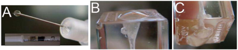
Simulated clinical setting: (A) Confirmed injection ability, (B) Mandibular teeth, and (C) Maxillary teeth. Unexpected solution drop or flow was not observed in a simulated dental setting.
Discussion
In this study we tested a hydrogel solution for tooth that solidifies upon light curing. Unlike low-viscosity hydrogel vehicles,26, 27 the viscosity of the proposed pre-hydrogel solution was suitable for both maxillary and mandibular teeth. Low-viscosity materials are not suitable for maxillary injections because the solution will leak out due to gravity before the light-curing procedure can be conducted. As a result of the gelation time test, the predicted light-curing times at 25 µL and 12.5 µL were 68.3 seconds and 63.1 seconds, respectively. The preferred light-curing times for composite resin materials in current dental practice range from 30 to 90 seconds; therefore, hydrogel gelation time should fall within that range. Significant cell viability was observed with exposure over this time frame. In general, PEG-based hydrogels typically swell considerably after polymerization in moist environments; therefore, the polymerized hydrogel is expected to cover by restorative material to minimize polymerization contraction.26, 27
After the injection ability and gelation time were examined, the cytotoxicity of the material was tested on L929 fibroblasts. The toxicity was generally high because we tested all the hydrogel materials directly on cells at very high concentrations of material components. However, in the clinical setting, all the materials will be used up in the gelling process when the hydrogel is formed (set). Accordingly, as hydrogels form in real time, there will be a negligible amount of unreacted components, which will not make the system cytotoxic. This was evident from the results of PPEGMC gel toxicity, where the cell viability of over 80% was observed, suggesting the hydrogel system can be successfully employed in clinical settings.
The current initiator selection (AAPH) was appropriate for the proposed visible light-curing system as evidenced by the clinically acceptable curing times and low cytotoxicity observed in the results. When designing new bioengineered materials/hydrogels for dental pulp, AAPH concentration of 1% or below is recommended. When redesigning a hydrogel vehicle for dental pulp, its cytotoxicity and the individual component amounts within their biocompatibility range should be considered.
The hydrogel also has the advantage that it can be used as a drug delivery system. The disadvantage of traditional root canal drug delivery systems is that only a small percentage of the drug within the root canal placement material actually reaches the affected area. The time course of drug delivery by these systems is also an issue. The lack of drug reaching canal tissue in the proper time course limits the current systems therapeutic value.28-30 However, state-of-the-art bioengineering techniques have recently led to maximizing therapeutic doses in various types of controlled drug delivery systems.29, 31-33 The advantages of these systems include improved targeted efficacy and local delivery, long-term drug release, and reduced overdoses and potential side effects.31, 33, 34 Normally the approach of simply mixing a drug such as Ca(OH)2 into a hydrogel precursor is expected to cause rapid burst-type release kinetics. In the present study, Ca2+ release by the hydrogel showed kinetics of a controlled release and thus suggests that hydrogel may be a superior drug delivery system than that are currently available. In future studies, other drugs, such as growth factors, anti-inflammatory medicine, will be tested.
The properties of the hydrogels will have clinical impact on regenerative endodontics in the following ways. First, when the hydrogels are incorporated with an anti-inflammatory drug and injected at the pulpotomy or direct pulp capping site they can release the drugs, reducing the pulp inflammation. The hydrogels could be designed to have their degradation rates parallel to the reparative dentin healing rate. Second, the hydrogel could function as a scaffold which could be used in conjunction with dental stem cells for pulp regeneration or to induce calcification to seal the capping site. In summary, the innovative aspects of this study includes 1) developing a hydrogel drug delivery system to dental treatment for direct pulp capping and regenerative endodontics, and 2) developing a new treatment modality to increase the successful rate of vital pulp therapy. A subsequent animal study is the next vital step towards the future clinical human trials.
Acknowledgments
This paper was supported in part by NIH UL1TR0000451.
Footnotes
The authors deny any conflicts of interest.
References
- 1.Zander H. Reaction of the pulp to calcium hydroxide. J Dent Res. 1939;12:373–9. [Google Scholar]
- 2.Schroder U. Effects of calcium hydroxide-containing pulp-capping agents on pulp cell migration, proliferation, and differentiation. J Dent Res. 1985;64:541–8. doi: 10.1177/002203458506400407. [DOI] [PubMed] [Google Scholar]
- 3.Hebling J, Giro EM, Costa CA. Biocompatibility of an adhesive system applied to exposed human dental pulp. J Endod. 1999;25:676–82. doi: 10.1016/s0099-2399(99)80354-9. [DOI] [PubMed] [Google Scholar]
- 4.Torabinejad M, Pitt Ford TR. Root end filling materials: a review. Endod Dent Traumatol. 1996;12:161–78. doi: 10.1111/j.1600-9657.1996.tb00510.x. [DOI] [PubMed] [Google Scholar]
- 5.Bergenholtz G, Mjor IA, Cotton WR, Hanks CT, Kim S, Torneck CD, et al. The biology of dentin and pulp. Consensus report. J Dent Res. 1985;64:631–3. doi: 10.1177/002203458506400422. [DOI] [PubMed] [Google Scholar]
- 6.Pashley DH. Dynamics of the pulpo-dentin complex. Crit Rev Oral Biol Med. 1996;7:104–33. doi: 10.1177/10454411960070020101. [DOI] [PubMed] [Google Scholar]
- 7.Couve E. Ultrastructural changes during the life cycle of human odontoblasts. Arch Oral Biol. 1986;31:643–51. doi: 10.1016/0003-9969(86)90093-2. [DOI] [PubMed] [Google Scholar]
- 8.Zander HA, Glass RL. The healing of phenolized pulp exposures. Oral Surg Oral Med Oral Pathol. 1949;2:803–10. doi: 10.1016/0030-4220(49)90116-4. [DOI] [PubMed] [Google Scholar]
- 9.Bergenholtz G. Advances since the paper by Zander and Glass (1949) on the pursuit of healing methods for pulpal exposures: historical perspectives. Oral Surg Oral Med Oral Pathol Oral Radiol Endod. 2005;100:S102–8. doi: 10.1016/j.tripleo.2005.03.032. [DOI] [PubMed] [Google Scholar]
- 10.Al-Hiyasat AS, Barrieshi-Nusair KM, Al-Omari MA. The radiographic outcomes of direct pulp-capping procedures performed by dental students: a retrospective study. J Am Dent Assoc. 2006;137:1699–705. doi: 10.14219/jada.archive.2006.0116. [DOI] [PubMed] [Google Scholar]
- 11.Tronstad L, Mjor IA. Capping of the inflamed pulp. Oral Surg Oral Med Oral Pathol. 1972;34:477–85. doi: 10.1016/0030-4220(72)90327-1. [DOI] [PubMed] [Google Scholar]
- 12.Barthel CR, Rosenkranz B, Leuenberg A, Roulet JF. Pulp capping of carious exposures: treatment outcome after 5 and 10 years: a retrospective study. J Endod. 2000;26:525–8. doi: 10.1097/00004770-200009000-00010. [DOI] [PubMed] [Google Scholar]
- 13.Langeland K, Dowden WE, Tronstad L, Langeland LK. Human pulp changes of iatrogenic origin. Oral Surg Oral Med Oral Pathol. 1971;32:943–80. doi: 10.1016/0030-4220(71)90183-6. [DOI] [PubMed] [Google Scholar]
- 14.Lin L, Langeland K. Light and electron microscopic study of teeth with carious pulp exposures. Oral Surg Oral Med Oral Pathol. 1981;51:292–316. doi: 10.1016/0030-4220(81)90060-8. [DOI] [PubMed] [Google Scholar]
- 15.Paterson RC. Corticosteroids and the exposed pulp. Br Dent J. 1976;140:174–7. doi: 10.1038/sj.bdj.4803726. [DOI] [PubMed] [Google Scholar]
- 16.Langeland K. Management of the inflamed pulp associated with deep carious lesion. J Endod. 1981;7:169–81. doi: 10.1016/S0099-2399(81)80231-2. [DOI] [PubMed] [Google Scholar]
- 17.Watts A, Paterson RC. The response of the mechanically exposed pulp to prednisolone and triamcinolone acetonide. Int Endod J. 1988;21:9–16. doi: 10.1111/j.1365-2591.1988.tb00948.x. [DOI] [PubMed] [Google Scholar]
- 18.Rutherford RB, Spangberg L, Tucker M, Rueger D, Charette M. The time-course of the induction of reparative dentine formation in monkeys by recombinant human osteogenic protein-1. Arch Oral Biol. 1994;39:833–8. doi: 10.1016/0003-9969(94)90014-0. [DOI] [PubMed] [Google Scholar]
- 19.Hu CC, Zhang C, Qian Q, Tatum NB. Reparative dentin formation in rat molars after direct pulp capping with growth factors. J Endod. 1998;24:744–51. doi: 10.1016/S0099-2399(98)80166-0. [DOI] [PubMed] [Google Scholar]
- 20.Zhang W, Walboomers XF, Jansen JA. The formation of tertiary dentin after pulp capping with a calcium phosphate cement, loaded with PLGA microparticles containing TGF-beta1. J Biomed Mater Res A. 2008;85:439–44. doi: 10.1002/jbm.a.31558. [DOI] [PubMed] [Google Scholar]
- 21.Gyawali D, Nair P, Zhang Y, Tran RT, Zhang C, Samchukov M, et al. Citric acid-derived in situ crosslinkable biodegradable polymers for cell delivery. Biomaterials. 2010;31:9092–105. doi: 10.1016/j.biomaterials.2010.08.022. [DOI] [PMC free article] [PubMed] [Google Scholar]
- 22.Ramanan RM, Chellamuthu P, Tang L, Nguyen KT. Development of a temperature-sensitive composite hydrogel for drug delivery applications. Biotechnology progress. 2006;22:118–25. doi: 10.1021/bp0501367. [DOI] [PubMed] [Google Scholar]
- 23.Sabnis A, Rahimi M, Chapman C, Nguyen KT. Cytocompatibility studies of an in situ photopolymerized thermoresponsive hydrogel nanoparticle system using human aortic smooth muscle cells. J Biomed Mater Res A. 2009;91:52–59. doi: 10.1002/jbm.a.32194. [DOI] [PMC free article] [PubMed] [Google Scholar]
- 24.Sabnis A, Wadajkar AS, Aswath P, Nguyen KT. Factorial analyses of photopolymerizable thermoresponsive composite hydrogels for protein delivery. Nanomedicine. 2009;5:305–315. doi: 10.1016/j.nano.2008.11.003. [DOI] [PMC free article] [PubMed] [Google Scholar]
- 25.Wadajkar AS, Koppolu B, Rahimi M, Nguyen KT. Cytotoxic evaluation of N-isopropylacrylamide monomers and temperature-sensitive poly(N-isopropylacrylamide) nanoparticles. J Nanopart Res. 2009;11:1375–82. [Google Scholar]
- 26.Peppas NA, Sahlin JJ. Hydrogels as mucoadhesive and bioadhesive materials: a review. Biomaterials. 1996;17:1553–61. doi: 10.1016/0142-9612(95)00307-x. [DOI] [PubMed] [Google Scholar]
- 27.Peppas NA, Bures P, Leobandung W, Ichikawa H. Hydrogels in pharmaceutical formulations. Eur J Pharm Biopharm. 2000;50:27–46. doi: 10.1016/s0939-6411(00)00090-4. [DOI] [PubMed] [Google Scholar]
- 28.Gupta P, Vermani K, Garg S. Hydrogels: from controlled release to pH-responsive drug delivery. Drug Discov Today. 2002;7:569–79. doi: 10.1016/s1359-6446(02)02255-9. [DOI] [PubMed] [Google Scholar]
- 29.Qiu Y, Park K. Environment-sensitive hydrogels for drug delivery. Adv Drug Deliv Rev. 2001;53:321–39. doi: 10.1016/s0169-409x(01)00203-4. [DOI] [PubMed] [Google Scholar]
- 30.Hoffman AS. Hydrogels for biomedical applications. Ann N Y Acad Sci. 2001;944:62–73. doi: 10.1111/j.1749-6632.2001.tb03823.x. [DOI] [PubMed] [Google Scholar]
- 31.Sawhney AS, Pathak CP, Hubbell JA. Bioerodible hydrogels based on photopolymerized poly(ethylene glycol)-co-poly(.alpha.-hydroxy acid) diacrylate macromers. Macromolecules. 1993;26:581–7. [Google Scholar]
- 32.Quick DJ, Anseth KS. DNA delivery from photocrosslinked PEG hydrogels: encapsulation efficiency, release profiles, and DNA quality. J Control Release. 2004;96:341–51. doi: 10.1016/j.jconrel.2004.01.021. [DOI] [PubMed] [Google Scholar]
- 33.Hamidi M, Azadi A, Rafiei P. Hydrogel nanoparticles in drug delivery. Adv Drug Deliv Rev. 2008;60:1638–49. doi: 10.1016/j.addr.2008.08.002. [DOI] [PubMed] [Google Scholar]
- 34.Gehrke SH, Fisher JP, Palasis M, Lund ME. Factors determining hydrogel permeability. Ann N Y Acad Sci. 1997;831:179–207. doi: 10.1111/j.1749-6632.1997.tb52194.x. [DOI] [PubMed] [Google Scholar]