Figure 8.
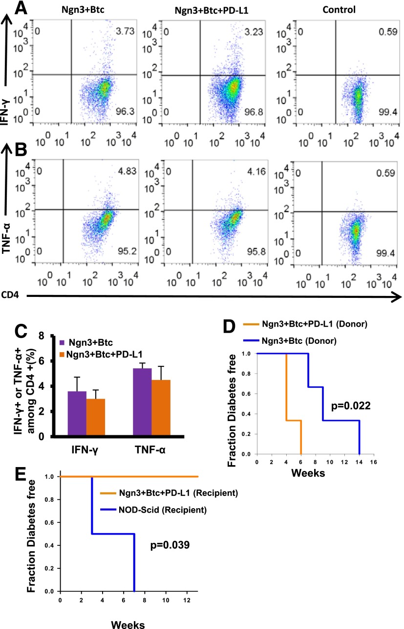
No peripheral immunosuppression in the mice treated with Ngn3-Btc+PD-L1. A–C: IFN-γ– and TNF-α–positive CD4+ T cells isolated from spleens of treated mice after ex vivo stimulation with anti-CD3 and anti-CD28 on FACS analysis. Representative dot plot from one set of mice is shown in A and B, and quantification from three to five separate mice is shown in C. D: Diabetes induction in NOD-Scid mice after adoptive transfer of splenocytes from Ngn3-Btc+PD-L1 or controls. E: Diabetes induction in Ngn3-Btc+PD-L1 or control mice after adoptive transfer of splenocytes from newly diabetic NOD mice. Data are mean ± SEM (n = 3–5 per group).

