Abstract Abstract
Brazilian cave diversity, especially of invertebrates, is poorly known. The Bodoquena Plateau, which is located in the Cerrado Biome in central Brazil, has approximately 200 recorded caves with a rich system of subterranean water resources and high troglobitic diversity. Herein we describe a new troglobitic species of Girardia that represents the first obligate cave-dwelling species of the suborder Continenticola in South America. Specimens of the new species, which occur in a limestone cave in the Bodoquena Plateau, in the Cerrado biome, are unpigmented and eyeless. Species recognition in the genus Girardia is difficult, due to their great morphological resemblance. However, the new species can be easily recognized by a unique feature in its copulatory apparatus, namely a large, branched bulbar cavity with multiple diverticula.
Keywords: New cave-dwelling species, subterranean diversity, Brazilian savannah, planarians, triclads
Introduction
Despite a significant development of the speleobiology in Brazil over the last two decades, species diversity of Brazilian cave fauna has been highly underestimated (Ferreira 2005, Trajano and Bichuette 2010). More than 10,000 caves have been documented in Brazil, but this may represent only 10% of the total number of Brazilian caves, especially considering the extensive karst regions and other potential areas in the country (Auler et al. 2001, Galvão and Cruz 2012). There is much heterogeneity in the degree of knowledge about different karst areas and associated taxa. More troglobitic species have been described in well-known areas from southeast Brazil, such as Alto Ribeira, São Paulo, than in central and north Brazil (Trajano 2000, Trajano and Bichuette 2010, Cordeiro et al. 2014).
The Bodoquena Plateau, in central Brazil (Mato Grosso do Sul), has approximately 200 recorded caves with a rich system of subterranean water resources from the phreatic level (Sallun et al. 2010, Neto 2010). The region has high troglobitic diversity, especially in freshwater ecosystems (Trajano et al. 2000, Trajano and Bichuette 2010; Cordeiro et al. 2014). A total of 34 species of obligate cave-dwelling animals has been recorded from the Bodoquena Plateau, including catfishes and many invertebrates, such as amphipods, spelaeogriphaceans and oligochaetes (Godoy 1986, Gnaspini and Trajano 1994; Gnaspini et al. 1994, Pinto da Rocha 1995, Moracchioli 2002; Costa-Junior 2004, Pires-Vanin 2012, Cordeiro et al. 2013, Cordeiro et. al. 2014). Among the invertebrates, a triclad species hereby assigned to the suborder Continenticola was found in one of the caves.
The diversity of freshwater triclads of the suborder Continenticola in the Neotropical region is considered to be low, and most of the species belong to the Dugesiidae genus Girardia Ball (Sluys et al. 2005). However, extensive areas of South America remain unexplored, such as the Cerrado Biome in central Brazil. The genus Girardia ranges from South to North America and contains 46 species (Sluys 2005, Tyler et al. 2006-2013). According to several authors, species recognition is difficult in this genus, due to their great morphological resemblance (Hyman 1939, Du Bois-Reymond Marcus 1953, Sluys 1996, Sluys et al. 1997, 2005). Most species are recognized on the basis of a combination of morphological characters rather than unique features (Sluys 1996, Sluys et al. 1997).
Triclad diversity in South American subterranean habitats is largely unknown. Kawakatsu and Froehlich (1992) and Trajano and Bichuette (2010) recorded unidentified Dugesiidae species in caves from three different locations, one of which is herein described as new. In addition, Kawakatsu and Froehlich (1992) documented the presence of troglophilous specimens of the suborder Continenticola in three Brazilian caves in Pará State, northern Brazil that they assigned to Girardia paramensis (Fuhrmann). Recently, the first troglobitic triclad of the suborder Cavernicola in South America was recorded in a limestone cave in northeastern Brazil (Leal-Zanchet et al. 2014). Herein we describe a new species of freshwater triclad, the first troglobitic representative of the suborder Continenticola in South America that can be recognized by a unique feature of its copulatory apparatus.
Material and methods
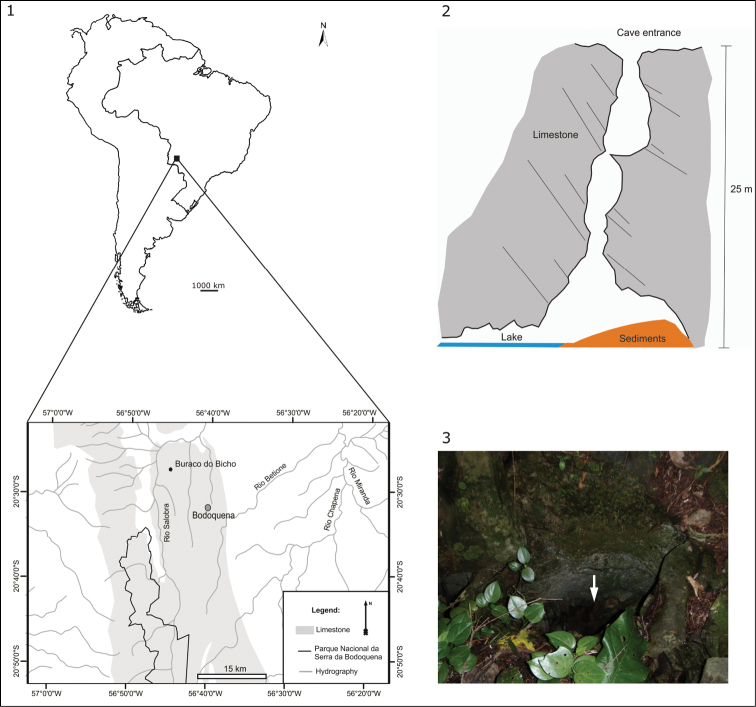
Specimens were collected from the limestone cave “Buraco do Bicho”, located at 266 m a.s.l. in the karst area of Bodoquena Plateau (20°33’50”S and 56°43’50”W), Mato Grosso do Sul, Brazil (Fig. 1). The type-locality is situated in the southern part of the Cerrado biome. The Brazilian savanna is dominated by a tropical climate with a dry winter (type Aw of Köppen’s classification), but the southern portion has a tropical humid climate with warm winter (type Cfa of Köppen’s classification). The mean annual rainfall is approximately 1,400 mm year–1, and the mean annual temperature is about 22 °C to 24 °C (Sallun et al. 2010).
Figures 1–3.
Type-locality of Girardia multidiverticulata: 1 location of the “Buraco do Bicho” cave, in Bodoquena Plateau, Mato Grosso do Sul, Brazil, showing the range of limestone outcrops and the adjacent “Serra da Bodoquena” National Park 2 schematical drawing of the “Buraco do Bicho” cave from where the flatworms were sampled 3 cave entrance (arrow).
The flatworms were directly sampled from a lake (10 m2) in the cave, at a depth of 25 m from the narrow entrance of the cave (Figs 2–3). The lake has a maximum depth of 1.60 m and shows clear waters over a clayey bottom with the parent rock exposed in some places.
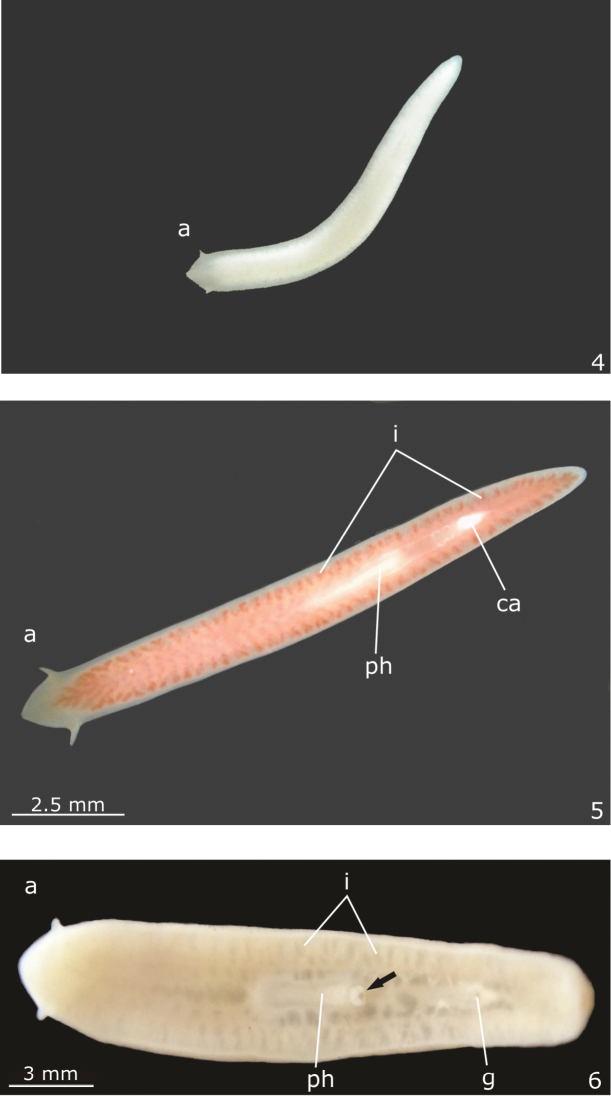
Live specimens were photographed in the field and in the laboratory (Figs 4–6). Specimens were analysed under a stereomicroscope and fixed with 10% Formalin. They were dehydrated and embedded in Paraplast. This material was sectioned at 5−7 µm and stained with hematoxyline/eosine or Goldner’s Masson (Romeis 1989). Colour descriptors, based on the uptake of dyes of particular colours, were used for classifying secretions with trichrome methods.
Figures 4–6.
Girardia multidiverticulata: 4 photograph of a live specimen in ventral view soon after sampling 5 photograph of a live specimen, in ventral view, fed at the laboratory 6 photograph of a preserved specimen in ventral view. The tip of the pharynx is protruded (arrow) through the mouth. Scale bar for the Fig. 4 not available.
Type-material was deposited in the following reference collections: Museu de Zoologia da Universidade do Vale do Rio dos Sinos, São Leopoldo, Rio Grande do Sul, Brazil (MZU), and the Helminthological Collection of Museu de Zoologia da Universidade de São Paulo, São Paulo, São Paulo State, Brazil (MZUSP).
The flatworms were maintained in a permanently dark laboratory under a temperature of 24 °C for three years. They were kept in small tanks and fed weekly with live Artemia salina.
Abbreviations used in the figures
a; bc; br; ca; cb; cbc; cg; ceg; cf; ci; cm; cs; de; di; eg; ej; em; es; fa; g; go; i; im; in; lf; lm; lu; m; ma; mm; n; o; om; ov; pb; pg; ph; phg; php; pp; r; rg; s; sd; sg; sv; t; ve; vi; xg.
Systematic acount
Order Tricladida Lang, 1884: Suborder Continenticola Carranza et al., 1998: Family Dugesiidae Ball, 1974: Genus Girardia Ball, 1974
Girardia multidiverticulata sp. n.
http://zoobank.org/147CB963-DECB-4125-985D-124B306B5EA0
Material examined.
Holotype. MZUSP PL.1573: “Buraco do Bicho” cave, Bodoquena Plateau, Mato Grosso do Sul (MS), Brazil, July 2011, coll. L. M. Cordeiro & R. Borghezan, sagittal sections on 18 slides.
Paratypes. “Buraco do Bicho” cave, Bodoquena Plateau, MS, Brazil, July 2011, coll. L. M. Cordeiro & R. Borghezan. MZU PL.00184: sagittal sections on 61 slides; MZU PL.00185: sagittal sections on 8 slides; MZU PL.00186: transverse sections on 16 slides.
Etymology.
The species name refers to the multiple diverticula of the bulbar cavity.
Diagnosis.
Blind and unpigmented Girardia species characterized by a branched bulbar cavity with multiple irregular diverticula.
Description.
Live specimens unpigmented and eyeless (Figs 4–6). Head highly triangular with long and pointed auricles, which become moderately sized and almost rounded after fixation (Fig. 6); posterior tip rounded (Figs 4–6). Preserved specimens up to 20 mm long and 3 mm wide (Table 1). Mouth and gonopore located at the posterior half of the body (Table 1, Fig. 6).
Table 1.
Measurements, in mm, of specimens of Girardia multidiverticulata, sp. n. DG: distance of gonopore from anterior end; DM: distance of mouth from anterior end. The numbers given in parentheses represent the position relative to body length. * Measurements after fixation; ** Measurements after histological processing; -: not measured.
| Holotype MZUSP PL.1573 | Paratype MZU PL.00184 | Paratype MZU PL.00185 | Paratype MZU PL.00186 | |
|---|---|---|---|---|
| Length* | 16 | 20 | 12 | 14 |
| Length** | 12.5 | 15 | 9 | 12 |
| Width* | 2 | 3 | 2 | 3 |
| DM | 9 (72%)* | 9 (60%)** | 6 (67%)** | - |
| DG | 11 (88%)* | 10.5 (70%)** | 7 (78%)** | - |
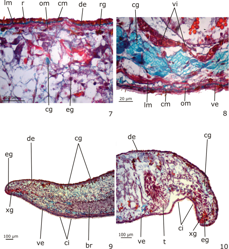
Epidermis (Figs 7–10). Columnar epithelium, ciliated on the ventral body surface (Figs 7, 10). The whole epidermis receives secretions of three types of glands: (1) xanthophil, rhabidtogen secretion (rhammites); (2) erythrophil, fine granular secretion; (3) cyanophil amorphous secretion (Figs 7–10). Rhammites are more densely distributed at the dorsal surface (Fig. 7). The erythrophil glands and a fourth type of gland, with xanthophil, granular secretion, concentrate their openings medially at the anterior and posterior tips of the body (Figs 9–10) as well as at the body margins. Cyanophil glands become numerous towards the anterior tip (Fig. 9).
Figures 7–10.
Girardia multidiverticulata, holotype in sagittal section: 7–8 dorsal and ventral surfaces of the body, respectively 9–10 anterior and posterior tips of the body, respectively.
Cutaneous musculature (Figs 7–8). Three layers, viz. a thin subepithelial circular layer, followed by an oblique layer with decussate fibers and a thicker layer of longitudinal muscle. Dorsal and ventral cutaneous musculatures show similar height in the pre-pharyngeal region (10–13 µm thick in the holotype).
Sensory organs. The auricular sensory organs are lined with densely ciliated, low cuboidal epithelium, with insunk nuclei. Few secretory cells open through this epithelium. The cutaneous musculature is very thin at the level of the sensory organs.
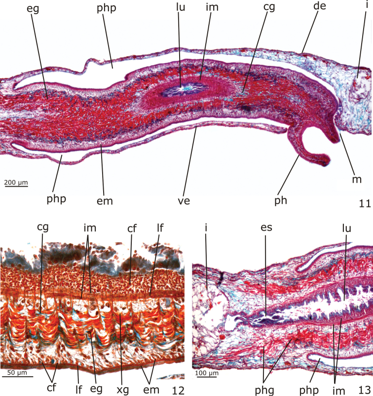
Digestive system (Figs 5–6, 11–13). Pharynx cylindrical, non-pigmented; between about 1/4th and 1/6th of the body length. It is located in the posterior half or in the median third of the body (Figs 5–6). Mouth at the posterior end of the pharyngeal pouch (Fig. 11). Pharynx lined by cuboidal ciliated epithelium with insunk nuclei; pharyngeal lumen lined by non-ciliated, columnar epithelium with some insunk nuclei. Pharyngeal glands of the usual three types (xanthophil, cyanophil and erythrophil glands). Outer musculature of the pharynx constituted of a thin subepithelial layer of longitudinal muscle, followed by a thin layer of circular muscle, each about 4 µm thick in the holotype. Inner pharyngeal musculature composed of a thick subepithelial layer of circular muscle (30–60 µm thick in the holotype), followed by a layer of longitudinal muscle (15–20 µm thick in the holotype) (Figs 11–13). An esophagus, about 1/6 of the pharyngeal length, connects the pharynx with the intestine (Fig. 13). The esophagus is lined by a flat to cuboidal epithelium with insunk nuclei; it is coated with a thin muscularis containing circular fibers near the intestine, gradually becoming thicker towards the pharynx and similar to the inner pharyngeal musculature. Intestine with the usual tricladid form (Fig. 5), with the anterior intestinal trunk extending onto the posterior part of the brain.
Figures 11–13.
Girardia multidiverticulata, holotype in sagittal section: 11 pharynx in general view 12 detail of pharyngeal musculature and glands 13 detail of the esophagus.
Male reproductive system (Figs 10, 14–19, 24–26).
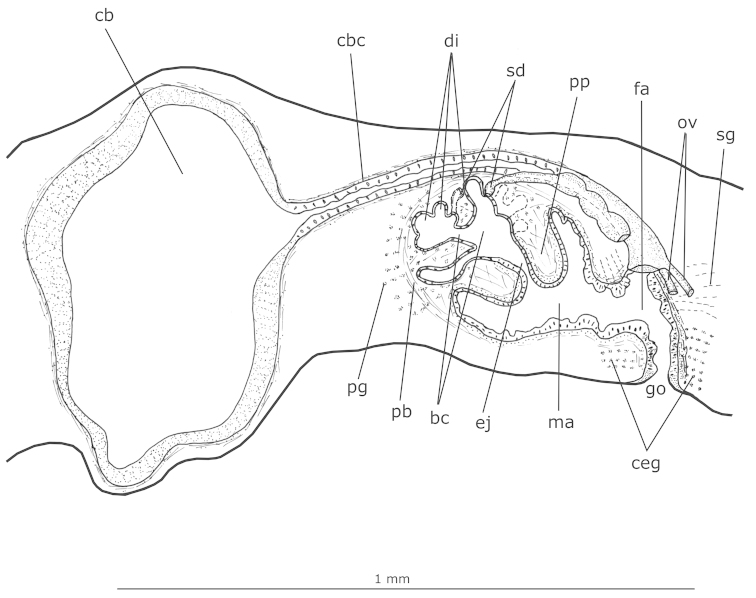
Figure 14.
Girardia multidiverticulata: sagittal composite reconstruction of the copulatory apparatus of the holotype.
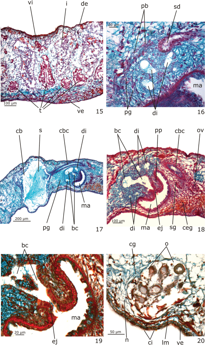
Figures 15–20.
Girardia multidiverticulata, holotype in sagittal section: 15 testes in the anterior body region 16 detail of the opening of a sperm duct into a diverticulum of the bulbar cavity 17–18 copulatory apparatus in general view 19 detail of the male copulatory organs 20 ovary.
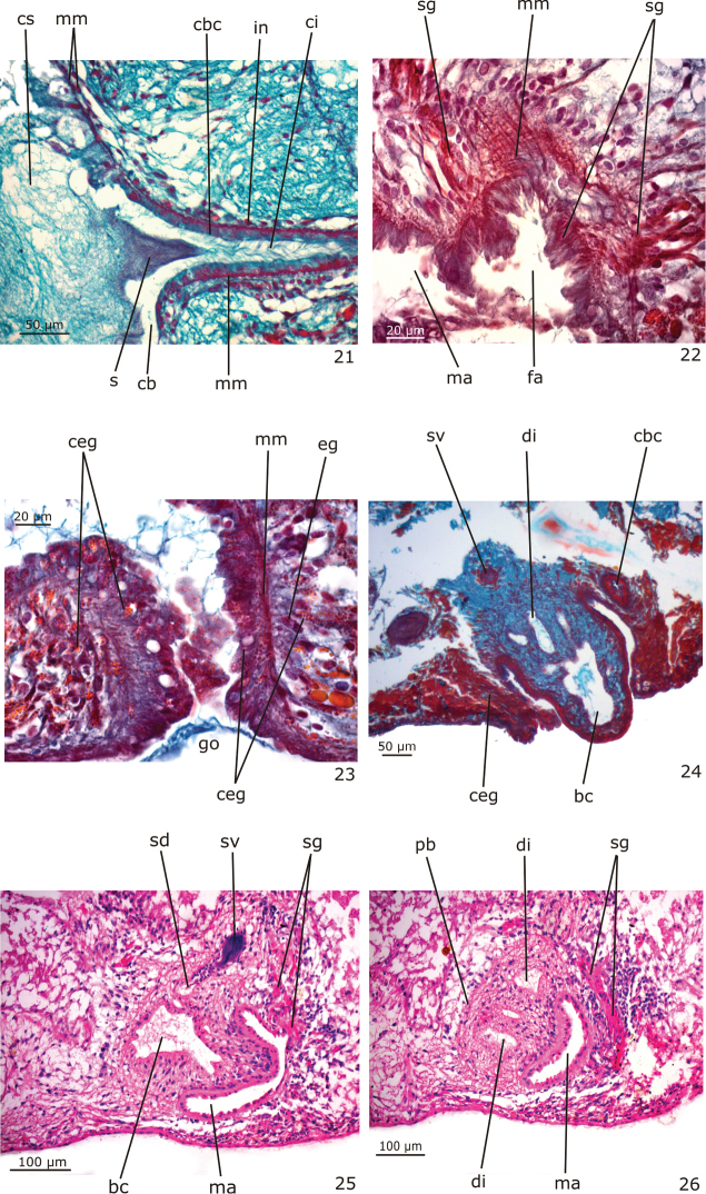
Figures 21–26.
Girardia multidiverticulata, holotype in sagittal section (21–23); paratypes MZU PL.00186 in transverse section (24) and MZU PL.00184 in sagittal section (25–26): 21 detail of the copulatory bursa and its canal 22 detail of the proximal part of the female atrium 23 gonoduct 24 protruded penis papilla 25–26 male copulatory organs.
Numerous testicular follicles, 100–200 µm in diameter in the holotype, arranged in one irregular row on each side of the body. They are situated mainly ventrally (Fig. 15), but may occupy the whole body height; some are situated dorsally. Testes extend from about 2 mm from the anterior tip in the holotype (equal to 16% of body length), just behind the brain, to the posterior end of the body (Fig. 10). Sperm ducts form spermiducal vesicles laterally to the pharynx, diminishing in diameter close to their opening into the bulbar cavity (Fig. 16). Laterally to the copulatory apparatus, they ascend, forming a loop, and turn anteriad. Sperm ducts separately penetrate the penis bulb, and open laterally into the large, branched bulbar cavity which contains various irregular diverticula (Figs 14, 16, 18, 24–26). The short ejaculatory duct narrows towards its opening at the tip of the penis papilla. The latter is a stubby, symmetrical cone, obliquely oriented in the male atrium (Figs 14, 18–19, 25–26).
Sperm ducts lined with a ciliated, cuboidal epithelium, becoming flattened in the spermiducal vesicles; they are coated with a circular muscle layer (3 µm thick in the holotype). The large penis bulb consists of a loose connective tissue containing abundant gland necks of penis glands and interwoven muscle fibers (Figs 14, 16). Bulbar cavity lined with a non-ciliated, cuboidal to flat epithelium, underlain with a weak and inconspicuous muscle layer. Numerous penis glands with extrabulbar cell bodies and mixed secretion open into the bulbar cavity (Figs 14, 16–17). This secretion has a cyanophil external part and an erythrophil internal core. In addition, few erythrophil penis glands with extrabulbar cell bodies open into the bulbar cavity. Ejaculatory duct lined with non-ciliated, columnar epithelium, and surrounded by a thin muscularis (about 3 µm thick in the holotype) composed of a subepithelial layer of circular muscle and a layer of longitudinal muscle. Erythrophil glands have abundant openings into the distal, narrow portion of this duct (Fig. 19). Penis papilla covered with a non-ciliated, columnar epithelium that becomes flat towards the tip of the papilla. Muscularis of penis papilla (5–9 µm thick in the holotype) composed of a thick subepithelial layer of circular fibres and a thin subjacent layer of longitudinal fibres (Fig. 19). Few penis glands with amorphous, cyanophil secretion and with fine granular, erythrophil secretion open through the epithelium of the penis papilla. Cyanophil glands with extrabulbar cell bodies; erythrophil glands with intrapapillar cell bodies. Male atrium lined with a non-ciliated, cuboidal to columnar epithelium, the cells of which have an irregular height and cyanophil cytoplasm (Fig. 19). The male atrial muscularis (4–5 µm thick in the holotype) is constituted of a thick subepithelial layer of circular fibres, followed by a thin layer of longitudinal fibres. Glands with cyanophil amorphous secretion and erythrophil glands with fine granular secretion open into the male atrium. Cyanophil glands with extrabulbar cell bodies, and erythrophil glands with subepithelial cell bodies.
Female reproductive system (Figs 8, 14, 17, 20–23).
Vitellaria well developed (Fig. 8), located between intestinal branches. Ovaries ovoid (Fig. 20), 150–200 µm in diameter in the holotype. They are situated dorsally to the ventral nerve cords, at about the same transversal level as the anteriormost testes and in close proximity to the brain (about 0.9 mm behind it in the holotype). Ovovitelline ducts arising from the lateral surface of the ovaries and running backwards dorsally to the nerve cords. At about the level of the gonoduct, the ovovitelline ducts turn medially, and separately open into the most distal, postero-ventral part of the bursal canal, in close proximity to each other. Copulatory bursa large and ovoid (Figs 14, 17). Bursal canal long, curving towards the ventral surface of the body and opening into the short female atrium (Figs 14, 17). Gonoduct almost straight (Fig. 14, 23).
Ovovitelline ducts lined with ciliated, cuboidal epithelium with insunk nuclei and covered mainly by circular muscle fibres (2–3 µm thick in the holotype). Copulatory bursa lined with non-ciliated, columnar epithelium composed of cells with erythrophil secretion and cells with heavily stained, cyanophil secretion; it is covered by a thin muscle coat constituted by interwoven longitudinal and circular muscle fibres (5–8 µm thick in the holotype). The bursa of the holotype contains sperm and cyanophil secretion in its lumen (Figs 17, 21); some spermatozoids are absorbed by its epithelial cells. Bursal canal lined with a ciliated, cuboidal to columnar epithelium with cyanophil cytoplasm (Fig. 21). The muscularis of the bursal canal (3–4 µm thick in the holotype) is constituted of interwoven circular and longitudinal muscle fibres (Fig. 21). There are some insunk nuclei and cell bodies of xanthophil glands around the copulatory bursa and bursal canal. Female atrium lined with a ciliated, tall columnar epithelium, the cells of which show irregular height. The muscularis of the female atrium (6 µm thick in the holotype) is constituted of a subepithelial layer of circular fibres, followed by a layer of longitudinal fibres (Fig. 22). Numerous glands with fine granular, erythrophil secretion (shell glands) and few cyanophil glands open into the female atrium. Gonoduct lined by ciliated, tall columnar epithelium, and coated with a subepithelial layer of circular muscle, followed by a layer of longitudinal muscle (about 9 µm thick in the holotype) (Fig. 23). Abundant cement glands with coarse granular, xanthophil secretion (Fig. 23) and numerous glands with heavily stained, cyanophil amorphous secretion discharge into the gonoduct. Both cell types have long cell necks and their cell bodies are located in the mesenchyme. Few glands with fine, erythrophil secretion and subepithelial cell bodies also open into the gonoduct (Fig. 23).
Geographical distribution.
Known only from the type-locality (“Buraco do Bicho” cave), Bodoquena Plateau, Mato Grosso do Sul, Brazil.
Variability.
In paratype MZU PL.00186 with contracted body, the penis papilla protrudes into the gonoduct and the bulbar cavity formes two main proximal chambers and one large distal one (Fig. 24). The distal portion of the bursal canal and the female atrium of this paratype were elongated and protruded towards the ventral surface of the body (Fig. 24). Paratype MZU PL.00184 has a more elongate, conical and truncated penis papilla occupying almost the whole cavity of the male atrium (Fig. 25). Despite the fact that this specimen is mature, it has a small copulatory bursa with narrow cavity, probably due to a different physiological state in relation to the other specimens. In this paratype, stained with Hematoxyline/Eosine, the penis glands showed an amorphous, chromophobous secretion, shell glands were stained deep pink (Figs 25–26) and cement glands showed chromophobous, coarse granular secretion.
Ecology
There was a density of about 5 to 10 flatworms per m2 in the lake that constitutes the type-locality of Girardia multidiverticulata. Other invertebrates, such as the spelaeogriphacean Potiicoara brasiliensis Pires, the amphipod Megagidiella sp. and an undetermined species of troglomorphic oligochaete, were also observed. The water level did not vary between June and August 2011, when field work was performed. The recorded values of temperature and conductivity were 23.1 °C and 0.55 mS.cm-1, respectively.
Flatworms maintained in the laboratory reproduced sexually and produced stalked egg capsules. Usually 2 to 3 specimens hatched from each egg capsule.
Discussion
Due to the lack of eyes and body pigmentation, the troglobitic Girardia multidiverticulata differs from the majority of its congeners, which are pigmented, epigean organisms. It can be differentiated from the hipogean Girardia mckenziei (Mitchell & Kawakatsu) from Chiapas, Mexico, which has a smaller body length, dorsal surface with a slight, microscopic pigmentation and minute eyes (Mitchell and Kawakatsu 1973). The new species herein described is similar to two other troglobite dugesiids, Girardia typhlomexicana (Mitchell & Kawakatsu) and Girardia barbarae (Mitchell & Kawakatsu), from Tamaulipas, Mexico, which are blind and eyeless (Mitchell and Kawakatsu 1972). However, both have a small body length, up to 8 mm, whereas mature specimens of the new species are between 12 mm and 20 mm long after fixation. In addition, live specimens of Girardia multidiverticulata show long and pointed auricles in contrast to the moderate-sized auricles of Girardia typhlomexicana and Girardia barbarae. Girardia multidiverticulata also differs from the troglophilous species Girardia guatemalensis (Mitchell & Kawakatsu), from Tamaulipas, Mexico, which has a pigmented body with two small eyes (Mitchell and Kawakatsu 1972, Kawakatsu and Mitchell 1981), and from the troglophilous specimens of Girardia paramensis, with pigmented body and a pair of eyes, recorded in Pará State, northern Brazil by Kawakatsu and Froehlich (1992).
Regarding the reproductive system, Girardia multidiverticulata has large, mainly ventral testes that occupy most of the dorso-ventral space of the body height, a large, branched bulbar cavity with multiple diverticula, and a stubby penis papilla. This combination of characteristics cannot be found in other species of Girardia from epigean or hipogean environments. The epigean species Girardia anderlani (Kawakatsu & Hauser) from the vicinity of São Leopoldo, southern Brazil, also has a large bulbar cavity, but with only two main chambers. In addition, this species has mainly ventral testes in two or three longitudinal rows and a conical and asymmetrical penis papilla (Kawakatsu et al. 1983). Girardia multidiverticulata shares an intermingled muscle coat around the copulatory bursa with the epigean species Girardia bursalacertosa Sluys (Sluys et al. 2005), but this feature is more developed in the latter than in the new species. In addition, Girardia bursalacertosa has an almost tubular bulbar cavity and a small copulatory bursa (Sluys et al. 2005), among other distinctive features.
Concluding, in comparison to other species of Girardia, most of which with very similar reproductive systems, the troglobitic Girardia multidiverticulata shows a unique feature in its copulatory apparatus, namely a large and branched bulbar cavity with multiple diverticula. Additionally, the new species has a combination of other characteristics of its external and internal morphology that differentiate it from its congeners.
Supplementary Material
Acknowledgements
We thank Prof Dr Eleonora Trajano (Universidade de São Paulo - USP, Brazil) for providing the laboratory conditions to keep live specimens. We acknowledge Rodrigo Borghezan (USP, Brazil), who first noticed the troglobitic planarians, for his help with field work and the technician Rafaela Canello (UNISINOS, Brazil) for her help in histological preparation. We also thank the Brazilian Research Council (CNPq) for the grants 477712/2006-1 and 143379/2009-7, and the Coordenação de Aperfeiçoamento de Pessoal de Nível Superior (CAPES) for grants and fellowships in support of this study. We acknowledge MSc Emily Toriani and MSc Edward Benya for the English review of the text. We gratefully thank Dr Leigh Winsor, Australia, for suggestions regarding anatomical descriptors. We are grateful to Prof Dr Masaharu Kawakatsu, Japan, and an anonymous reviewer for their valuable suggestions on an early draft of the paper.
Citation
de Souza ST, Morais ALM, Cordeiro LM, Leal-Zanchet AM (2015) The first troglobitic species of freshwater flatworm of the suborder Continenticola (Platyhelminthes) from South America. ZooKeys 470: 1–16. doi: 10.3897/zookeys.470.8728
References
- Auler A, Rubbioli E, Brandi R. (2001) As grandes cavernas do Brasil. Grupo Bambuí de Pesquisas Espeleológicas, Belo Horizonte, Brazil. [Google Scholar]
- Cordeiro LM, Borghezan R, Trajano E. (2014) Subterranean biodiversity in the Serra da Bodoquena karst area, Paraguay River basin, Mato Grosso do Sul, Southwestern Brazil. Biota Neotropica 14(3): 1–28. doi: 10.1590/1676-06032014011414 [Google Scholar]
- Cordeiro LM, Borghezan R, Trajano E. (2013) Distribuição, riqueza e conservação dos peixes troglóbios da Serra da Bodoquena, MS (Teleostei: Siluriformes). Revista da Biologia 10: 21–27. doi: 10.7594/revbio.10.02.04 [Google Scholar]
- Costa Jr E. (2004) Fish from the underwater caves of Bodoquena Plateau, Mato Grosso do Sul, Southwestern Brazil. DIR Lifestyle & Underwater Adventure Magazine 5: 8–12. doi: 10.1590/1676-06032014011414 [Google Scholar]
- Du Bois-Reymond Marcus E. (1953) Some South American triclads. Anais da Academia Brasileira de Ciências 25: 65–78. [Google Scholar]
- Ferreira RL. (2005) A vida subterrânea nos campos ferruginosos. O Carste 3: 106–115. [Google Scholar]
- Galvão ALCO, Cruz JB. (2012) Brasil ultrapassa mais de 10.000 cavernas conhecidas. ESPELEOINFO | CECAV - Centro Nacional de Pesquisa e Conservação de Cavernas 3: 1–8. [Google Scholar]
- Gnaspini P, Trajano E. (1994) Brazilian cave invertebrates, with a checklist of troglomorphic taxa. Revista Brasileira de Entomologia 38: 549–584. doi: 10.1590/S0031-10492003000500001 [Google Scholar]
- Gnaspini P, Trajano E, Sanchez LE. (1994) Província Espeleológica da Serra da Bodoquena, MS: exploração, topografia e biologia. Espeleo-tema 17: 19–44. [Google Scholar]
- Godoy NM. (1986) Nota sobre a fauna cavernícola de Bonito, MS. Espeleo-tema 15: 80–92. [Google Scholar]
- Hyman LH. (1939) New species of flatworms from North, Central, and South America. Proceedings of the United States Natural Museum 86: 419–439. doi: 10.5479/si.00963801.86-3055.419 [Google Scholar]
- Kawakatsu M, Froehlich EM. (1992) Freshwater Planarians from Four Caves of Brazil: Dugesia paramensis (Fuhrmann, 1914) and Dugesia sp. (Turbellaria, Tricladida, Paludicola). Journal of the Speleological Society of Japan 17: 1–19. [Google Scholar]
- Kawakatsu M, Mitchell RW. (1981) An additional note on Dugesia guatemalensis Mitchell et Kawakatsu (Turbellaria: Tricladida: Paludicola), a troglophilic planarian from México. Annales de Spéléologie 6: 37–41. [Google Scholar]
- Kawakatsu M, Hauser J, Friedrich SMG. (1983) Morphological, karyological and taxonomic studies of freshwater planarians from South Brazil. IV. Dugesia anderlani sp. nov. (Turbellaria: Tricladida: Paludicola), a new species from São Leopoldo in estado de Rio Grande do Sul. Annotationes Zoologicae Japoneses 56(3): 196–208. [Google Scholar]
- Leal-Zanchet AM, Souza ST, Ferreira RL. (2014) A new genus and species for the first recorded cave-dwelling Cavernicola (Platyhelminthes) from South America. ZooKeys 442: 1–15. doi: 10.3897/zookeys.442.8199 [DOI] [PMC free article] [PubMed] [Google Scholar]
- Mitchell RW, Kawakatsu M. (1972) Freshwater cavernicole planarians from Mexico: new troglobitic and troglophilic Dugesia from caves of the Sierra de Guatemala. Annales de Spéléologie 27: 639–681 [Printed in May 1973] [Google Scholar]
- Mitchell RW, Kawakatsu M. (1973) A new cave-adapted planarian (Tricladida, Paludicola, Planariidae) from Chiapas, Mexico. Association for Mexican Cave Studies, Bulletin 5: 165–170. [Google Scholar]
- Moracchioli N. (2002) Estudo dos Spelaeogriphacea brasileiros, crustáceos Peracarida subterrâneos. PhD thesis, Universidade de São Paulo, São Paulo, Brazil. [Google Scholar]
- Neto JLB. (2010) Cavernas inundadas na Serra da Bodoquena. O Carste 2: 34–38. [Google Scholar]
- Pinto-da-Rocha R. (1995) Sinopse da fauna cavernícola do Brasil (1907-1994). Papéis Avulsos de Zoologia 39: 61–173. [Google Scholar]
- Pires-Vanin AMS. (2012) The discovery of male Potiicoara brasiliensis (Crustacea, Spelaeogriphacea) with notes on biology and distribution. Zootaxa 3421: 61–68 http://www.mapress.com/zootaxa/2012/f/z03421p068f.pdf [Google Scholar]
- Romeis B. (1989) Mikroskopische Technik. Urban und Schwarzenberg, München, Germany. [Google Scholar]
- Sallum WF, Karmann I, Lobo HAS. (2010) Cavernas da Serra da Bodoquena. O Carste 22: 27–33. [Google Scholar]
- Sluys R. (1996) Reconsiderations of the species status of some South American planarians (Platyhelminthes: Tricladida: Paludicola). Proceedings of the Biological Society of Washington 109: 229–235 http://biostor.org/reference/74375 [Google Scholar]
- Sluys R, Hauser J, Wirth QJ. (1997) Deviation from the Groundplan: a unique new species of freshwater planarian from South Brazil (Platyhelminthes, Tricladida, Paludicola). Journal of Zoology 241: 593–601. doi: 10.1111/j.1469-7998.1997.tb04851.x [Google Scholar]
- Sluys R, Kawakatsu M, Ponce de León R. (2005) Morphological stasis in an old and widespread group of species: Contribution to the taxonomy and biogeography of the genus Girardia (Platyhelminthes, Tricladida, Paludicola). Studies on Neotropical Fauna and Environment 40(2): 155–180. doi: 10.1080/01650520500070220 [Google Scholar]
- Trajano E. (2000) Cave Faunas in the Atlantic Tropical Rain Forest: Composition, Ecology, and Conservation. Biotropica 32: 882–893. doi: 10.1111/j.1744-7429.2000.tb00626.x [Google Scholar]
- Trajano E, Bichuette ME. (2010) Diversity of Brazilian subterranean invertebrates, with a list of troglomorphic taxa. Subterranean Biology 7: 1–16. [Google Scholar]
- Tyler S, Schilling S, Hooge M, Bush LF. (comp.) (2006–2013) Turbellarian taxonomic database. Version 1.5. http://turbellaria.umaine.edu [accessed on September 2014]
Associated Data
This section collects any data citations, data availability statements, or supplementary materials included in this article.


