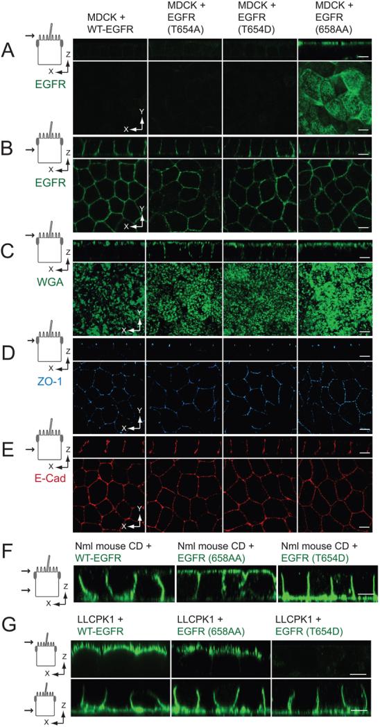
Figure 2. Thr654 regulates EGFR sorting independently of AP1B.
(A - B) Vertical x-z and horizontal x-y confocal sections (see schematics) from fixed MDCK cell monolayers expressing EGFR proteins stained with human-specific EGFR1 antibody (green) added to Ap (A) or BL (B) surface. (C – E) Confocal sections from fixed and permeabilized MDCK cell monolayers stained with Alexa Fluor 488 WGA (green; C) and antibodies to ZO-1 (blue; D) and E-cadherin (red; E). (A-E) Schematics to left show the plane of the vertical confocal image. (F) Confocal images of AP1B positive normal mouse collecting duct (CD) cell monolayers expressing human EGFRs listed in the figure fixed and stained with EGFR1 (green) added to both surfaces (monolayer leakiness precluded domain-specific EGFR1 staining). (G) Confocal images of AP1B negative LLCPK1 pig epithelial cell monolayers expressing human EGFRs listed in the figure fixed and stained with EGFR1 (green) added to Ap (top) or BL side of the filter. Size bars = 5 μm.

