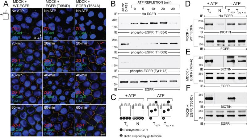
Figure 4. EGFR Thr654 regulates EGFR sorting in ATP depletion-repletion renal injury model.
ATP was depleted by incubating mature MDCK monolayers in PBS supplemented with 2-deoxy-d-glucose and antimycin A for 30 min, and then replenished by re-feeding cells with complete media for times indicated in the figure. (A) Confocal images from just beneath the Ap membrane in cells stained with antibodies to ZO-1 (red) and E-cadherin (green) and counterstained with DAPI (blue). Size bar = 5 μm. (B) Cells were lysed to recover human EGFR immune complexes for immunoblotting with phospho-specific EGFR antibodies listed in the figure and an activation-independent EGFR antibody. Control cells were incubated in Dulbecco's PBS (D-PBS) for 30 min without ATP depletion. (C) Schematic showing experimental procedure for evaluating BL EGFR distribution in ATP-depleted cells. Established MDCK cell monolayers were labeled from the BL side with a cleavable biotin reagent. T0, total biotinylated EGFR (positive control); N, biotin stripped from BL side immediately after biotinylation (negative control); T-ATP, total biotinylated EGFR from ATP-depleted cells (control for stability of biotinylated EGFR); TAp + In, biotin stripped from BL surface in ATP-depleted cells (internalized biotinylated EGFR and biotinylated EGFR reinserted in Ap membrane). (D - F) Cells expressing WT-EGFR (D), EGFR (T654A) (E), or EGFR (T654D) (F) were treated according to the scheme in (C). EGFR immune complexes were resolved by non-reducing SDS-PAGE for immunoblotting with a biotin antibody (top panels) or an EGFR antibody to control for protein loading (bottom panels). Representative immunoblots from at least two independent experiments.

