

Abstract
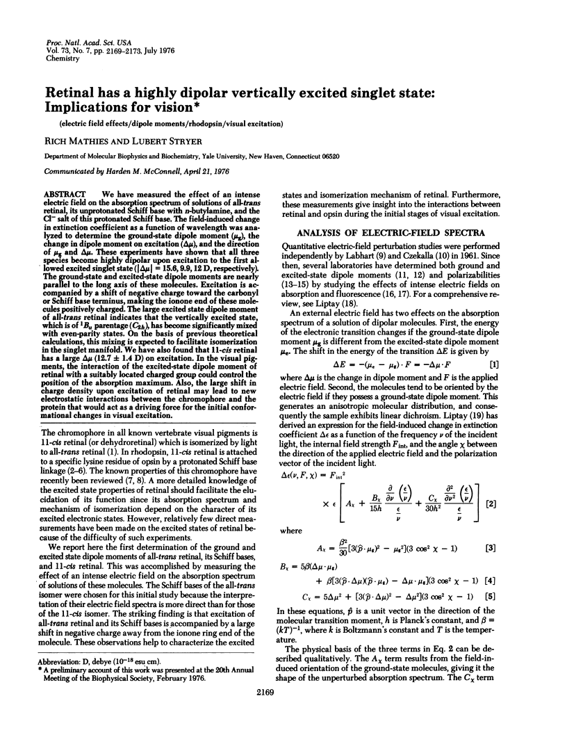
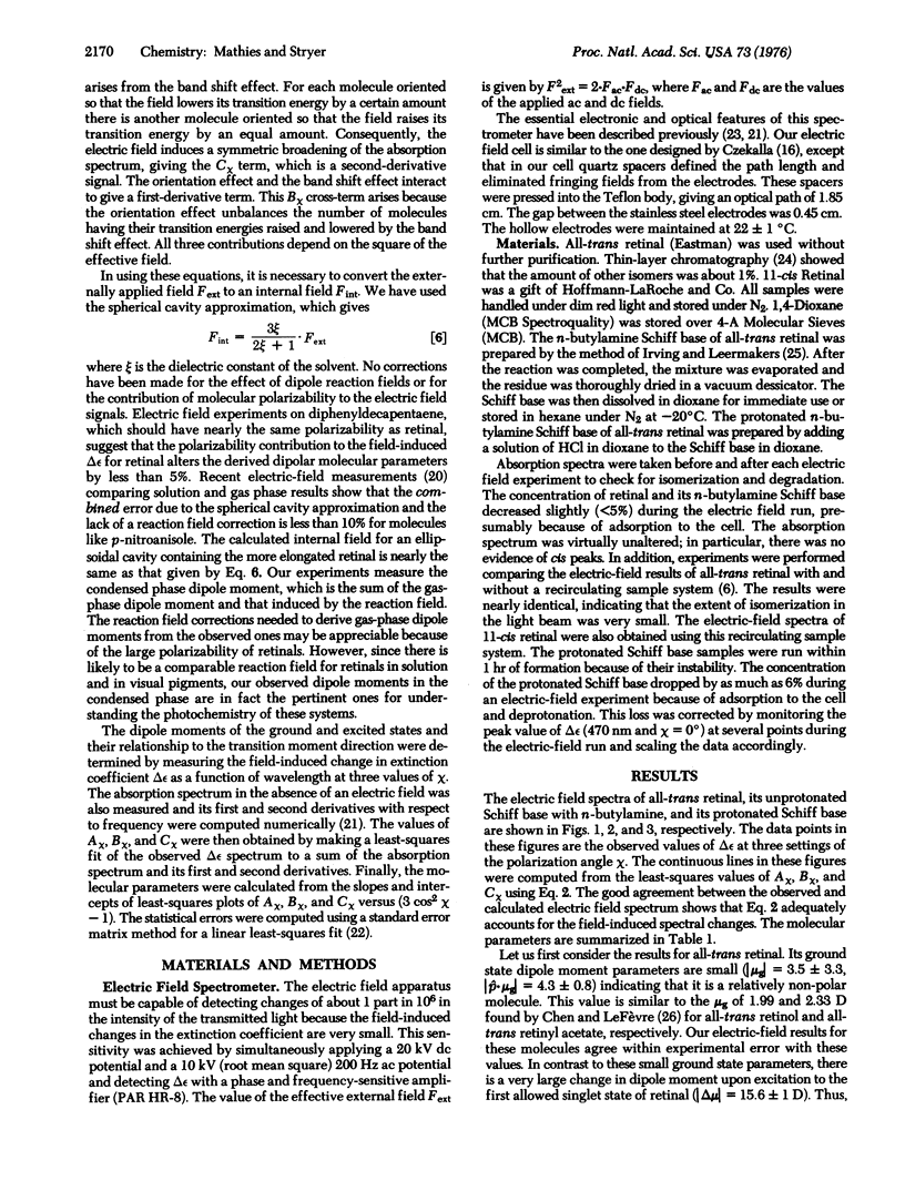
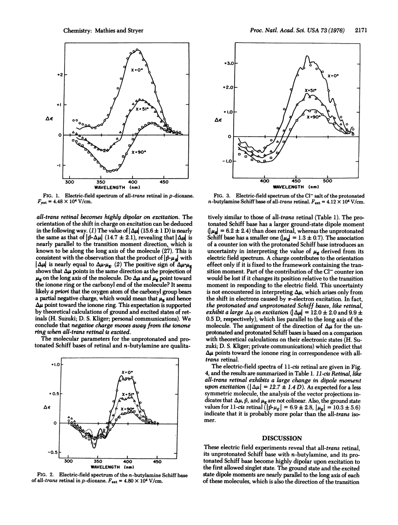
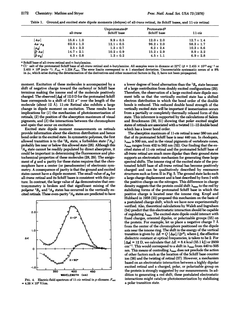
We have measured the effect of an intense electric field on the absorption spectrum of solutions of all-trans retinal, its unprotonated Schiff base with n-butylamine, and the Cl- salt of this protonated Schiff base. The field-induced change in extinction coefficient as a function of wavelength was analyzed to determine the ground-state dipole moment (mug), the change in dipole moment on excitation (deltamu), and the direction of mug and deltamu). These experiments have shown that all three species become highly dipolar upon excitation to the first allowed excited singlet state (deltamu = 15.6, 9.9, 12D, respectively). The ground-state and excited-state dipole moments are nearly parallel to the long axis of these molecules. Excitation is accompanied by a shift of negative charge toward the carbonyl or Schiff base terminus, making the ionone end of these molecules positively charged. The large excited state dipole moment of all-trans retinal indicates that the vertically excited state, which is of 1Bu parentage (C2h), has become significantly mixed with even-parity states. On the basis of previous theoretical calculations, this mixing is expected to facilitate isomerization in the singlet manifold. We have also found that 11-cis retinal has a large deltamu (12.7 +/- 1.4 D) on excitation. In the visual pigments, the interaction of the excited-state dipole moment of retinal with a suitably located charged group could control the position of the absorption maximum. Also, the large shift in charge density upon excitation of retinal may lead to new electrostatic interactions between the chromophore and the protein that would act as a driving force for the initial conformational changes in visual excitation.
Full text
PDF
Selected References
These references are in PubMed. This may not be the complete list of references from this article.
- Akhtar M., Blosse P. T., Dewhurst P. B. Studies on vision. The nature of the retinal-opsin linkage. Biochem J. 1968 Dec;110(4):693–702. doi: 10.1042/bj1100693. [DOI] [PMC free article] [PubMed] [Google Scholar]
- Blatz P. E., Mohler J. H. Effect of selected anions and solvents on the electron absorption, nuclear magnetic resonance, and infrared spectra of the N-retinylidene-n-butylammonium cation. Biochemistry. 1975 Jun 3;14(11):2304–2309. doi: 10.1021/bi00682a005. [DOI] [PubMed] [Google Scholar]
- Bownds D. Site of attachment of retinal in rhodopsin. Nature. 1967 Dec 23;216(5121):1178–1181. doi: 10.1038/2161178a0. [DOI] [PubMed] [Google Scholar]
- Dartnall H. J., Lythgoe J. N. The spectral clustering of visual pigments. Vision Res. 1965 Apr;5(3):81–100. doi: 10.1016/0042-6989(65)90057-x. [DOI] [PubMed] [Google Scholar]
- Ebrey T. G., Honig B. Molecular aspects of photoreceptor function. Q Rev Biophys. 1975 May;8(2):129–184. doi: 10.1017/s0033583500001785. [DOI] [PubMed] [Google Scholar]
- Fröhlich H. The extraordinary dielectric properties of biological materials and the action of enzymes. Proc Natl Acad Sci U S A. 1975 Nov;72(11):4211–4215. doi: 10.1073/pnas.72.11.4211. [DOI] [PMC free article] [PubMed] [Google Scholar]
- Green E., Reible S. Paired moving charges in mitochondrial energy coupling. II. Universality of the principles for energy coupling in biological systems. Proc Natl Acad Sci U S A. 1975 Jan;72(1):253–257. doi: 10.1073/pnas.72.1.253. [DOI] [PMC free article] [PubMed] [Google Scholar]
- Honig B., Ebrey T. G. The structure and spectra of the chromophore of the visual pigments. Annu Rev Biophys Bioeng. 1974;3(0):151–177. doi: 10.1146/annurev.bb.03.060174.001055. [DOI] [PubMed] [Google Scholar]
- Irving C. S., Byers G. W., Leermakers P. A. Spectroscopic model for the visual pigments. Influence of microenvironmental polarizability. Biochemistry. 1970 Feb 17;9(4):858–864. doi: 10.1021/bi00806a020. [DOI] [PubMed] [Google Scholar]
- KROPF A., HUBBARD R. The mechanism of bleaching rhodopsin. Ann N Y Acad Sci. 1959 Nov 12;74(2):266–280. doi: 10.1111/j.1749-6632.1958.tb39550.x. [DOI] [PubMed] [Google Scholar]
- MORTON R. A., PITT G. A. Studies on rhodopsin. IX. pH and the hydrolysis of indicator yellow. Biochem J. 1955 Jan;59(1):128–134. doi: 10.1042/bj0590128. [DOI] [PMC free article] [PubMed] [Google Scholar]
- Mathies R., Oseroff A. R., Stryer L. Rapid-flow resonance Raman spectroscopy of photolabile molecules: rhodopsin and isorhodopsin. Proc Natl Acad Sci U S A. 1976 Jan;73(1):1–5. doi: 10.1073/pnas.73.1.1. [DOI] [PMC free article] [PubMed] [Google Scholar]
- Oseroff A. R., Callender R. H. Resonance Raman spectroscopy of rhodopsin in retinal disk membranes. Biochemistry. 1974 Sep 24;13(20):4243–4248. doi: 10.1021/bi00717a027. [DOI] [PubMed] [Google Scholar]
- Salem L., Bruckmann P. Conversion of a photon to an electrical signal by sudden polarisation in the N-retinylidene visual chromophore. Nature. 1975 Dec 11;258(5535):526–528. doi: 10.1038/258526a0. [DOI] [PubMed] [Google Scholar]
- Salem L. Theory of photochemical reactions. Science. 1976 Feb 27;191(4229):822–830. doi: 10.1126/science.1251196. [DOI] [PubMed] [Google Scholar]
- Sperling W., Rafferty C. N. Relationship between absorption spectrum and molecular conformations of 11-cis-retinal. Nature. 1969 Nov 8;224(5219):590–594. doi: 10.1038/224591a0. [DOI] [PubMed] [Google Scholar]
- Wald G. The molecular basis of visual excitation. Nature. 1968 Aug 24;219(5156):800–807. doi: 10.1038/219800a0. [DOI] [PubMed] [Google Scholar]
- Waleh A., Ingraham L. L. A molecular orbital study of the protein-controlled bathochromic shift in a model of rhodopsin. Arch Biochem Biophys. 1973 May;156(1):261–266. doi: 10.1016/0003-9861(73)90364-0. [DOI] [PubMed] [Google Scholar]