Abstract
AIM: To assess the feasibility and usefulness of multi-detector CT enterography with orally administered iso-osmotic mannitol as negative contrast in demonstrating small bowel disease.
METHODS: Thirteen volunteers and 38 patients with various kinds of small bowel disease were examined. We administered about 1500 mL iso-osmotic mannitol as negative contrast agent and then proceeded with helical CT scanning on a Siemens Sensation 16 scanner. All volunteers and patients were interviewed about their tolerance of the procedure. Two radiologists post-processed imaging data with MPR, thin MIP, VRT and INSPACE when necessary and then interpreted the scans, and adequacy of luminal distention was evaluated on a four-point scale. Demonstration of features of various kinds of small bowel disease was analyzed.
RESULTS: The taste of iso-osmotic mannitol is good (slightly sweet) and acceptable by all. Small bowel distention was excellent and moderate in most volunteers and patients. CT features of many kinds of diseases such as tumors, Crohn’s disease,and small bowel obstruction, etc. were clearly displayed.
CONCLUSION: Multi-detector CT enterography with iso-osmotic mannitol as negative contrast to distend the small bowel is a simple, rapid, noninvasive and effective method of evaluating small bowel disease.
Keywords: Small bowel, Contrast, Enterography
INTRODUCTION
The small bowel remains the most challenging segment of the alimentary tube to examine, due to its length, caliber, and overlap of loops. Barium enteroclysis is the conventional radiographic examination method which shows intraluminal lesions, while abdominal CT displays extraintestinal manifestations of small bowel disease. CT enteroclysis (CT-E) is a method of examining the small bowel that combines the advantages of both the the above-mentioned methods[1].
Due to the ability of multidetector CT scanners to scan large volumes at faster speed with the ability to perform reconstruction following the examination, CT-E has become a more feasible extension of the conventional enteroclysis and CT methods of examining the small intestine. A well distended small bowel is mandatory to prevent diagnostic error due to empty loops, so placement of a nasojejunal tube is usually needed which is uncomfortable. The study is to assess the feasibility and usefulness of CT enterography using iso-osmotic mannitol as a orally administered negative contrast agent.
MATERIALS AND METHODS
Materials
Fifty-one subjects (female 21, male 30, mean age 52.3 years, age range = 19-81 years) were included, 13 of which were normal volunteers and 38 were patients with various kinds of diseases proved surgically, endoscopically, pathologically and clinically. There were 15 cases of small-bowel tumors, nine cases of Crohn’s disease, four cases of small-bowel adhesions after surgery, five cases of mesenteric ischemia, one case of intussusception, one case of eosinophilic gastroenteroitis, one case of volvulus, one case of duodenal stasis and one case of ileal chronic ulcer with multiple inflammatory polyps.
Protocol
The cleansing preparation usually requires a low-residue diet, ample fluids, and 500 mL 20% mannitol administered orally as laxative on the day prior to the exam, and nothing to eat on the day of the examination. About 1500 mL of iso-osmotic mannitol was drunk continuously in 40-60 min, and then 20 mg of Raceanisodamine hydrochloride was injected intravenously. Five to ten minutes later began the helical CT scanning on a Siemens Sensation 16 scanner (Siemens, Erlanger, Germany) with CARE DOSE 4D software and the following parameters: 120 KVP, 130 effective mAs, 0.5 S rotation time, 7.0 mm thickness, and Kernel B 20 f smooth. After plain scan, arterial phase and venous phase were scanned 20-30 s and 50-70 s after the start of the intravenous injection of 100 mL Omnipaque (300 mgI/mL) at a rate of 3.5 mL/s. Raw data were retrospectively reconstructed with a thickness of 0.75 mm and a gap of 0.7 mm, which was post-processed into MPR, thin MIP and VRT at a WIZARD workstation, using Inspace when necessary.
Acceptance of the procedure was interviewed, and the cost of the examination was also evaluated.
Image evaluation
Imaging data were independently interpreted by three experienced abdominal radiologists together. Small bowel was divided into three segments: duodenum, jejunum and ileum. Adequacy of luminal distention was assessed with a four-point scale in each segment. A distention score 0, 1, 2, 3 represents less than 30%, 30-50%, 50-80% and more than 80% respectively of the evaluated segment was adequately distended. The maximun outer diameter and wall thickness of duodenum, jejunum and ileum were measured and the CT values of bowel wall in different phase were measured with a ROI of 2 mm2. The truncated areas of superior mesenteric artery and superior mesenteric vein were measured in front of the unicate of pancreas. The demonstration of mesenteric vasculature was evaluated. MDCT-E features and characteristics of various kinds of diseases were also observed and analyzed including the changes of lumen, wall, vascular, mesenteric and other organs.
Statistical analysis
The results of maximun outer diameter of the small bowel, CT values of bowel wall, and the truncated areas of superior mesenteric artery and superior mesenteric vein were all expressed as mean±SD, and the difference of distension scores among three radiologists was analyzed by χ2 test. P<0.05 was considered statistically significant.
RESULTS
Acceptance of the procedure
All volunteers and patients considered the taste of iso-osmotic mannitol as good or acceptable, and no discomfort and complication were found. In our hospital, it costs about 1000 yuan RMB, just equal to the charge of the CT examination of the whole abdomen.
Luminal distention and measurements of volunteers
Luminal distention was satisfactory and adequate especially in the ileum in most normal volunteers, the distention score being 2.52±0.22, 2.33±0.19 and 2.81±0.31 in duodenum, jejunum and ileum respectively, and difference of distension scores among three radiologists was not statistically significant (χ2 = 1.464, P = 0.486). The normal outer diameter and wall thickness of small bowel were generally less than 30 and 3 mm in all volunteers (Table 1). The truncated area of superior mesenteric artery (SMA) and superior mesenteric vessel (SMV) were 0.43±0.14 and 1.34±0.31 cm2. Normal branching of SMA, SMV, and inferior mesenteric artery and inferior mesenteric vein was demonstrated clearly even to the vasa recta (Figure 1).
Table 1.
Distension, diameter, wall thickness and enhancement of normal small intestine.
| Distension | Diameter/wall thickness | Plain/artery phase/venous phase enhancement (Hu) | |
| Duodenum | 2.52±0.22 | 19.4±7.01/1.68±0.83 | 36.65±4.15/70.65±11.95/78.6±12.75 |
| Jejunum | 2.33±0.19 | 20.68±3.47/1.1±0.51 | 33.26±5.06/65.43±8.15/73.31±11.74 |
| Ileum | 2.81±0.31 | 18.10±4.11/0.90±0.34 | 6.96±4.51/65.29±8.06/72.17±12.11 |
Figure 1.
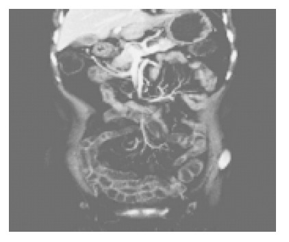
Coronal section shows adequate small bowel luminal distention, uniform wall enhancement and normal superior mesenteric vessels and their branching.
MDCT-E features of various kinds of diseases
Small bowel tumors Three cases of adenocarcinoma, two in the duodenum and one in jejunum, manifested as lobular small mass and small bowel obstruction (Figures 2 and 3A). The mean diameter of the masses is 3.05±1.23 cm and enhanced markedly (mean CT value being 42.03±8.70, 91.30±11.51 and 95.30±9.08 HU at plain scan phase, artery phase and venous phase, respectively). No obviously dilated feeding artery and draining vein were found. Six cases of gastrointestinal stromal tumors (GISTs), one in duodenum and four in jejunum (Figure 3B) and one in ileum, characterized as bigger mass and no bowel obstruction. The masses are larger than adenocarcinoma (mean diameter = 5.13±2.91 cm) and enhanced more significantly (mean CT value being 36.30±12.33, 124.50±12.34, and 136.78±9.12 HU at plain scan phase, artery phase and venous phase, respectively). There were dilated feeding arteries and draining veins in two cases, ring-like calcification in two cases, punctuated calcification in one case, internal necrosis in three cases, hepatic metestasis in one case. All masses are subserously located. One case of T-cell lymphoma demonstrated as thickened wall with ulcer. One case of mucosa associated lymphoid tissue lymphoma in ileum was featured as aneurysmal dilatation of a long segment of thickened ileal loop that cannot be seperated from the bladder and multiple mesenteric nodes (Figure 3C). One case of fibromyxoid sarcoma manifested as a intraluminal mass enhanced markedly combined with retroperitoneal masses. One case of jejunal lipoma displayed as an intraluminal fat density mass with a long stalk. Two cases of peritoneal metastatic adenocarcinoma, which were from pancreatic carcinoma and colon carcinoma respectively, showed peritoneal masses and jejunal obstruction.
Figure 2.
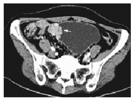
Axial CT-E image of lower abdomen shows an irregular mass at the obstruction point (arrow) which is supposed to be at the ileum.
Figure 3.

Value of coronal images. A: Cronal projection of the same patiet as fig 2 vividly displays the enhanced mass (arrow) at the dilated and elongated proximal jejunum; B: Coronal projection of a jejunal GIST clearly exhibits subserously located, markedly enhanced the mass (thin arrow) and its feeding artery and draining vein (thick arrow). C: Coronal projection of a MALT lymphoma shows aneurysmal dilatation of a long segment of thickened ileal loop that cannot be seperated from the bladder and multiple mesenteric nodes. D: Coronal projection of a patient with Crohn’s disease shows segmental ileal wall thickening, mucosal enhancement, prominent perienteric vasculacture and cutaneous orificium fistulae (arrow).
Crohn’s disease In nine cases of Crohn’s disease, 14 segments were involved. The common CT-E signs (Figures 3D and 4) were mucosal enhancement, bowel thickening, luminal stricture and prestenotic dilatation, mesenteric fibrofatty change, prominent perienteric vasculature, mural stratification, enlarged lymph nodes. Other signs include ascites (four cases), abscess anal fistula (one case), enterocutaneous fistula (1 case), gall bladder wall enhancement (two cases) and pelvic bone destruction (one case).
Figure 4.
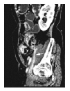
Sagittal projection of the same patient as figure 3D shows a markedly enhanced enterocutaneous fistula (arrow).
Post-operative adhesions In four cases of post-operative adhesions, common CT-E signs were abnormally distributed loops. Internal hernia and then strangulation (intramural gas) was found in one case. A band and a closed loop were found in the second patient with obstruction. Chronic ulcer in an adhered jejunal loop was displayed clearly in the third patient. Small-bowel loops surrounding a peritoneal catheter was found in the fourth patient.
Mesenteric ischemia In five cases of mesenteric ischemia the common signs were thickened bowel wall and fuzzy mesenteric fat. Two cases were atherosclerotic disease of the superior mesenteric artery (SMA), and luminal narrowing, fewer branching and calcified atherosclerotic plaque were found on the thin MIP. Pneumatosis was found in one patient who combined with pyemia. One case of acute mesenteric ischemia was caused by thrombosis in the SMA which showed the filling defect in the SMA and small bowel mural stratification (Figures 5 and 6). One case of thrombosis in the proximal superior mesenteric vein and one case of hematoma in mesenteric root were clearly demonstrated as intraluminal defect and high density mass around the proximal superior mesenteric vessel.
Figure 5.
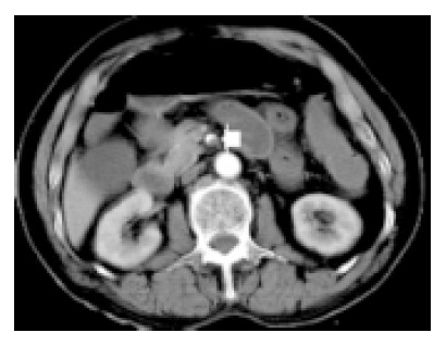
Axial section of an acute mensenteric ischemia caused by thrombosis shows an filling defect in SMA (arrow).
Figure 6.
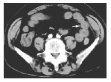
Lower section of the same patient as Figure 5 shows small bowel wall thickening with mural stratification.
Others One case of ileum-ileum intussusception looked like sausage-like mass at the obstruction point, the intussuscipiens and intussusceptum being clearly identified. One case of eosinophilic gastroenteritis had diffusely thickened bowel wall that resolved very soon after the administration of cortical hormone. One case of small bowel volvulus after gastric operation showed the whirl sign of the vessels in the mesentery and the mesenteric density increased. One case of duodenal stasis was characterized as dilated proximal duodenum and compressed transverse segment of duodenum by aorta and superior mesenteric artery. One case of chronic ileal ulcer with multiple inflammatory polyps were clearly defined on CT enterography, and the ulcer demonstrated as a regional eccentric thickened bowel wall with irregular inner border and polyps as brightly enhanced nodules.
DISCUSSION
Owing to its length, caliber, and overlap of loops, small bowel remains the most challenging part to be assessed. Barium enteroclysis shows the intraluminal lesion clearly but cannot depict mural and extraintestinal changes, while conventional abdominal CT without adequate distention of the bowel can display the outside of the bowel but usually overlook the intraluminal disease and mural change. MDCT-E combines the advantages of the above two methods and overcomes each other’s disadvantages. Currently, data on the technical and clinical application of MDCT-E is fragmentary and preliminary[1]. The adequate distention of small bowel is mandatory to CT-E, and numerous techniques have been designed to optimize visualization of the small bowel[1-3], and the majority of these studies have employed fluoroscopic placement of a nasojejunal tube to infuse contrast material. A few recent reports described CT enterography with orally administered negative contrast, which had satisfactory results[4,5].
Clinical applications of CT-E included: (1) small bowel obstruction - conventional CT has high sensitivity in diagnosing high-grade obstruction and is of value in confirming the presence or absence of strangulation. Contrast examination is not indicated in these cases[6-8]. CT-E has been reported to have greater sensitivity (89%) and specificity (100%) than conventional CT (50% and 94%, respectively) in patients suspected of having a partial small bowel obstruction, and this difference was even greater when abdominal malignancy was known or suspected[1]. The precise localization and classification of adhesions, the most common cause of small bowel obstruction, is readily made with CT-E. Analysis of axial images aided by coronal and sagittal reformatting allows categorization of small bowel adhesions into parietal and visceral adhesions. This ability is especially useful in complicated cases and often increase the confidence of the radiologists[9]. In addition, clinicians and surgeons appreciate the availability of additional planes, which helps them to better understand complex cases[10]. Single or multiple points of obstruction are readily demonstrated similar to barium enteroclysis but their precise location is made easier with CT-E. Obstruction from primary malignancy or metastatic disease is also diagnosed readily with CT-E[11]. (2) Crohn’s disease - CT has come to play an increasingly important role in the evaluation of patients with Crohn’s disease because of its ability to accurately demonstrate the bowel wall as well as adjacent structures and extraluminal extension of disease[12]. For acutely ill patients, CT is often the only study required, providing crucial information for both the accurate diagnosis and treatment of the many complications associated with Crohn’s disease[13-15]. Patients with Crohn’s disease often have complex disease that involves multiple bowel loops or adjacent organs. The ability of CT-E to visualize the scan in more than the axial plane is essential for complete understanding of the extent of the disease. Multiplanar reconstructions significantly improved the observer’s confidence in their interpretation of the imaging and in their ability to detect the extent of the bowel involvement[16]. We also found that multiplanar reformation is very useful in vividly displaying the complex involvement of Crohn’s disease. CT-E can be highly accurate in depicting mucosal abnormalities, bowel thickening, fistulae, and extraintestinal complications. Activity of Crohn’s disease was evaluated on CT-E by the presence of mural stratification, mucosal and mural hyperenhancement, edema in the perienteric mesenteric fat, and prominent pericolic or perienteric vasculature[2,17-19]. (3) Neoplasms - CT plays a more active role in detection and staging of small bowel neoplasms. Even subtle adenocarcinomas can be visualized by CT-E, which most frequently appears as eccentric or as circumferential wall thickening involving a short segment of the small bowel and results in “apple core” appearance[20]. Gastrointestinal stromal tumors (GISTs) can be seen as a large mass on CT-E, which may be helpful to define the site of the tumor; benign GISTs cannot be reliably differentiated from malignant ones on CT unless there is obvious metastasis or local extension. GISTs can ulcerate or calcify and usually are not associated with significant adenopathy[20]. In our group, GISTs usually enhanced significantly and have dilated feeding arteries and draining veins. Carcinoid tumor has a propensity for ileum[21], which has not been very successfully detected by traditional CT examinations and now can be visualized when water is given as oral contrast and a good IV contrast bolus is administered and thin-collimation MDCT is performed[22]. Carcinoids that have infiltrated the mesentery demonstrate a characteristic CT appearance as an infiltrating mesenteric mass that contains calcification in upto 70% of cases. MDCT-E well demonstrate the relationship of the mass to the mesenteric vessels, which is crucial for surgical planning[23]. CT has played an important role in evaluation and follow-up of patients with lymphoma[24]. Primary small bowel lymphoma often appears as focally thickened loop, which usually does not result in obstruction. CT-E offers the advantage of simultaneously assessing adenopathy, extraluminal extent and small bowel lesions. CT-E, because of the large surface rendering with a volume challenge, allows delineation of lesions as small as 0.5 cm in size[25]. (4) Obscure gastrointestinal bleeding - in patients with an obscure gastrointestinal bleeding, the conventional small bowel follow through has no place in clearing the small bowel after negative endoscopy, angiography, or a tagged red cell scan[26,27]. Traditional doublecontrast enteroclysis is estimated to be successful in 21% of such cases[28]. CT-E maybe useful in patients with anemia of unknown origin. Arteriovenous malformations are the most common vascular etiology. CT-E using methylcellulose as a neutral luminal contrast material with an IV contrast bolus of 150 mL at 4 mL/s can potentially identify the source of unexplained gastrointestinal bleeding. Vascular malformations have been diagnosed on helical CT with IV contrast material[29,30]. Duodenal varices have been reported to be successfully demonstrated by multislice helical CT[31].
To our knowledge, this is the first report of CT enterography using iso-osmotic mannitol as orally administered negative contrast to distend the small bowel. Iso-osmotic mannitol is slightly sweet and acceptable, which cannot be absorbed by the intestine and a large volume administered orally in a short time can distend the small bowel adequately. This method freed the patients from the placement of nasojejunal tube and acceptable by most patients without important complications. The lumen, wall, extraintestinal structure, and other organs are satisfactorily demonstrated in both volunteers and patients. In our group, adenocarcinoma manifested as small mass with obstruction while GIST as bigger mass enhanced significantly without obstruction. Crohn’s disease characterized as thickened wall, enhanced mucosae, fuzzy mesenteric fat, enlarged lymph nodes, prominent perienteric vasculature and extraintestinal complications. Other diseases have also been shown clearly.
With comparison to conventional CT, we found that MDCT-E has many advantages: (1) lesion can be localized more precisely and characterized accurately. Two cases of adenocarcinoma associated with bowel obstruction had been mis-interpreted as distal small bowel obstruction or colon obstruction on conventional axial CT scans, while CT enterography accurately showed the mass causing proximal small bowel obstruction. (2) CT enterography can display mesenteric lymphadenopathy which usually be overlooked at conventional CT. (3) CT enterography with its 3-D display can more precisely explain the relationship between lesions and neighboring structure that is helpful to surgery. (4) CT enterography with its coronal projections that is similar to conventional enteroclysis is much more welcomed by surgeons and physicians.
Although our experience is limited, we believe that CT enterography with iso-osmotic mannitol as orally administered negative contrast is a simple, noninvasive, economic, effective method for assessing small bowel diseases and others, which should be further studied.
Footnotes
Science Editor Guo SY Language Editor Elsevier HK
References
- 1.Bender GN, Maglinte DD, Klöppel VR, Timmons JH. CT enteroclysis: a superfluous diagnostic procedure or valuable when investigating small-bowel disease? AJR Am J Roentgenol. 1999;172:373–378. doi: 10.2214/ajr.172.2.9930786. [DOI] [PubMed] [Google Scholar]
- 2.Makó EK, Mester AR, Tarján Z, Karlinger K, Tóth G. Enteroclysis and spiral CT examination in diagnosis and evaluation of small bowel Crohn's disease. Eur J Radiol. 2000;35:168–175. [PubMed] [Google Scholar]
- 3.Bender GN, Timmons JH, Williard WC, Carter J. Computed tomographic enteroclysis: one methodology. Invest Radiol. 1996;31:43–49. doi: 10.1097/00004424-199601000-00007. [DOI] [PubMed] [Google Scholar]
- 4.Doerfler OC, Ruppert-Kohlmayr AJ, Reittner P, Hinterleitner T, Petritsch W, Szolar DH. Helical CT of the small bowel with an alternative oral contrast material in patients with Crohn disease. Abdom Imaging. 2003;28:313–318. doi: 10.1007/s00261-002-0040-4. [DOI] [PubMed] [Google Scholar]
- 5.Wold PB, Fletcher JG, Johnson CD, Sandborn WJ. Assessment of small bowel Crohn disease: noninvasive peroral CT enterography compared with other imaging methods and endoscopy--feasibility study. Radiology. 2003;229:275–281. doi: 10.1148/radiol.2291020877. [DOI] [PubMed] [Google Scholar]
- 6.Maglinte DD, Gage SN, Harmon BH, Kelvin FM, Hage JP, Chua GT, Ng AC, Graffis RF, Chernish SM. Obstruction of the small intestine: accuracy and role of CT in diagnosis. Radiology. 1993;188:61–64. doi: 10.1148/radiology.188.1.8511318. [DOI] [PubMed] [Google Scholar]
- 7.Megibow AJ, Balthazar EJ, Cho KC, Medwid SW, Birnbaum BA, Noz ME. Bowel obstruction: evaluation with CT. Radiology. 1991;180:313–318. doi: 10.1148/radiology.180.2.2068291. [DOI] [PubMed] [Google Scholar]
- 8.Maglinte DD, Balthazar EJ, Kelvin FM, Megibow AJ. The role of radiology in the diagnosis of small-bowel obstruction. AJR Am J Roentgenol. 1997;168:1171–1180. doi: 10.2214/ajr.168.5.9129407. [DOI] [PubMed] [Google Scholar]
- 9.Caoili EM, Paulson EK. CT of small-bowel obstruction: another perspective using multiplanar reformations. AJR Am J Roentgenol. 2000;174:993–998. doi: 10.2214/ajr.174.4.1740993. [DOI] [PubMed] [Google Scholar]
- 10.Khurana B, Ledbetter S, McTavish J, Wiesner W, Ros PR. Bowel obstruction revealed by multidetector CT. AJR Am J Roentgenol. 2002;178:1139–1144. doi: 10.2214/ajr.178.5.1781139. [DOI] [PubMed] [Google Scholar]
- 11.Maglinte DD, Bender GN, Heitkamp DE, Lappas JC, Kelvin FM. Multidetector-row helical CT enteroclysis. Radiol Clin North Am. 2003;41:249–262. doi: 10.1016/s0033-8389(02)00115-x. [DOI] [PubMed] [Google Scholar]
- 12.Fishman EK, Wolf EJ, Jones B, Bayless TM, Siegelman SS. CT evaluation of Crohn's disease: effect on patient management. AJR Am J Roentgenol. 1987;148:537–540. doi: 10.2214/ajr.148.3.537. [DOI] [PubMed] [Google Scholar]
- 13.Gore RM, Ghahremani GG. Radiologic investigation of acute inflammatory and infectious bowel disease. Gastroenterol Clin North Am. 1995;24:353–384. [PubMed] [Google Scholar]
- 14.Wills JS, Lobis IF, Denstman FJ. Crohn disease: state of the art. Radiology. 1997;202:597–610. doi: 10.1148/radiology.202.3.9051003. [DOI] [PubMed] [Google Scholar]
- 15.Gore RM, Balthazar EJ, Ghahremani GG, Miller FH. CT features of ulcerative colitis and Crohn's disease. AJR Am J Roentgenol. 1996;167:3–15. doi: 10.2214/ajr.167.1.8659415. [DOI] [PubMed] [Google Scholar]
- 16.Raptopoulos V, Schwartz RK, McNicholas MM, Movson J, Pearlman J, Joffe N. Multiplanar helical CT enterography in patients with Crohn's disease. AJR Am J Roentgenol. 1997;169:1545–1550. doi: 10.2214/ajr.169.6.9393162. [DOI] [PubMed] [Google Scholar]
- 17.Goldberg HI, Gore RM, Margulis AR, Moss AA, Baker EL. Computed tomography in the evaluation of Crohn disease. AJR Am J Roentgenol. 1983;140:277–282. doi: 10.2214/ajr.140.2.277. [DOI] [PubMed] [Google Scholar]
- 18.Del Campo L, Arribas I, Valbuena M, Maté J, Moreno-Otero R. Spiral CT findings in active and remission phases in patients with Crohn disease. J Comput Assist Tomogr. 2001;25:792–797. doi: 10.1097/00004728-200109000-00020. [DOI] [PubMed] [Google Scholar]
- 19.Meyers MA, McGuire PV. Spiral CT demonstration of hypervascularity in Crohn disease: "vascular jejunization of the ileum" or the "comb sign". Abdom Imaging. 1995;20:327–332. doi: 10.1007/BF00203365. [DOI] [PubMed] [Google Scholar]
- 20.Buckley JA, Jones B, Fishman EK. Small bowel cancer. Imaging features and staging. Radiol Clin North Am. 1997;35:381–402. [PubMed] [Google Scholar]
- 21.Maglinte DT, Reyes BL. Small bowel cancer. Radiologic diagnosis. Radiol Clin North Am. 1997;35:361–380. [PubMed] [Google Scholar]
- 22.Wallace S, Ajani JA, Charnsangavej C, DuBrow R, Yang DJ, Chuang VP, Carrasco CH, Dodd GD. Carcinoid tumors: imaging procedures and interventional radiology. World J Surg. 1996;20:147–156. doi: 10.1007/s002689900023. [DOI] [PubMed] [Google Scholar]
- 23.Horton KM, Fishman EK. The current status of multidetector row CT and three-dimensional imaging of the small bowel. Radiol Clin North Am. 2003;41:199–212. doi: 10.1016/s0033-8389(02)00121-5. [DOI] [PubMed] [Google Scholar]
- 24.Rubesin SE, Gilchrist AM, Bronner M, Saul SH, Herlinger H, Grumbach K, Levine MS, Laufer I. Non-Hodgkin lymphoma of the small intestine. Radiographics. 1990;10:985–998. doi: 10.1148/radiographics.10.6.2259769. [DOI] [PubMed] [Google Scholar]
- 25.Bender GN, Maglinte DD, McLarney JH, Rex D, Kelvin FM. Malignant melanoma: patterns of metastasis to the small bowel, reliability of imaging studies, and clinical relevance. Am J Gastroenterol. 2001;96:2392–2400. doi: 10.1111/j.1572-0241.2001.04041.x. [DOI] [PubMed] [Google Scholar]
- 26.Bender GN. Radiographic examination of the small bowel. An application of odds ratio analysis to help attain an appropriate mix of small bowel follow through and enteroclysis in a working-clinical environment. Invest Radiol. 1997;32:357–362. doi: 10.1097/00004424-199706000-00007. [DOI] [PubMed] [Google Scholar]
- 27.Maglinte DD, Lappas JC, Kelvin FM, Rex D, Chernish SM. Small bowel radiography: how, when, and why? Radiology. 1987;163:297–305. doi: 10.1148/radiology.163.2.3550876. [DOI] [PubMed] [Google Scholar]
- 28.Moch A, Herlinger H, Kochman ML, Levine MS, Rubesin SE, Laufer I. Enteroclysis in the evaluation of obscure gastrointestinal bleeding. AJR Am J Roentgenol. 1994;163:1381–1384. doi: 10.2214/ajr.163.6.7992733. [DOI] [PubMed] [Google Scholar]
- 29.Ettorre GC, Francioso G, Garribba AP, Fracella MR, Greco A, Farchi G. Helical CT angiography in gastrointestinal bleeding of obscure origin. AJR Am J Roentgenol. 1997;168:727–731. doi: 10.2214/ajr.168.3.9057524. [DOI] [PubMed] [Google Scholar]
- 30.Mindelzun RE, Beaulieu CF. Using biphasic CT to reveal gastrointestinal arteriovenous malformations. AJR Am J Roentgenol. 1997;168:437–438. doi: 10.2214/ajr.168.2.9016222. [DOI] [PubMed] [Google Scholar]
- 31.Weishaupt D, Pfammatter T, Hilfiker PR, Wolfensberger U, Marincek B. Detecting bleeding duodenal varices with multislice helical CT. AJR Am J Roentgenol. 2002;178:399–401. doi: 10.2214/ajr.178.2.1780399. [DOI] [PubMed] [Google Scholar]


