

Abstract
Purpose
To compare the outcome of arthrocentesis alone and arthrocentesis with sodium hyaluronate in internal derangement of TMJ.
Materials and methods
20 patients of disc displacement with reduction of TMJ were randomized into 2 experimental groups. Control group of patients received arthrocentesis alone and study group of patients received arthrocentesis with sodium hyaluronate. Patients TMJ status and clinical symptoms were evaluated for 6 months follow up. The clinical parameter recorded were maximal mouth opening (MMO), lateral excursions (right and left side), protrusive movement, joint noises, and pain (at rest, at function).
Results
When 2 groups are compared there was no statistically significant difference in terms of inter-incisal opening, lateral excursion, protrusive movement, joint noises and pain however the group with sodium hyaluronidase has shown better results than the control group.
Conclusion
Both study and control group found to be statistically insignificant but patient who were in the group of arthrocentesis with admission of sodium hyaluronidase had better results.
Keywords: TMJ internal derangements, Arthrocentesis, Sodium hyaluronidase
1. Introduction
Internal derangement (ID) of temporomandibular joint is described as an abnormal positional relationship of the articular disc to the mandibular condyle and the articular eminence. The disorder has been associated with characteristic clinical findings such as pain, joint sounds, and irregular or deviating jaw function.1 In the subset of patients with TMJ disorders who do not respond to conservative treatment, arthrocentesis considered as a first line of surgical intervention. Arthrocentesis is thought to break down adhesions within the joint and remove inflammatory mediators including cytokines and interleukins, which result in chronic pain. Relief of TMJ pain also leads to improvement in both mouth opening and dysfunction.2
Arthrocentesis is traditionally defined as procedure in which the fluid in the joint cavity is aspirated in the needle and therapeutic substance is injected.3 Sodium hyaluronate is largely responsible for the viscosity of normal synovial joint. Its capacity to function as a molecular sieve is thought to be important both in regulating the nutrition of articular cartilage and in physical interactions with the macromolecules of the articular surfaces. Hyaluronic acid is a linear polysaccharides consisting of poly-disaccharide units of glucuronic acid and N-acetyl glucosamine linked by B1-3 & B1-4, glycosidic bonds.4
The objective of the study is to evaluate the efficacy of arthrocentesis with or without sodium hyaluronate in treatment of internal derangement of TMJ.
2. Materials and methods
The present study comprised of twenty patients with internal derangement visiting the Department of Oral &Maxillofacial Surgery, Institute of Dental Studies and Technologies, Modinagar. The study was randomized, non-blinded with 6 months follow up comparing the efficacy of arthrocentesis with or without sodium hyaluronate injection. The thorough history and clinical examination was done.
Inclusion criteria
-
-
Age more than 15 years
-
-
Wilkes stage 2 disease for atleast 2 months
-
-
TMJ pain >3 cm in visual analog scale
-
-
Patient who were resistant to conservative treatment (Physical therapy, Muscle relaxants) for atleast 2 months.
Exclusion criteria
-
-
Infection of the affected joint.
-
-
Previous surgery of the affected joint.
-
-
Injection of sodium hyaluronate or corticosteroids into the target TMJ during previous 6 months.
-
-
Any drug allergy.
-
-
Pregnant and lactating lady.
Preoperatively, the baseline data in the form of maximal mouth opening (MMO), lateral excursions, protrusive movement, TMJ pain and click were measured.
The data were collected after 1st week after 1st intervention and on subsequent follow up visits (1st week after 2nd intervention, 1 month, 3 month, and 6 month) in order to gauge the effectiveness.
MMO was measured as the distance in millimeters between the incisal edge of central incisors at maximum pain free mouth opening. One VAS (VAS I) ranging from level 0–10 is used to assess the level of pain and other VAS (VAS II) ranging from level 0–10 is used to assess the click. Zero (0) in VAS scale means no pain (VAS I), no click (VAS II). Ten (10) in VAS scale means marked intense pain (VAS I), audible click (VAS II).
These patients were diagnosed as having internal derangement of temporomandibular joint, and were divided randomly into two groups irrespective of age, sex & religion.
-
1
Group A: arthrocentesis with normal saline solution.
-
2
Group B: arthrocentesis with normal saline solution plus intra-articular injection of sodium hyaluronate immediately after arthrocentesis.
2.1. Group A: arthrocentesis
2.1.1. Technique
This procedure was done under local anesthesia to block auriculo–temporal nerve. The patient is seated inclined at a 45° angle with the head turned contralateral side to provide an easy approach to the joint to be treated. After proper preparation of target site, the external auditory meatus is blocked with moist cotton.
The points of needle insertion are marked on the skin according to the method suggested by McCain (1988) for the performance for arthroscopy, is as follows: A line is drawn from the middle of the tragus to the outer canthus of the eye (Fig. 1). The posterior entrance point is located along the cantho–tragal line, 10 mm from middle of the tragus line and 2 mm below (Point A); the anterior entrance point placed 10 mm further forward along the line and 10 mm below it (Point B). These marking over the skin indicate the location of the articular fossa and the eminence of the TMJ. 200 ml of normal saline solution is passed through the joint space (Fig. 2). During the lavage, Fig. 2 mandible is moved through opening, excursive, and protrusive movements to facilitate lysis of adhesions. This procedure was repeated after 1 week as 2nd intervention.
Fig. 1.

Points on canthotragal line.
Fig. 2.
Arthrocentesis.
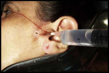
2.2. Group B: arthrocentesis plus injection sodium hyaluronate
In this group arthrocentesis was performed as described in the previous group and 2 ml intra-articular injection sodium hyaluronate was given (20 mg per ml). 1 ml was injected in superior joint cavity as 1st intervention and other 1 ml was injected after 1 week as 2nd intervention (Fig. 3).
Fig. 3.

Injection sodium hyaluronate.
These patients were followed at regular interval of 1 week after 1st intervention, 1st week after 2nd intervention, 1st month, 3rd month, 6th month and assessed in terms of relief of symptoms, achievement of maximal mouth opening and pain free movements and complication if any.
Pain (visual analog scale 0–10)
TMJ sounds (visual analog scale 0–10)
Inter-incisal opening (in mm)
Lateral excursions (in mm)
Protrusive movement (in mm)
3. Results
A total of 20 patients with internal derangement were enrolled in the present study. Out of these 10 subjects were managed with temporomandibular lavage arthrocentesis alone (Control group) and the remaining 10 subjects were managed with arthrocentesis followed by intra-articular injection of sodium hyaluronate (Study group).
Majority of subjects in both the groups were in age group 15–25 years. Three (30%) subjects in control group and 3 (30%). subjects in study group were aged above 25–35 years. Three (30%) subjects in control group and 2 (20%) subjects in study group were aged more than 35 years. Statistically, there was no significant difference between two groups (p = 0.815).
In control group, 4 (40%) subjects were male and 6 (60%) subjects were female. In study group, there were 2 (20%) males and 8 (80%) females. Statistically, there was no significant difference between two groups (p = 0.639). When inter-incisal opening between two groups were compared, it found to be statistically insignificant (p = 0.258) – Graph 1.
Graph 1.

Inter-incisal opening.
Lateral excursions (right and left side) in control group was higher (8.72) as compared to study group (7.91) at all-time intervals. Both the groups were statistically insignificant (p = 0.284) – Graphs 2 and 3
Graph 2.

Lateral excursions (Right side).
Graph 3.

Lateral excursions (Left side).
The mean change in study group was higher (p = 7.05) as compared to control group (p = 6.61) at all-time intervals in terms of protrusive movements. Both the groups were statistically insignificant (p = 0.490) – Graph 4
Graph 4.

Protrusive movement.
The mean change in joint noises in study group was higher (p = 0.89) as compared to control group (0.70) at all-time intervals. Both the groups were statistically insignificant (p = 0.616) – Graph 5
Graph 5.

Joint noises.
There was no statistically significant difference in pain (at rest) of two groups (p = 0.357), pain (at function) groups (p = 0.847) – Graphs 6 and 7
Graph 6.

TMJ PAIN (at rest).
Graph 7.

TMJ PAIN (at function).
4. Discussion
An internal derangement occurs when there is a disturbance in the normal anatomic relationship between the articular disc and condyle that interferes with smooth movement of the joint and causes momentary catching, clicking, popping or locking.
Internal derangement is defined as any interference with smooth joint movement. Although the term therefore includes all types of intracapsular interferences that impede smooth functional joint movements, with regard to the temporomandibular joint (TMJ) the term is typically used interchangeably with disc displacement.5
Murakami et al (1987) was first to offer a systematic description of TMJ arthrocentesis and found excellent results in releasing closed lock by arthrocentesis lavage and lysis.6 Arthrocentesis as described by Nitzan et al is another simpler modification of arthroscopic lavage and lysis.7 Parafunctional habits such as clenching associated with high TMJ impact loading that convert shearing stresses to compressive stresses.8 Intra and extra articular overloading which exerts effects on synovial joint (eg interruption of blood supply) is a major reason for the collapse of lubrication system.9 When joint is overloaded, the hypoxia reperfusion cycle evokes non-enzymatic release of radical oxygen species (ROS) such as superoxide and hydroxyl anions. The highly reactive radical oxygen species (ROS) degrade hyaluronic acid, causing marked decrease in synovial fluid viscosity. Thus, the degraded form of hyaluronic acid indirectly affects the joint lubrication.10 In the absence of lubricant, there is increased adhesiveness, friction, shear and rupture of articular surfaces.11
Lavage of upper compartment through TMJ arthrocentesis forces apart the flexible disc from fossa, washes away the degraded particles and inflammatory components (Radical Oxygen Species (ROS), interleukin, substance P, tumor necrosis factor, Bradykinin, Prostaglandin E2) and decreases the intra-articular pressure whenever the joint is inflamed.12
Inflammatory response is not only a significant source of pain, but also it may lead to the development of synovitis, capsulitis and ultimately fibrous adhesions and pseudowalls.13 Internal derangement may develop secondary to this, or it may be the inciting event. The relation between inflammatory changes and internal derangement is, however, not well defined. The development of arthrocentesis as a therapeutic procedure has also allowed routine synovial fluid sampling. The success of arthrocentesis in many patients with nonspecific arthralgia, internal derangement, and osteoarthritis may be the result of decreasing inflammation, eliminating adhesions, or re-establishing a normal disc–fossa relation.14
Moses and Poker15 (1989) reported that arthrocentesis treat both mechanical and inflammatory aspects of TMJ internal derangement. Sato et al16(2001) reported that improvement in maximal inter-incisal opening was significantly better when extensive techniques involving anterior release of the disc and lateral capsular release were used, than when only conventional Arthrocentesis Lysis and Lavage (ALL) was used.
Kopp et al17 (1985) first published the intra-articular hyaluronic acid injections as a new approach in treatment of TMJ disorders. In various studies Kopp etal,17 Bertolami et al18 (1993) found the significant improvement in subjective symptoms after sodium hyaluronate injection.
Hyaluronic acid is a linear polysaccharides consisting of repeating disaccharide units of glucurornic acid and N-acetyl glucosamine linked by B1-3 & B1-4 glycosidic bonds. It is available in the extracellular matrix of various mammalian tissues including skin, cartilage, umbilical cord, & synovial fluid. Sodium hyaluronate is largely responsible for viscosity and rheological properties of synovial fluid. Its capacity to function as molecular sieve is thought to be important both in regulating the nutrition of articular cartilage and in physical interactions with macromolecules of articular surfaces.19
The precise mechanism of injection sodium hyaluronate is unknown. It provides lubrication for the articular surfaces and is largely responsible for synovial fluid viscosity.18
According to our study average age of patients of internal derangement who attended the clinic ranges from 15 to 25 years.
In our study higher incidence of female patients were diagnosed with internal derangement of Temporomandibular joint. This finding is in accordance with the finding of Dolwick & Nitzan20 (1991) & Kit et al21 (2006).
When Inter-Incisal Opening in study and control groups was compared shows statistically insignificant difference. This result is in accordance with the study of Dolwick and Nitzan20 (1991), Dimitroulis et al22 (2000).
When parameters lateral excursions, protrusive movements, joint noises in both groups were compared with each other the difference found is statistically insignificant. This is in accordance with the result of the study done by Gokhan H Alpaslan and Cansu Alpaslan23 (2001).
When pain at rest and at function was compared with baseline, study group was found to be more statistically significant in all-time intervals (p = 0.000) than control group (p = 0.003). Although there was statistically insignificant difference within these groups when compared for pain. Thus, arthrocentesis with injection sodium hyaluronate have a faster and longer effect than arthrocentesis alone. This effect can be explained by long term lubricating effect of injection sodium hyaluronate, which prevents the onset of inflammatory mediators those are responsible for pain which is in the correlation with the study done by Gokhan H Alpaslan and Cansu Alpaslan23 (2001).
Thus, in our study when two intra-articular injections of sodium hyaluronate 1 week apart were given after arthrocentesis, a significant improvement at 1 month and 6 month was detected when compared with baseline for all outcome measures including maximal inter-incisal opening, lateral excursions (right and left side), protrusive movement, joint pain, TMJ pain (at rest, at function).
Interesting finding was reported by Zardeneta et al24 (2000), when they compared arthrocentesis alone and arthrocentesis with sodium hyaluronate. In both the groups all patients had symptomatic improvement in their pain but when checked for biomarkers, those patients who received injection sodium hyaluronate, considerable decrease was found in these biomarkers. This may provide indirect evidence supporting the anti-inflammatory properties of sodium hyaluronate.
5. Conclusion
Therefore, both the groups found to be statistically insignificant when compared with each other, but found to be statistically significant when both groups were compared with baseline. The limitation of this study is less number of patients, and non-blinded study. Thus, a study with large sample size is required for further speculation.
Conflicts of interest
All authors have none to declare.
References
- 1.Marguelles R.E., Carpentier P., Yung L.P. Clinical diagnosis compared with findings of magnetic resonance in 242 patients with internal derangement of the TMJ. J Orofac Pain. 1995;9:244. [PubMed] [Google Scholar]
- 2.Nitzan D.W., Dolwick M.F., Martinez G.A. Temporomandibular joint arthrocentesis: a simplified treatment for severe, limited mouth opening. J Oral Maxillofac Surg. 1991;49:1163–1165. doi: 10.1016/0278-2391(91)90409-f. [DOI] [PubMed] [Google Scholar]
- 3.Abantagelo G., Oregan M. Biological role and function in articular joint. European Journal of Rheumatology & Inflammation. 1995:10–15. [Google Scholar]
- 4.Samuelson C.O., Jr., Cannon G.W., Ward J.R. Arthrocentesis. J Fam Pract. 1985;20:179–184. [PubMed] [Google Scholar]
- 5.Stegenga B., de Bont L.G.M. TMJ disk derangements. In: Laskin D.M., Greene C.S., Hylander W.L., editors. Temporomandibular Disorders. An Evidence-based Approach to Diagnosis and Treatment. Quintessence; Chicago: 2006. pp. 125–136. [Google Scholar]
- 6.Murakami K.I., Matsuki M., Iizuka T. Recapturing the persistent anterior displaced by mandibular manipulation after pumping and hydraulic pressure to the upper joint cavity of the temporomandibular joint. J Craniomandibular Pract. 1987;5:18–20. doi: 10.1080/08869634.1987.11678169. [DOI] [PubMed] [Google Scholar]
- 7.Nitzan Dorrit W., Franklin Dolwick M., Alejanero Garza. Martinez: temporomandibular joint arthrocentesis, a simplified treatment for severe, limited mouth opening. J Oral Maxillofac Surg. 1991;48:1163–1167. doi: 10.1016/0278-2391(91)90409-f. [DOI] [PubMed] [Google Scholar]
- 8.Nitzan D.W. Intraarticular pressure in the functioning human temporo-mandibular joint and its alteration by uniform elevation of the occlusal plane. J Oral Maxillofac Surg. 1994;52(7):671–679. doi: 10.1016/0278-2391(94)90476-6. [DOI] [PubMed] [Google Scholar]
- 9.Mapp P.I., Grootveld M.C., Blake D.R. Hypoxia, oxidative stress and rheumatoid arthritis. Br Med Bull. 1995;51(2):419–436. doi: 10.1093/oxfordjournals.bmb.a072970. [DOI] [PubMed] [Google Scholar]
- 10.Grootveld M., Henderson E.B., Farrell A. Oxidative damage to hyaluronate and glucose in synovial fluid during exercise of the inflamed rheumatoid joint. Detection of abnormal low molecular-mass metabolites by proton-NMR. spectroscopy. Biochem J. 1991;273(2):459–467. doi: 10.1042/bj2730459. [DOI] [PMC free article] [PubMed] [Google Scholar]
- 11.Stanley H.M., Etzion I., Boggy D.B. Adhesion of contacting rough surfaces in the presence of sub-boundary lubrication. J Tribol. 1990:98–112. [Google Scholar]
- 12.Arinci A., Ademoglu E., Aslan A. Molecular correlates of temporo-mandibular joint disease. Oral Surg Oral Med Oral Pathol Oral Radiol Endod. 2005;99(6):666–670. doi: 10.1016/j.tripleo.2004.08.029. [DOI] [PubMed] [Google Scholar]
- 13.Israel H., Langevin J., Singer M. The relationship between temporomandibular joint synovitis and adhesions: pathogenic mechanisms and clinical implications for surgical management. J Oral Maxillofac Surg. 2006;64:1066–1068. doi: 10.1016/j.joms.2006.03.012. [DOI] [PubMed] [Google Scholar]
- 14.Nitzan D. Rationale and indications for arthrocentesis of the temporomandibular joint. Alpha Omegan. 2003;96:57–61. [PubMed] [Google Scholar]
- 15.Moses J.J., Poker I.D. TMJ arthroscopic surgery: an analysis of 237 patients. J Oral Maxillofac Surg. 1989;47:790. doi: 10.1016/s0278-2391(89)80035-7. [DOI] [PubMed] [Google Scholar]
- 16.Sato S., Oguri S., Yamaguchi K., Kawamura H., Motegi K. Pumping action of sodium hyaluronate for patients with non reducing disc displacement of the temporomandibular joint two year follow up. J Cranio Maxillofacial Surgery. 2001;29(2):89–93. doi: 10.1054/jcms.2000.0189. [DOI] [PubMed] [Google Scholar]
- 17.Kopp S., Wenneberg B., Haraldson T., Carlsson G.E. The short-term effect of intra-articular injections of sodium hyaluronate and corticosteroid on temporomandibular joint pain and dysfunction. J Oral Maxillofac Surg. 1985;43:429–430. doi: 10.1016/s0278-2391(85)80050-1. [DOI] [PubMed] [Google Scholar]
- 18.Bertolami C.N., Gay T., Clark G.T. Use of Sodium Hyaluronate in treating temporomandibular joint disorders: a randomized, double-blind, placebo-controlled clinical trial. J Oral Maxillofac Surg. 1993;51:232–325. doi: 10.1016/s0278-2391(10)80163-6. [DOI] [PubMed] [Google Scholar]
- 19.Abantagelo G., Oregan M. Biological role and function in articular joints. Eur J Rheumatol Inflamm. 1995;15:10–15. [Google Scholar]
- 20.Nitzan D.W., Dolwick M.F. an alternative explanation of the genesis of closed-lock symptoms in the internal derangement process. J Oral Maxillofac Surg. 1991;49:810. doi: 10.1016/0278-2391(91)90008-a. [DOI] [PubMed] [Google Scholar]
- 21.Yeung Richie Wai Kit, Chow Raymond Lop Keung, Samman Nabil, Chin Khen. Short term therapeutic outcome of intra-articular high molecular weight hyaluronic acid injection for non reducing disc displacement of temporomandibular joint. Oral Surg Oral Med Oral Pathol. 2006;102:453–461. doi: 10.1016/j.tripleo.2005.09.018. [DOI] [PubMed] [Google Scholar]
- 22.Dimitroulis G., Dolwick M.F., Martinez A. Temporomandibular joint arthrocentesis and lavage for the treatment of closed lock: a follow up study. Br J Oral Maxillofac Surg. 2000;4:450–454. doi: 10.1016/0266-4356(95)90081-0. [DOI] [PubMed] [Google Scholar]
- 23.Alpaslan Gokhan H., Alpaslan Cansu. Efficacy of temporomandibular joint arthrocentesis with and without injection of sodium hyaluronate in treatment of internal derangements. J Oral Maxillofac Surg. 2001;59:613–618. doi: 10.1053/joms.2001.23368. [DOI] [PubMed] [Google Scholar]
- 24.Zardeneta G., Milam S., Schmitz J. Iron-dependent generation of free radicals: plausible mechanisms in the progressive deterioration of the temporomandibular joint. J Oral Maxillofac Surg. 2000;58:302. doi: 10.1016/s0278-2391(00)90060-0. [DOI] [PubMed] [Google Scholar]