

Abstract
Introduction
The aim of this in vitro study was to evaluate and compare the antimicrobial substantivity of different concentrations of chlorohexidine as root canal irrigant by microbiological assay using mitis salivarius bacitracin streptomycin, MSBS agar plate.
Methods
Extracted single rooted permanent human teeth were selected as samples and randomly divided into four groups (three experimental, one control). The samples of the three experimental groups used different concentration of chlorhexidine, CHX (0.1%, 1% and 2%), deionized water used as an irrigant served as control. In each group the apices of teeth were sealed with composite and mounted on plaster blocks. Root canals were prepared using step back technique and enlarged upto no 80. With each change in the file size the corresponding irrigant was used and final irrigation was done with deionized water. Samples were taken with paper points at 12 h, 1 day, 2 days and 3 days respectively and stored in sterile phials which were then arranged on MSBS agar plates for microbiological assay.
Results and conclusion
Results were analysed by ANOVA and Tukey's HSD test showed that antibacterial substantivity of 2% CHX was best followed by 1% CHX and 0.1% CHX in decreasing order respectively.
Keywords: Endodontics, Irrigants, Chlorhexidine, Medicaments, Substantivity
1. Introduction
The aim of endodontic treatment is to preserve the tooth in the dental arch in healthy/return the tooth to health, therefore allowing it to be functional. Endodontic treatment can be divided into three main phase: biomechanical preparation of the root canal (cleaning and shaping), disinfection and obturation. The initial step for cleaning and shaping the root canal is proper access to the pulp chamber that leads to straight-line penetration of the root canal orifices. The next step is exploration of root canal, extirpation of the remaining pulp tissue or gross debridement of necrotic pulp tissue, and verification of the instrument depth. This step is followed by proper instrumentation, irrigation, debridment and disinfection (sanitation) of root canal. Obturation usually completes the procedure, if any of above described step is faulty, root canal treatment may fail. During biochemical preparation, the use of an irrigant is mandatory to wipe out the remnants of pulp tissue and resultant organic debris.1
An ideal irrigant should be biocompatible, antimicrobial, be able to lubricate the canal walls, prevent smear layer formation and have substantivity.2 The most widely used irrigating solutions are physiological saline solution, 30% urea, sodium hypochlorite (NaOCl), urea-peroxide, 15% EDTA, Endo PTC combined with Dakin's solution, a mix of sodium hypochlorite (NaOCl) and hydrogen peroxide, 2% chlorhexidine and many more but no single irrigant meets all these requirements.1,3
Chlorhexidine gluconate is an effective oral antimicrobial agent. It has been used in periodontal therapy in caries prevention and as a therapeutic agent for oral infections in general. Chlorhexidine possesses many properties i.e. a broad-spectrum, substantivity (extended residual activity) and a relative absence of toxicity, that suggest it may be useful as an endodontic irrigant.4
2. Methods and aims
The aim of the study was to evaluate and compare the antimicrobial substantivity of different concentrations of chlorohexidine (CHX) as root canal irrigant by microbiological assay using mitis salivarius bacitracin streptomycin (MSBS) agar plate on extracted human permanent teeth.
This study used forty single rooted human permanent teeth with close apices were randomly divided into four different groups viz. three experimental (0.1%, 1%, 2% CHX) and one control (sterile deionized water) having ten samples each as shown in Table 1. In each tooth of Group 1, before biomechanical preparation the apex was sealed with composite material (Filtek, 3M ESPE St Paul, U.S.A) using light cure as per manufacturer's instructions and mounted on plaster blocks. The access opening was done using a high speed hand piece and root canal instrumented using a step-back technique with K-file (Denstply India pvt. Ltd) to a size of no. 80 and the canal was irrigated with the 1 ml of corresponding irrigant with each change in file size. Final irrigation was done with sterile deionized water.
Table 1.
Distribution of samples and groups.
| Group | Irrigants | Number of samples |
|---|---|---|
| 1 [Experimental] | 0.1% Chlorhexidine | 10 |
| 2 [Experimental] | 1% Chlorhexidine | 10 |
| 3 [Experimental] | 2% Chlorhexidine | 10 |
| 4 [Control] | Sterile deionized water | 10 |
Canal was dried with absorbent paper points and filled with sterile deionized water and teeth placed in the humidifier (NuAire, Plymouth, Minnesota (MN)) for 6 h. After 6 h, paper point kept in the canals for 2 min, removed and stored in sterile phials. Same procedure was repeated at 12 h, 1 day, 2 days and 3 days respectively and samples stored in phials. Similarly the procedure was done for the other groups (2, 3 and 4).
Within 24 h of the last sample taken from each tooth, paper points were tested for antimicrobial activity. Freshly prepared Todd Hewitt broth culture of Streptococcus mutans (S. mutans) strain ATCC 25175 (American Type Culture Collection) was used for cultivation. Bacitracin (200 units/ml, HiMedia) and streptomycin (200 mg/ml, HiMedia) were added in Mitis Salivarius agar base to form MSBS agar plate before cultivation. S. mutans was spread over MSBS agar plate with sterile swab and allowed to dry for 30 min at room temperature. Then paper points were removed from the phials and placed on the MSBS agar plate in clock-face pattern, which were then incubated in an anaerobic jar (McIntosh Jar) at 37 °C for 48 h. The metallic scale calibrated in millimeters and centimeters was kept at 90° to the absorbent paper point and inhibitory zones were measured in millimeters, with the help of magnifying lens. The data collected was tabulated and subjected to statistical analysis using Analysis of Variance [ANOVA] and Tukey's HSD multiple comparison test (Figs. 1–4).
Fig. 1.

Group 1 (0.1% CHX).
Fig. 2.
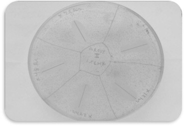
Group 2 (1% CHX).
Fig. 3.

Group 3 (2% CHX).
Fig. 4.

Group 4 (Deionized water).
3. Results
The effect of three groups (CHX concentrations) on zone of inhibition (mm) were observed over the periods (0–6 h, 6–12 h, 12–24 h, 24–48 h and 48–72 h). A parallel control (Group 4) i.e. of deionized water was done which showed 0.00 mm zone of inhibition at all periods therefore was not included in the analysis of variance (ANOVA).
An antimicrobial activity was detected in all experimental groups upto 72 h. In all the groups the decrease zone of inhibition with the increase in time period, indicating decrease in the antimicrobial substantivity (Table 2).
Table 2.
Zone of inhibition (Mean ± SD) of four groups at different periods.
| Groups | 0–6 h (n = 10) | 6–12 h (n = 10) | 12–24 h (n = 10) | 24–48 h (n = 10) | 48–72 h (n = 10) | % Change (0–6 h to 48–72 h) |
|---|---|---|---|---|---|---|
| Group 1 | 2.80 ± 0.25 | 1.94 ± 0.13 | 1.49 ± 0.19 | 0.99 ± 0.14 | 0.50 ± 0.11 | 82.1% |
| Group 2 | 3.93 ± 0.37 | 3.49 ± 0.23 | 2.81 ± 0.23 | 2.41 ± 0.19 | 2.03 ± 0.18 | 48.3% |
| Group 3 | 6.13 ± 0.21 | 5.60 ± 0.46 | 4.91 ± 0.61 | 4.59 ± 0.56 | 4.08 ± 0.28 | 33.4% |
| Group 4 | 0.00 ± 0.00 | 0.00 ± 0.00 | 0.00 ± 0.00 | 0.00 ± 0.00 | 0.00 ± 0.00 | 0.00% |
Tukey test revealed significantly (p < 0.001) different and higher zone of inhibition at 0–6 h of both Group 2 (28.8%) and Group 3 (54.3%) as compared to Group 1. Further, the mean of zone of inhibition of Group 3 was also found significantly (p < 0.001) different and 35.9% higher as compared to Group 2.
Similarly, at 6–12 h, comparing the mean zone of inhibition between the groups (Table 3), Tukey test revealed significantly (p < 0.001) different and higher zone of inhibition of both Group 2 (44.4%) and Group 3 (65.4%) as compared to Group 1. Further, the mean of zone of inhibition of Group 3 was also found significantly (p < 0.001) different and 37.7% higher as compared to Group 2.
Table 3.
For each period, comparison (p value) of mean zone of inhibition between the groups by Tukey test.
| Comparisons | 0–6 h | 6–12 h | 12–24 h | 24–48 h | 48–72 h |
|---|---|---|---|---|---|
| Group 1 vs. Group 2 | <0.001 | <0.001 | <0.001 | <0.001 | <0.001 |
| Group 1 vs. Group 3 | <0.001 | <0.001 | <0.001 | <0.001 | <0.001 |
| Group 2 vs. Group 3 | <0.001 | <0.001 | <0.001 | <0.001 | <0.001 |
Similarly, at 12–24 h, comparing the mean zone of inhibition between the groups (Table 3), Tukey test revealed significantly (p < 0.001) different and higher zone of inhibition of both Group 2 (47.0%) and Group 3 (69.7%) as compared to Group 1. Further, the mean of zone of inhibition of Group 3 was also found significantly (p < 0.001) different and 42.8% higher as compared to Group 2.
Similarly, at 24–48 h, comparing the mean zone of inhibition between the groups (Table 3), Tukey test revealed significantly (p < 0.001) different and higher zone of inhibition of both Group 2 (58.9%) and Group 3 (78.4%) as compared to Group 1. Further, the mean of zone of inhibition of Group 3 was also found significantly (p < 0.001) different and 47.5% higher as compared to Group 2.
Similarly, at 48–72 h, comparing the mean zone of inhibition between the groups (Table 3), Tukey test revealed significantly (p < 0.001) different and higher zone of inhibition of both Group 2 (75.4%) and Group 3 (87.7%) as compared to Group 1. Further, the mean of zone of inhibition of Group 3 was also found significantly (p < 0.001) different and 50.2% higher as compared to Group 2.
Result concluded that antibacterial substantivity of Group 3 was best followed by Group 2 followed by Group 1 as overall decrease in the zone of inhibition was minimum 33.4%, 48.3%, 82% respectively (0–6 to 48–72 h).
4. Discussion
The primary objective in root canal treatment is to remove the infected pulpal tissue and infectious microbial pathogens. This is accomplished with mechanical debridement via instrumentation and intracanal irrigants. Mechanical instrumentation with files and reamers widens the root canal space and removes the bulk of the pulp tissue and pathogenic microbial agents. However, some fragments of tissues and pathogenic microorganisms will remain even after the most stringent or aggressive mechanical instrumentation of the canal space, which can be eliminated only after the introduction of appropriate intracanal irrigants.1
The infection of the root canal system is considered to be a polymicrobial infection, consisting of both aerobic and anaerobic bacteria.5 These microorganisms are very susceptible to chlorhexidine, Parsons and associates who first studied the uptake and release of 0.02% and 1% solutions of chlorhexidine bovine pulp and dentin specimens and concluded that chlorhexidine was a potent antibacterial agent and may serve as an effective endodontic irrigant. Literature shows microbial properties of chlorhexidine gluconate as an effective antimicrobial agent similar as sodium hypochlorite in vitro,6 and 2.0% chlorhexidine gluconate a more effective antimicrobial agent than 5.25% sodium hypochlorite.4
Chlorhexidine gluconate (chlorhexidine) is a broad-spectrum antimicrobial drug. Acting as an antiseptic, it is an effective bactericidal agent against all categories of microbes, including bacteria, yeast, and viruses. The substantivity (residual antimicrobial activity) of chlorhexidine enhances this bactericidal effect, which allows for the retention of chlorhexidine in the oral cavity and a prolonged residual antimicrobial effect for up to 12 h or longer depending on the dosage and form.7
CHX is a synthetic cationic bis-guanide consists of two symmetric 4-cholorophenyl rings and two biguanide groups connected by a central hexamethylene chain. CHX is a positively charged hydrophobic and lipophilic molecule that interacts with phospholipids and lipopolysaccharides on the cell membrane of bacteria and then enters the cell through some type of active or passive transport mechanism. Its efficacy is due to the interaction of positive charge of the molecule and negatively charged phosphate groups on the microbial cell walls, thereby altering the cells' osmotic equilibrium. This increases the permeability of the cell wall, which allows the CHX molecule penetrates into the bacteria. CHX is a base and is stable as a salt. The most common oral preparation, chlorhexidine gluconate, is water-soluble and at physiologic pH, readily dissociates and releases the positively charged CHX component. At low concentration (0.2%), low molecular weight substances specifically potassium and phosphorous will leak out. On the other hand, at higher concentration (2%), CHX is bactericidal; precipitation of cytoplasmic contents occurs resulting in cell death.8
To prevent reinfection of the root canals and for the successful outcomes of endodontic treatment irrigants with having the property of the substantivity ex. CHX could be utilised. Substantivity is the continuing therapeutic action despite removal of vehicle for an extended period. Chlorhexidine can be adsorbed onto hydroxyapatite in tooth. The adsorbed chlorhexidine acts as a reservoir and is gradually released as its concentration in the environment decreases. The adsorption is related to the concentration of chlorhexidine. At low concentration of 0.005%–0.01% a stable monolayer of chlorhexidine is observed, which might change the physical and chemical properties of the tooth surface and prevent or reduce bacterial colonization. At higher concentration, multi-layers of chlorhexidine are present and the excess of chlorhexidine can be released rapidly into the surrounding environment. Chlorhexidine is normally used in concentration between 0.12% and 2%.At these concentrations, it has an extremely low level of tissue toxicity, both locally and systemically.9
Considering this feature the present study was carried out to evaluate and compare the substantivity of different concentrations of chlorhexidine (0.1%, 1% and 2%) and deionized water at varying periods of time.
The zone of inhibition was seen in all experimental groups but absent in Group 4 (control) because deionized water has no antibacterial activity. The maximum mean zone of inhibition was seen in Group 3 (2% CHX-6.13) as compared to other groups (0.1% CHX, 1% CHX and deionized water) at 0–6 h which decreased with increase in time period and lasted upto 48–72 h (4.59), this means although zone of inhibition decreased with time, still substantivity remained till 72 h the best result with 2% CHX could be related with the concentration of CHX because at higher concentration multilayer of CHX are formed which releases gradually. The positively charged molecule of CHX interact with bacteria and precipitate the cytoplasmic contents resulting in cell death. White et al10 reported best outcomes with 2% CHX and instills substantive antimicrobial activity up to 72 h, when compared with 0.12% CHX. Shalini et al3 also reported antimicrobial substantivity with 2% CHX which lasted up to 72 h. Oncag et al11 evaluated the antibacterial properties of 2% CHX and 0.2% CHX and reported the best efficacy with 2% CHX. Leonardo et al12 evaluated the antimicrobial substantivity of 2% CHX and they found residual antimicrobial activity up to 48 h. However, some studies revealed that the substantivity of CHX for longer period like Khandani et al13 found that 5 min treated with 2% CHX solution induced substantivity upto 4weeks. Rosenthal et al14 indicated that substantivity of CHX was extended for up to12 weeks. The number of interacted CHX molecule with dentine affects the substantivity. Therefore more concentrated CHX preparation decrease the microbial colonization. This conforms the direct relationship between concentration of CHX and substantivity.
Smaller inhibitory zones were observed with Group 1 (0.1% CHX) and Group 2 (1% CHX) when compared to 2% CHX and lasted up to 72 h. It could be due to lower concentration of CHX in groups 1 and 2 i.e. 0.1% and 1% causing availability of less number of CHX molecules to interact with bacteria9 and resulting in reduced antimicrobial efficacy when compared to Group 3 having 2% CHX as reported by Katherine et al,15 Swaty Jhamb et al,16 Caio C. et al.17
In the present study comparing all four irrigants, it was found that the substantivity of the 2% CHX was best and directly related to the concentration of chlorhexedine. Hence, the 2% CHX was better than 1% followed by 0.1%.
5. Conclusion
2% CHX is a preferred endodontic irrigant than 1% and 0.1% CHX as it showed substantive antimicrobial activity for 72 h. The substantivity of CHX is directly proportional to its concentration. Antimicrobial substantivity observed in this study which is effective in those areas that are inaccessible to instrumentation especially in primary teeth.
Conflicts of interest
All authors have none to declare.
References
- 1.Grossman L. 11th ed. 1974. Endodontic Practice. [Google Scholar]
- 2.Evanov Chris, Liewehr Frederick, Buxton Thomas B., Joyce Anthony P. Antibacterial efficacy of calcium hydroxide and chlorhexidine gluconate irrigants at 37 °C and 46 °C. J Endod. 2004;30:653–657. doi: 10.1097/01.don.0000121620.11272.22. [DOI] [PubMed] [Google Scholar]
- 3.Shahani M.N., Subba Reddy V.V. Comparison of antimicrobial substantivity of root canal irrigants in instrumented root canals up to 72 h: an in vitro study. J Indian Soc Pedod Prev Dent. 2011;29:28–33. doi: 10.4103/0970-4388.79925. [DOI] [PubMed] [Google Scholar]
- 4.Jeansonne Michael J., White Robert R. A comparison of 2.0% chlorhexidine gluconate and 5,25% sodium hypochlorite as antimicrobial endodontic irrigants. J Endod. 1994;20:276–278. doi: 10.1016/s0099-2399(06)80815-0. [DOI] [PubMed] [Google Scholar]
- 5.Pinky C., Shashibhushan K.K., Subbareddy V.V. Endodontic treatment of necrosed primary teeth using two different combinations of antibacterial drugs: an in vivo study. J Indian Soc Pedod Prev Dent. 2011;29:121–127. doi: 10.4103/0970-4388.84684. [DOI] [PubMed] [Google Scholar]
- 6.Iqbal Musani, Varun Goyal, Asha Singh, Chetan Bhat. Evaluation and comparison of biological cleaning efficacy of two endofiles and irrigants as judged by microbial quantification in primary teeth – an in vivo study. Int J Clin Pediatr Dent. 2009;2:15–22. doi: 10.5005/jp-journals-10005-1013. [DOI] [PMC free article] [PubMed] [Google Scholar]
- 7.Cohen S., Burns R.C. 7th ed. St. Luis Mosby; 1994. Pathways of Pulp; pp. 633–671. [Google Scholar]
- 8.Zahed Mohammadi. Chlorhexidine gluconate, its properties and applications in endodontics. Iran Endod J. 2008;2:113–125. [PMC free article] [PubMed] [Google Scholar]
- 9.Kholi A. 2011. Text Book of Endodontics; p. 170. [Google Scholar]
- 10.White R.R., Hays G.L., Janer L.R. Residual antimicrobial activity after canal irrigation with chlorhexidine. J Endod. 1997;23 doi: 10.1016/S0099-2399(97)80052-0. [DOI] [PubMed] [Google Scholar]
- 11.Oncag O., Hosgor M., Hilmioglu S., Zekioglu O., Eronat C., Burhanoglu D. Comparison of antibacterial and toxic effects of various root canal irrigants. Int Endod J. 2003;36:423–432. doi: 10.1046/j.1365-2591.2003.00673.x. [DOI] [PubMed] [Google Scholar]
- 12.Leonardo M.R., Tanomaru Filho M., Silva L.A., Nelson Filho P., Bonifacio K.C. In-vivo antimicrobial activity of 2% chlorhexidine used as a root canal irrigating solution. J Endod. 1999;25:167–171. doi: 10.1016/s0099-2399(99)80135-6. [DOI] [PubMed] [Google Scholar]
- 13.Kandaswamy D., Venkateshbabu N. Root canal irrigants: review. J Conserv Dent. 2010;13:256–264. doi: 10.4103/0972-0707.73378. [DOI] [PMC free article] [PubMed] [Google Scholar]
- 14.Rosenthal S., Spangberg L., Safavi K.E. Chlorhexidine substantivity in root canal dentine. Oral Surg Oral Med Oral Pathol Oral Radiol Endod. 2004;98:488–492. doi: 10.1016/j.tripleo.2003.07.005. [DOI] [PubMed] [Google Scholar]
- 15.Carson Katherine R., Goodell Gary G., McClanahan Scott B. Comparison of the antimicrobial activity of six irrigants on primary endodontic pathogens. J Endod. 2005;31:471–473. doi: 10.1097/01.don.0000148868.72833.62. [DOI] [PubMed] [Google Scholar]
- 16.Swaty Jhamb, Vineeta Nikhil, Vijay Singh. An in vitro study of antibacterial effect of ca(OH)2 and chlorhexidine on enterococcus faecalis. Indian J Dent Res. 2010;21:512–514. doi: 10.4103/0970-9290.74222. [DOI] [PubMed] [Google Scholar]
- 17.Ferraz Caio CR., Gomes Brenda PFA., Zaia Alexandre A., Teixeira Fabrício B., Souza-Filho Francisco J. Comparative study of the antimicrobial efficacy of chlorhexidine gel, chlorhexidine solution and sodium hypochlorite as endodontic irrigants. Braz Dent J. 2007;18:294–298. doi: 10.1590/s0103-64402007000400004. [DOI] [PubMed] [Google Scholar]