

Abstract
Diamond Light Source, a third-generation synchrotron radiation (SR) facility in the UK, celebrated its 10th anniversary in 2012. A private limited company was set up in April 2002 to plan, construct and operate the new user-oriented SR facility, called in brief Diamond. It succeeded the Synchrotron Radiation Source in Daresbury, a second-generation synchrotron that opened in 1980 as the world's first dedicated X-ray-providing facility, closing finally in 2008, by which time Diamond's accelerators and first beamlines were operating and user experiments were under way. This theme issue of Philosophical Transactions of the Royal Society A gives some examples of the rich diversity of research done in the initial five years, with some glimpses of activity up to 2014. Speakers at the 10 year anniversary symposium were drawn from a small number of major thematic areas and each theme was elaborated by a few speakers whose contributions were placed into a broader context by a leading member of the UK academic community in the role of rapporteur. This introduction gives a summary of the design choices and strategic planning of Diamond as a coherent user facility, a snapshot of its present status and some consideration of future perspectives.
Keywords: Diamond Light Source, synchrotron radiation, X-ray physics
1. Introduction
Synchrotron radiation (SR) was discovered in 1946, and scientists realized very early that it ‘would be a boon in X-ray physics’ if these accelerators ever became available as stable sources [1]. It was several decades before the boon really took off. Following the initial dark period of parasitic use, the first dedicated, purpose-built SR sources came about in the 1980s, and it was another decade before specially designed, stable insertion device (ID) based storage rings started operation for a very large community of users. Their experiments covered a huge breadth of different applications, ranging from physical sciences across life sciences into medical, environmental and even archaeological studies.
This success can be attributed to the unique properties of the electromagnetic waves provided by these sources. Not only is the photon beam of superbly high brilliance, but it also covers a broad spectral range from the infrared (meV) to the hard gamma (500 keV) region, spanning almost eight orders of frequencies. In addition, these waves have a variable well-defined state of polarization and a variable stroboscopic time structure.
In the UK, the first X-ray-producing storage ring, dedicated solely to SR experiments, came into operation in 1980. The Synchrotron Radiation Source (SRS) at Daresbury was an accelerator with small (96 m) circumference and relatively low electron energy (2.0 GeV) and X-rays were produced in a wavelength shifter, which locally exposed the electrons to a higher magnetic field than the field of the regular bending magnets of the storage ring. This extended the radiation spectrum into the normal X-ray range. Stability was also much improved in comparison to particle physics accelerators; however, there was not enough space for IDs and the emittance was too large to use undulators to generate X-rays. By the end of the 1990s, it had become clear that a medium-energy (3–4 GeV) machine would be in many ways a perfect SR source. User requests in the UK (number of proposals versus photon energy) clearly pointed to a source optimized for the photon energy range of 4–20 keV. Furthermore, operation at a lower electron energy than that chosen for the first ID-based machines (6–8 GeV) reduces operational costs because of lower power consumption.
In the 1990s, the success of the first ID-based X-ray machine, at the European Synchrotron Radiation Facility (ESRF) at Grenoble, made it obvious that similar national sources would also be required. Undulators provided unprecedented brilliance, which were able to make a major impact in nanoscience by allowing the investigation of small structures at high spatial resolution. Superconducting (SC) wigglers gave intense high-energy photon beams, and finally the beams that were produced showed a remarkable coherence, which opened up exciting methodological possibilities. SPring-8 in Japan paved the way to exploiting these advantages in low-energy high-brilliance machines by making in-vacuum undulators with small gaps and short periods work in a low-emittance machine.
In the UK, a 3 GeV machine with an emittance of 2.7 nm rad was finally proposed. The questions of circumference, energy and thus possible minimum emittance and maximum number of users were answered by asking the user community for beamline proposals. Clearly, the most desired energy range was the region around 10 keV, and, with almost 40 proposals submitted, each with a strong and substantial user community, a lattice with 24 sectors seemed the best strategy. This choice not only took into account the user demands but also enabled a circumference that made a small-emittance lattice possible and provided a reasonable length and number of straight sections. Using one straight section for injection and a second for cavities left 22 sectors, which meant 22 straight sections, for the installation of IDs. The choice of a double bend achromat (DBA) lattice meant that additionally 22 bending magnet sources could be used for experimental stations, if the demand was there.
The political decision to site the source at Rutherford Appleton Laboratory, 20 miles south of Oxford, was taken in 2000, co-locating it with the 0.25 MW ISIS neutron spallation source and the central laser facility, operating lasers up to 1 petawatt of power. This strategic and forward-looking co-location today provides the ideal platform to build an internationally strong campus by adding an X-ray free electron laser (XFEL) and a 20 petawatt laser (figure 1).
Figure 1.

Aerial view of the Harwell Science and Innovation Campus in Oxfordshire.
A limited company, Diamond Light Source, was formed in April 2002 to design, build, operate and eventually decommission the facility. The UK Government, represented as shareholder through a Research Council (then CCLRC and since 2007 STFC), holds 86% of the shares and was joined by The Wellcome Trust as 14% shareholder partner.
Ground breaking was in 2003, first electrons were stored in May 2006 and the first experimental users arrived in January 2007. Phase I financed the buildings, the accelerator system and seven beamlines. These seven beamlines started operation for users in 2007 (of which three were operating for protein crystallography). In Phase II, an additional 15 beamlines were brought into user operation by 2012; and by 2018 Phase III will add 10 more beamlines to the portfolio.
2. Diamond's accelerator system
Diamond's accelerator system starts out with a 100 MeV linear accelerator, the only commercially bought turnkey component of the whole system. The electrons are then accelerated up to 3 GeV in a synchrotron with a circumference of 158 m before they are injected at full energy into the main storage ring, which has 24 sectors in a 561.6 m circumference. (Figure 2 shows this system.) Each sector is part of a DBA lattice, each with two bending magnets, 10 quadrupole magnets and seven sextupole magnets. This provides 24 straight sections, of which 22 are for experimental stations (18×5 m, four×8 m long), one for injection and one for the three SC, 500 MHz radio-frequency cavities.
Figure 2.
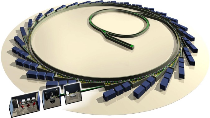
Diamond's synchrotron machine and layout of the beamlines.
Figure 3 shows for the years 2007–2013 the overall uptime of this system for experiments, with down periods being used for maintenance, machine studies and the installation of additional IDs. The stability and predictability of the whole system is established by a frequent topping-up of the electron current such that the current is kept almost constant. This in turn keeps the power load constant on all components, which is especially important for the optical components such as slits, monochromators, mirrors and radiation monitors. The resulting high level of performance and predictability forms the basis for a genuine user-oriented facility.
Figure 3.
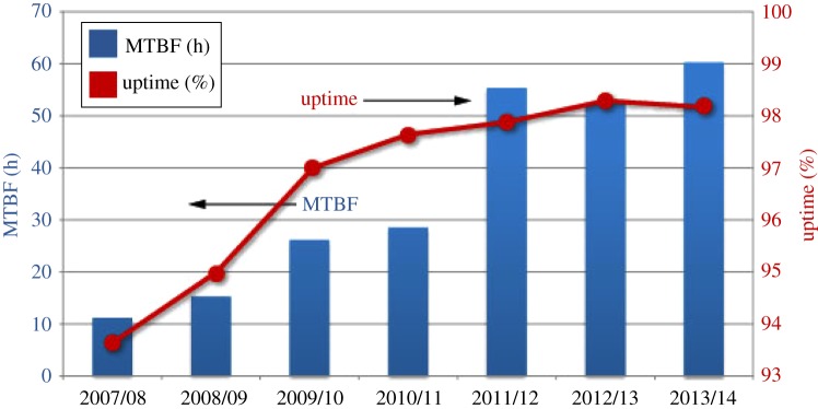
Machine uptime and mean time between failures (MTBF), 2007–2013.
This DBA lattice provides a horizontal emittance of 2.7 nm rad and the 0.3% coupling factor from the horizontal into the vertical plane enables a vertical emittance of 8 pm rad. This means that SR under these conditions is at 10 keV vertically fully spatially coherent.
The storage ring is filled in the normal mode with 900 bunches giving a current of 300 mA. Each bunch is around 10 mm long (FWHM), thus providing a corresponding flash in the experimental station of ca 30 ps. Special filling patterns include a hybrid mode consisting of a 686 bunch train plus a single bunch, a 156 regularly spaced bunch mode with 135 mA total current and a ‘low alpha’ mode to produce short ca 6 ps (FWHM) bunches for both time-resolved studies and terahertz range applications using coherent SR from the short electron bunch itself.
3. Insertion devices
Diamond's IDs were specified in a bespoke way, optimized for each experimental station around the ring. The low-photon-energy beamlines used either bending magnets (IR, circular dichroism (CD) and soft X-ray spectroscopy; note beamlines are defined in the tables for each village below) or a permanent magnet ex-vacuum undulator (angle-resolved photoemission spectroscopy beamline, ARPES). Experiments in the VUV range used ex-vacuum mostly circularly polarizing undulators (photoelectron emission microscope (PEEM), BLADE, IXS). For the normal X-ray range (around 10 keV) experiments used either bending magnets (e.g. core-EXAFS) or in-vacuum, small-gap undulators (e.g. macromolecular crystallography (MX), surface scattering, SISA). Finally, the hard X-ray beamlines, which require energies up to 150 keV, used SC wigglers with fields up to 4.3 T. In one case (BLADE), the beam can even be switched at 10 Hz between two undulators to swiftly change the state of polarization. Overall, 21 beamlines use undulators, four beamlines rely on wigglers and seven beamlines are installed with bending magnet sources. Already one beamline, I07, uses the next generation of in-vacuum undulators, where the peak field is increased by cryogenic cooling of the permanent magnets (so-called CPMUs), producing a significant increase in X-ray flux.
Figure 4 shows a few examples of spectra from such IDs in order to demonstrate the broad range of options used for the variety of experimental stations, all concentrating on different scientific missions.
Figure 4.

Insertion device spectra.
4. Diamond's beamline selection
The selection of beamlines and experimental stations had to take into account many different aspects, such as scientific potential, size and strength of UK user communities, technical novelty and feasibility, potential of industrial applications and many more. For each of the three phases, there was intense competition.
Phase I followed mostly demand and impact. The new facility needed to satisfy certain very strong communities in the UK, which used the SRS and the ESRF, while the smaller emittance, meaning higher brightness, would enable faster and stronger scientific impact. These were state-of-the-art, leading-edge projects.
The choice of beamlines for Phase II continued this selection process with broadly similar criteria, but also included some choices that clearly pushed the boundaries of instrumentation towards cutting edge, which means that some beamlines were built with concepts that were very new and innovative, enabling completely new methods and applications. For example, the high-resolution multi-crystal analyser for powder diffraction, the two-colour provision at SISA (providing VUV and X-radiation from two undulators on the sample simultaneously) and the installation of two consecutive Kirkpatrick–Baez mirror optics to reduce an intermediate focus further in the MX microfocus beamline are only three of these novelties.
Finally, in Phase III attention was paid not only to fill gaps in methods still not provided for but also to identify a few areas where advances beyond what was already in place at Diamond and at other SR facilities worldwide might be possible. In addition, the possibility of bridging length scales for analysis was exploited. Thus two X-ray microscopes were installed together with a nanofocus beamline, in a joint facility also housing state-of-the-art electron microscopy, bridging the ångström/nanometre scale to micrometre dimensions.
All beamlines were designed with a high level of automation and some even with robotic control of sample changes. This makes remote operation feasible and enables the users in many cases to operate a rather complex and sophisticated technical installation through a simple user interface. For coherence, Diamond beamlines use a common control system based on EPICS, a common data acquisition software layer (GDA) and a central high-performance compute and storage facility. In some areas of science, this has already led to ca 50% of all experiments being performed remotely, with first-cut data analysis being provided in close to real time.
5. Diamond's science villages
Construction of a new synchrotron facility on a green-field site brought the opportunity to create a coherent science-focused institution whose strength and capacity far exceeded the sum of its individual components, the beamlines. When the span of applications of SR was considered it became obvious that beamlines with common objectives or overlapping user communities should be grouped together. Wherever possible, the grouping would be geographical, similar beamlines should not be far apart and institutional structures within Diamond should encourage similar beamlines to learn from each other and go forward together. This led to the creation of Diamond's science villages (figure 5), an innovative concept at that time. Another important feature of this arrangement was to help make efficient shared use of the extensive off-line laboratories that were built into the periphery of the Experimental Hall. Naturally science moves on and by the end of Phase III the villages will need to be expanded to reflect the increasing focus on imaging. For instance, the correlative imaging in the life sciences, extending from super-resolution cryo-light microscopy and X-ray microscopy at B24 (table 1) to cryo-electron microscopy at the electron bio-imaging facility, co-located with I14, the X-ray nanoprobe.
Figure 5.

Diamond's beamline layout and village structure.
Table 1.
The macromolecular crystallography village and cryo-TXM beamlines.
| beamline name and number | main techniques | energy/wavelength range | status |
|---|---|---|---|
| I02 MC | macromolecular crystallography, multiwavelength anomalous diffraction (MAD) | 5–25 keV | operational |
| I02/I02-1 versatile MX (VMXi/VMXm) | VMX | construction/ technical design | |
| I03 macromolecular crystallography | macromolecular crystallography, MAD | 5–25 keV | operational |
| I04 macromolecular crystallography | macromolecular crystallography, MAD | 5–25 keV | operational |
| I04-1 monochromatic MX | macromolecular crystallography | 13.53 keV (0.9163 Å fixed wavelength) | operational |
| I23 long-wavelength MX | long-wavelength MX | optimized for 1.5 and 3.1 Å | commissioning |
| I24 microfocus MX | macromolecular crystallography, MAD | 6.5–25 keV | operational |
| B24 cryo-transmission X-ray microscopy (TXM) | full-field X-ray imaging | 0.2–2.6 keV | operational |
(a). Macromolecular crystallography village
The MX village aims to provide the UK and worldwide community with state-of-the-art facilities for the local or remote collection of the highest-quality diffraction data from macromolecular crystals for structure solution. This goal must be achieved in a rapidly developing landscape where significant increases in flux, decreases in beam size, advances in detector technology and automated sample handling have come about in the last few years. These, coupled with the rapid availability of beamtime, have changed the way users exploit the beamlines, and markedly improved throughput. The MX village has responded to these challenges and is highly productive, having already by November 2014 enabled the elucidation of over 2400 protein structures. The beamlines have enabled advances in leading-edge academic research, across a very broad range of applications, and the articles in this theme issue give a flavour of these. Waksman et al. [2] show how we can get to grips with the complex macromolecular motors that underpin secretion in bacteria, Jones [3] demonstrates how constantly changing interactions at the cell surface enable cellular communication in multicellular organisms, and Barford [4] shows how, by combining electron microscopy and crystallography, we can get to grips with the massive macromolecular complexes within our cells that regulate their proper functioning.
MX is a well-established technique, but the samples being studied are highly variable in size, diffraction quality, radiation hardness and phasing method required, and also in the biological hazard they constitute. To accommodate this broad range of requirements, a suite of beamlines with varied capabilities has been established, whose essential characteristics are summarized in table 1. The first three beamlines (I02, I03 and I04) were very similar to each other and built almost simultaneously as part of the first tranche of beamlines. They were designed to cater for the core requirements of MX in a high-throughput manner. Thus, they are tunable over a wide energy range (to facilitate anomalous dispersion methods, high-resolution diffraction, etc.), with intense, focused beams and reliable automated endstations. One, I03, has the capability to handle biological containment level 3 samples, the only such beamline outside of the USA. The use of microfocus beams for collection of diffraction data from protein crystals was pioneered by the ESRF, but in the mid-2000s nowhere yet had developed a dedicated tunable microfocus beamline for MX. To fill this gap, I24 was developed to provide a world-leading facility for structure solution from microcrystals and came online in 2008, with the innovative flexible focusing system described above. It has successfully collected data from crystals as small as 1 μm. I04-1 was designed to add capacity to the MX facilities without requiring another dedicated machine straight section. By using canted undulators, I04 and I04-1 used the same straight section and optics hutch, with a single-bounce monochromator separating the fixed energy beam of I04-1. The energy was carefully selected to be above the bromine K edge to cover many phasing opportunities and came online as an automated high-throughput beamline in 2009.
Heavy-atom soaking or incorporation of selenomethionine usually allows the eventual solution of the phase problem for unknown protein structures; however, most proteins naturally contain some sulfur atoms, which can, at longer wavelengths, provide anomalous phasing signal. In order to provide a general vehicle for phase determination using sulfur, phosphorus (for nucleic acids) and perhaps, for large complexes, the M absorption edges of heavier atoms such as uranium, a unique dedicated soft X-ray MX beamline, I23, is being built. This will be the first in-vacuum beamline for MX, designed to allow users to effectively exploit data collection with X-rays in the 1.5–4 Å wavelength range.
Currently at the construction stage, VMXi will replace I02 in 2016 for the automated collection of in situ data from crystallization media. This builds on the success and uptake of the implementation of this method on I03, I04-1 and I24. The final approved beamline, VMXm, aims to take MX at Diamond into the sub-micrometre regime for the exploitation of ever smaller crystals. This presents considerable design challenges but will build upon the experience available at Diamond.
(b). Materials village
A medium-energy synchrotron source offers a large number of opportunities for the study of solid materials by a wide variety of different techniques that span diffraction, imaging and spectroscopy. Spectroscopy has always been distinct from a number of standpoints: technical, data analysis and practitioner community. Thus, it was obvious that a village should be set aside for spectroscopy. However, the division of beamlines that offer diffraction and imaging is less clear, as we will see, from a practical or scientific standpoint. Not wishing to create a ‘small township’ within Diamond, beamlines whose focus is solid materials were grouped into a ‘materials’ village and an ‘engineering and environment’ village.
The materials village brings together four beamlines with a range of techniques for studying a diverse range of materials. The specifications of the beamlines are collected in table 2. The I16 materials and magnetism beamline was one of the beamlines able to claim ‘first users’ at Diamond in 2007, and since that time it has gone on to establish itself as a world-leading beamline for studies of electronic ordering in strongly correlated complex materials via high-resolution resonant diffraction. Radaelli & Dhesi [5] and also McMorrow [6] provide more details of how I16 has contributed to this field in their contributions. The use of SR for chemical crystallography was one of the outstanding successes of the SRS in its later years of operation—one of those ‘why didn't we build this earlier’ beamlines. Therefore, there was widespread support for I19, the small-molecule diffraction beamline at Diamond, and its successes most notably in energy materials are described in details by Blake et al. [7].
Table 2.
The materials village beamlines.
| beamline name and number | main techniques | energy/wavelength range | status |
|---|---|---|---|
| I13 X-ray imaging and coherence | phase contrast imaging, tomography, full-field microscopy (under commissioning), coherent diffraction and imaging (CXRD), photocorrelation spectroscopy (XPCS) (under commissioning), innovative microscopy and imaging | imaging branch: 8–30 keV | operational |
| coherence branch: 7–20 keV | |||
| I16 materials and magnetism | diffraction/scattering, spectroscopy | 3–15 keV | operational |
| B16 test beamline | diffraction, imaging, reflectometry | 4–20 keV monochromatic focused | operational |
| 4–45 keV monochromatic | |||
| unfocused | |||
| white beam | |||
| I19 small-molecule single-crystal diffraction | small-molecule single-crystal diffraction | 5–25 keV/0.5–2.5 Å | operational |
The construction of a test beamline and an optics and metrology laboratory was one of the most significant and farsighted investments in infrastructure made by Diamond. All incoming optics and many new detectors are trialled in this facility, greatly increasing the efficiency of beamline construction. In addition, the test beamline is remarkably productive in terms of publications, since it is one of the few places in Diamond where it is still possible to construct an entirely new type of experiment, more typical of the early pioneering era of SR. The last beamline in this village is the I13 imaging and coherence beamline, which is—at more than 200 m—Diamond's longest beamline, with one of its branchlines operated in collaboration with the University of Manchester. I13 facilitates non-destructive examination of internal features of materials under in situ conditions and is perhaps currently unique in the world because it can provide imaging solutions using medium-energy X-rays that span the 20 μm to 5 nm range.
(c). Engineering and environment village
SR is finding an increased number of applications in engineering and environmental science, and in the field of diffraction and imaging all of these applications benefit from an increase in the energy of photons used. Diamond currently has two SC wiggler IDs, which supply high-energy and highly penetrative X-rays that make the study of engineering materials and planetary interiors feasible. The engineering beamline, I12, is unique among high-energy beamlines worldwide thanks to a monochromator that produces a large (up to 100×30 mm) beam for imaging engineering-scale objects. This is combined with high-speed tomography to make feasible a vast range of experiments in materials engineering, which are further complemented by diffraction measurements. The key parameters for all of these beamlines are summarized in table 3. Withers [8] outlines the achievements and opportunities in this theme issue. High-energy diffraction is a key feature of the study of materials under extreme conditions. This is the goal of the Phase I beamline I15, and the successes of this beamline are further illustrated in the contribution by McMahon [9]. I15 was built to encompass a multi-beamline concept, which after many years is now being realized by the construction of a truly user facility for the emergent X-ray pair distribution function technique due to come online in late 2015.
Table 3.
The engineering and environment village beamlines.
| beamline name and number | main techniques | energy/wavelength range | status |
|---|---|---|---|
| I11 high-resolution powder diffraction | X-ray powder diffraction | 6–25(30) keV (0.5–2.1 Å) | operational |
| I12 JEEP: joint engineering, environmental and processing | imaging and tomography, single-crystal diffraction, powder diffraction, energy dispersive diffraction, high-energy small-angle scattering | 53–150 keV monochromatic or continuous white beam | operational |
| I15 extreme conditions | powder diffraction, single-crystal diffraction | 20–80 keV monochromatic focused; minimum beam size restrictions for E>30 keV | operational |
| white beam for special applications | |||
| I15-1 X-ray pair distribution function (XPDF) | XPDF | 40, 65 and 76 keV | construction |
Powder diffraction has been a stalwart of SR facilities for many years, because SR offers massively improved resolution compared with laboratory-based difractometers. The beamline, I11, was constructed to take advantage of medium energy and offers the resolution to match any worldwide; it is used for a wide range of energy materials and engineering materials studies. I11 has also become a tool valued by industry. In acknowledgement that faster measurements do not always provide the answers to all questions in science, perhaps the most surprising development to take place on this beamline is the upgrade to build a high-efficiency facility for long-term experiments. This offers a radical solution to the problem that many economically important phenomena needing SR, such as degradation in general, are not fast, but pursuit of these is in conflict with the economic need to maximize the throughput of all facilities at Diamond.
(d). Soft condensed matter village
The soft condensed matter village covers a wide range of methodologies, usually dealing with non-crystalline samples investigated by X-ray scattering, IR microspectroscopy or CD in the ultraviolet region. The wide range of photon energies demanded by this village are given in table 4.
Table 4.
The soft condensed matter village beamlines.
| beamline name and number | main techniques | energy/wavelength range | status |
|---|---|---|---|
| B21 high-throughput SAXS | small- and wide-angle X-ray scattering | currently 11–23 keV but set to 12.4 keV until variable camera installed | operational |
| I22 small-angle scattering and diffraction | non-crystalline diffraction: SAXS, WAXS, ASAXS, USAXS | 3.5–20 keV | operational |
| B22 MIRIAM: multimode infrared imaging and mircrospectroscopy | IR microspectroscopy and imaging, THz spectroscopy | 5–10 000 cm−1 (0.6–1200 meV) spectroscopy | operational |
| 100–10 000 cm−1 (12–1200 meV) microscopy | |||
| 900–10 000 cm−1 (0.1–1.2 eV) imaging | |||
| B23 CD | circular dichroism | module A: 125–500 nm (for multiplates and films) | operational |
| module B: 165–650 nm (for solutions) |
Small- and wide-angle X-ray scattering is used extensively in both the life and physical sciences to investigate structures, revealing detail in the nanometre to micrometre length range. Given the demand for this technique, the delivery of the first beamline, I22, a Phase II beamline, was accelerated to come very soon after the completion of the Phase I beamlines. This beamline has delivered results for a very diverse community of users and continues to extend its reach into microfocus and grazing-incidence applications. A second SAXS beamline, B21, came online more recently, optimized for ‘routine’ high-throughput experiments. At present, this beamline is being optimized for biological small-angle scattering, a method very much complementary to MX, and for which there is a considerable demand, which Diamond has, until now, not been able to meet. This beamline is already competitive, but plans are being developed to increase the X-ray flux by two orders of magnitude.
Synchrotron CD is a well-established technique and has had a strong UK community interested in the analysis especially of protein secondary structure. B23 came online for users in 2009 and has since built up a user community that extends beyond the traditional life science base, with particular potential in the field of polymers. The unique features of B23 are its small, very bright beam with an extended wavelength range. These features will enable higher-throughput plate-based experiments, of value for investigating protein–protein and protein–small molecule experiments, and formulation optimization, and also should enable the label-free investigation of dynamic effects.
B22, the MIRIAM beamline, came online at the end of 2009 and provided IR microspectroscopy, using commercial endstations (microscopes) in conjunction with highly intense synchrotron beams. Much of the initial drive for the beamline came from the life sciences, including clinicians seeking to identify IR signatures for cancer cells. However, the beamline now serves a much broader community, although the interest in mapping cell development using IR microspectroscopy continues strongly. The unique properties of the synchrotron beam are now being exploited for rapid imaging, using a focal-plane array detector, and even for the first reported IR spectroscopic imaging of live cells. As with other imaging modalities, there is considerable scope for further developments, including exciting possibilities of super-resolution via near-field microscopy.
(e). Spectroscopy village
The UK X-ray absorption spectroscopy (XAS) community was one of the strongest and diverse user groups at Daresbury's SRS. Provision of excellent detectors had allowed the SRS to remain competitive long after the emergence of the ESRF and had also allowed UK users to make extensive use of the ESRF. This community built a coherent strategy based around three beamlines, whose specifications are summarized in table 5. The plan was for a core XAS bending magnet beamline building on the best beamlines at the SRS: a microfocus beamline and a versatile beamline for ultradilute and time-resolved spectroscopy. A controversial decision was made to build the microfocus beamline I18 in Phase 1; controversial since this decision disenfranchized almost all existing UK users of XAS. However, the importance of building a new community for XAS in the UK overrode these concerns and the core XAS beamline came into operation quickly in 2010. Both of these beamlines have established themselves as world-competitive facilities and their presence has led to a steady growth of two particular communities—earth and environmental sciences and catalysis. Since the start of Diamond, XAS has become an integral part of the conservation of the Mary Rose warship. The most challenging project in this village has, without doubt, been the construction of a ‘double’ beamline for ultradilute and time-resolved spectroscopy. This project has pushed against the boundaries of the possible in beam optics design (two double-bounce crystal arrangement in anti-parallel arrangement combined with overall six mirrors) and in detector design. It benefited, however, the construction of other beamlines at Diamond by pushing and testing mechanical innovations. The initial results are tantalizingly outstanding, but optimization and development are still needed to make the beamline operate in a trouble-free and routine manner. Burke et al. [10] and Tromp [11] illustrate how spectroscopy is an invaluable tool in environmental and chemical sciences. Another beamline that has been added to the portfolio of spectroscopy beamlines at Diamond during 2014 is the soft X-ray spectromicroscopy beamline, I08. This complements the original strategy for spectroscopy by extending the resolution from 2 μm on I18 to less than 50 nm on day one, while still allowing the use of a similar range of sample environments. The final beamline in this village is I21, an inelastic X-ray scattering beamline, which will come into operation in 2017. It will complement the ARPES beamline in using photon-in/photon-out spectroscopy in the meV resolution range.
Table 5.
The spectroscopy village beamlines.
| beamline name and number | main techniques | energy/wavelength range | status |
|---|---|---|---|
| I08 soft X-ray microscopy | X-ray scanning microscopy | 250 eV–4.2 keV | operational |
| I14 hard X-ray nanoprobe | scanning X-ray fluorescence, X-ray spectroscopy and diffraction, small- and wide-angle X-ray scattering | 5–25 keV | construction |
| I18 microfocus spectroscopy | XAS, extended X-ray absorption fine structure (EXAFS), fluorescence tomography, micro XRD | 2.05–20.5 keV | operational |
| B18 core EXAFS | XAS | 2.05–35 keV | operational |
| I20 LOLA: X-ray spectroscopy | X-ray absorption spectroscopy (XAS), energy dispersive EXAFS (EDE), X-ray emission spectroscopy (XES) | dispersive branch: 6–26 keV | commissioning |
| scanning branch: 4–20 keV | |||
| I21 inelastic X-ray scattering | resonant/non-resonant inelastic scattering | 250–3000 eV | construction |
(f). Surfaces and interfaces village
SR was initially observed in low-energy electron accelerators and the first facilities using SR provided rather soft X-rays, which interacted strongly with matter and were ideal for the study of the surfaces of matter. Therefore, it is no surprise that before Diamond was built there was a strong user community involved in surface science in the UK. The technology involved in the exploitation of soft X-rays is substantially different from that needed for medium- and high-energy applications: grating spectrometer instead of crystal monochromators, electron detection and finally the ubiquitous use of ultra-high-vacuum conditions. All of these made it sensible to form a village around surfaces and interfaces. As in the case of spectroscopy, the first beamline, I06 nanoscience, built for this village, provided a PEEM as the principal endstation. This was a bold move since few if any UK researchers had any prior experience of this technique, and the surface and interface community had to learn both the opportunities and constraints of this technique. Following the shutdown of the SRS, I06 was the only source of soft X-rays in the UK and consequently has been one the most oversubscribed beamlines at Diamond. In recognition of this and the continuing opportunities that soft X-rays provide for studies of magnetism, Diamond constructed I10 (BLADE), a beamline dedicated for studies of magnetism by circular and linear dichroism. This beamline offers a 14 T magnet, one of the highest fields currently available, and in addition has a soft X-ray diffractometer built in collaboration with STFC and the University of Durham. The contributions from Radaelli & Dhesi [5] and from McMorrow [6] provide a more detailed perspective on the achievements and opportunities in this field.
One of the most surprising features of hard synchrotron X-rays is that they make a superb tool for the study of interfaces via diffraction, and I07 was therefore built to exploit this, building upon pioneering studies at SRS. Early studies using this technique focused upon surfaces painstakingly prepared using surface science techniques—these need a lot of time. In the past decades, parallel development has taken place to use X-rays to study ‘real’ and soft surfaces: liquids, electrolytes, polymers and membranes, for example. The beamline has been built to exploit both opportunities and in recent years industrial use of the beamline has also increased.
Table 6 gives a succinct summary of the wide range of beamlines needed to satisfy the requirements of this scientific community. The need for the initial three surface and interface beamlines was very clear and in some sense they filled an obvious gap. However, the final four are innovative and take the UK into entirely new directions.
Table 6.
The surfaces and interfaces village beamlines.
| beamline name and number | main techniques | energy/wavelength range | status |
|---|---|---|---|
| I05 ARPES | angle-resolved photoemission spectroscopy (ARPES) | 18–240 eV | operational |
| I06 nanoscience | X-ray absorption spectroscopy (XAS), X-ray photoemission microscopy and X-ray magnetic circular and linear dichroism | first harmonic circular: 106–1300 eV; linear horizontal: 80–2100 eV; linear vertical: 130–1500 eV | operational |
| I07 surface and interface diffraction | surface X-ray diffraction, grazing-incidence X-ray diffraction (GIXD), grazing-incidence small-angle X-ray scattering (GISAXS), X-ray reflectivity (XRR) | 6–30 keV | operational |
| B07 VERSOX: versatile soft X-ray | spectroscopic and scanned-probe imaging | 50–2800 eV | construction |
| I09 SISA: surface and interface structural analysis | XPS (including HAXPES), X-ray standing waves (XSW), near-edge X-ray absorption fine structure (NEXAFS), energy-scanned photoelectron diffraction | hard X-rays: 2.1–18+ keV (currently 2.35–18+ keV) | operational |
| soft X-rays: 0.1–2.1 keV (currently 0.1–1 keV) | |||
| I10 BLADE: beamline for advanced dichroism experiments | soft X-ray resonant scattering, XAS and X-ray magnetic circular and linear dichroism | 500–1600 eV | operational |
Perhaps the most bold and creative idea for a Phase II beamline is encapsulated in the surface and interface analysis beamline, I09, where it was proposed to let both hard and soft X-rays impinge at the same point on a sample. This has involved building hard and soft X-ray beamlines side by side—in fact, they are intertwined so that, when fully optimized, there will be three endstations: one soft, one hard and one combined. The combined energy endstation is complete and already producing spectacular results. The concept of the I09 beamline is already being copied at other facilities.
From the early days of SR, the provision of tunable and polarized light has been seen as an invaluable tool for the study of the band structure of solids. High-resolution ARPES is now one of the most powerful tools available for the study of the exotic materials created by condensed matter physics. Inelastic X-ray scattering is an emergent complementary technique that has been proved by the Swiss Light Source to have enormous promise. In view of these, a strategic decision was taken to build the I05 ARPES beamline and the I21 inelastic X-ray scattering beamline. I05 became operational after the 10-year Anniversary Symposium had been held, but within its first year it has attracted the attention of leading practitioners in the field worldwide and is producing outstanding quality publications. The inelastic scattering beamline is designed for a 17 m radius spectrometer arm and as such is housed in an external building. First users of this beamline are eagerly anticipated in spring 2017.
Finally, and as a complete contrast to the other beamlines, a versatile soft X-ray beamline, VERSOX, is being built on a bending magnet source to take advantage of the fact that it is now possible to perform XPS in non-UHV conditions. This beamline will allow experiments to be carried out on wet surfaces and will be of interest to researchers studying ice crystals in climate studies, to catalyst chemists and for studies of corrosion. Eventually, this beamline will develop a strong industrial portfolio because more realistic surfaces can be studied.
6. Industrial research and development
Diamond has engaged at a very early stage with industry through establishing the Diamond Industry Science Committee (DISCo), which has representatives from 12 different companies in various industries, including the pharmaceuticals and aerospace sectors.
With Diamond firmly into its third phase of construction, the availability of new beamlines is allowing the industrial usage to thrive. The scope for industrial research and development is greater than ever before, allowing researchers to probe their systems of interest with greater resolution or under closer to realistic operating conditions.
An Industrial Liaison Group at Diamond supports commercial users of the synchrotron and associated facilities throughout their research programme. The team of scientists covers a wide range of specialisms, including MX, X-ray absorption spectroscopy, small-angle X-ray scattering, X-ray powder diffraction and small-molecule crystallography. The myriad ways in which they support industrial scientists include running a mail-in service across a number of techniques, providing a full analytical service so that research problems are translated into solutions, and collaborating on large research projects through either peer review or grant funded projects.
While the pharmaceutical sector is still the predominant user of Diamond beamlines (mainly for MX, as described in the contribution of Brown & Shotton [12]), an increase in usage is being seen throughout the physical sciences. As of November 2014, over 70 companies from a range of sectors have purchased beamtime through the proprietary route, using a broad range of beamlines and techniques. The current amount of proprietary use of Diamond beamlines (on beamlines able to accept industrial users) is approximately 5%. However, an analysis of questionnaires sent to non-MX users accessing Diamond through peer review revealed that approximately 17% of the users had direct industrial involvement in the research undertaken at Diamond, with an additional 17% having indirect industrial involvement.
7. Summary and forward look
Diamond was constructed after many years of discussion with the user community. The choice of beamlines constructed in Phases I and II leaned very heavily towards the priorities of the majority of existing SR users in the UK. Now, Diamond has matured (figure 6) and a number of innovative and challenging beamlines have been delivered, or are poised for delivery. Some of these are leading strong existing user communities into new territory, such as I23, the in-vacuum MX beamline; however, it is also clear that Diamond should take a wider view of what could be of value to those communities who are not currently users. This poses two questions: What is best for UK science? And who could Diamond serve if an entirely new beamline were to be built for which there is no current user base? The success of the ARPES beamline shows the wisdom of building what is outstanding and then building the community around it. Conversely, Diamond now has to address the problem of updating beamlines and the machine, to keep the facility as a whole at the leading edge. This requires a constant process of detector, endstation, optics and ID evaluation, and a prioritized process of upgrade and replacement, while bearing in mind the possibility of, in time, upgrading the machine lattice to dramatically reduce the horizontal emittance. All of this goes on in the knowledge that some communities are now reliant on SR, and so resilience of the source is critical, and any plans for upgrades must consider how these communities will cope with the resultant dark period.
Figure 6.

Aerial view of Diamond in 2014. Max Alexander/Diamond.
The 10-year Anniversary Symposium looked back over the successes of the initial 10 years 2002–2012. The process of looking forward to the next 10 years has already started and is partly reflected in the contributions of this theme issue!
References
- 1.Parratt LG. 1959. Use of synchrotron orbit-radiation in X-ray physics. Rev. Sci. Instrum. 30, 297–299. ( 10.1063/1.1716610) [DOI] [Google Scholar]
- 2.Busch A, Phan G, Waksman G. 2015. Molecular mechanism of bacterial type 1 and P pili assembly. Phil. Trans. R. Soc. A 373, 20130153 ( 10.1098/rsta.2013.0153) [DOI] [PubMed] [Google Scholar]
- 3.Jones EY. 2015. Understanding cell signalling systems: paving the way for new therapies. Phil. Trans. R. Soc. A 373, 20130155 ( 10.1098/rsta.2013.0155) [DOI] [PMC free article] [PubMed] [Google Scholar]
- 4.Barford D. 2015. Understanding the structural basis for controlling chromosome division. Phil. Trans. R. Soc. A 373, 20130392 ( 10.1098/rsta.2013.0392) [DOI] [PMC free article] [PubMed] [Google Scholar]
- 5.Radaelli PG, Dhesi SS. 2015. The contribution of Diamond Light Source to the study of strongly correlated electron systems and complex magnetic structures. Phil. Trans. R. Soc. A 373, 20130148 ( 10.1098/rsta.2013.0148) [DOI] [PubMed] [Google Scholar]
- 6.McMorrow DF. 2015. Physical sciences at Diamond: past achievements and future opportunities. Phil. Trans. R. Soc. A 373, 20130150 ( 10.1098/rsta.2013.0150) [DOI] [PubMed] [Google Scholar]
- 7.Allan DR, Blake AJ, Schröder M, Tang CC, Yang S. 2015. Structural aspects of metal-organic framework-based energy materials research at Diamond. Phil. Trans. R. Soc. A 373, 20130149 ( 10.1098/rsta.2013.0149) [DOI] [PMC free article] [PubMed] [Google Scholar]
- 8.Withers PJ. 2015. Fracture mechanics by three-dimensional crack-tip synchrotron X-ray microscopy. Phil. Trans. R. Soc. A 373, 20130157 ( 10.1098/rsta.2013.0157) [DOI] [PMC free article] [PubMed] [Google Scholar]
- 9.McMahon MI. 2015. Diamonds on Diamond: structural studies at extreme conditions on the Diamond Light Source. Phil. Trans. R. Soc. A 373, 20130158 ( 10.1098/rsta.2013.0158) [DOI] [PubMed] [Google Scholar]
- 10.Burke IT, Mosselmans JFW, Shaw S, Peacock CL, Benning LG, Coker VS. 2015. Impact of the Diamond Light Source on research in Earth and environmental sciences: current work and future perspectives. Phil. Trans. R. Soc. A 373, 20130151 ( 10.1098/rsta.2013.0151) [DOI] [PMC free article] [PubMed] [Google Scholar]
- 11.Tromp M. 2015. Catalysis seen in action. Phil. Trans. R. Soc. A 373, 20130152 ( 10.1098/rsta.2013.0152) [DOI] [PubMed] [Google Scholar]
- 12.Brown DG, Shotton EJ. 2015. Diamond: shedding light on structure-based drug discovery. Phil. Trans. R. Soc. A 373, 20140468 ( 10.1098/rsta.2013.0468) [DOI] [PubMed] [Google Scholar]