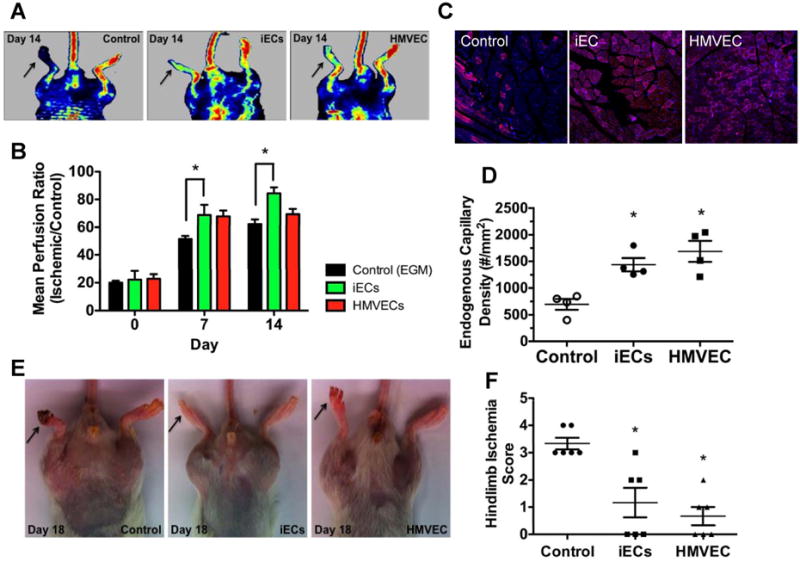
Figure 4.

Blood flow and capillary density is improved in ischemic hindlimbs by iEC transplantation. (A) Representative images of laser Doppler perfusion imaging. (B) Summarized data of perfusion ratio (value of the ischemic limb divided by that of non-ischemic limb) at day 0, 7 and 14 post-treatment (n=5 each group, *P<0.05, Repeated-measures ANOVA followed by multiple comparisons with Bonferroni's method). (C) Immunofluorescence CD31 staining of ischemic tissues from mice treated with iECs, vehicle or HMVEC. (D) Quantification of total capillary density in the ischemic limbs (n=3, *P<0.05 – Control vs iECs; #P<0.05 – Control vs HMVECs, 1-way ANOVA corrected with Bonferroni method). (E) Representative images of ischemic limbs at day 18 from iECs, vehicle and HMVEC treated animals. (F) Hindlimb ischemia score obtained by two blinded observers (n=3, *P<0.05 – Control vs iECs; #P<0.05 – Control vs HMVECs, 1-way ANOVA corrected with Bonferroni method).
