Abstract
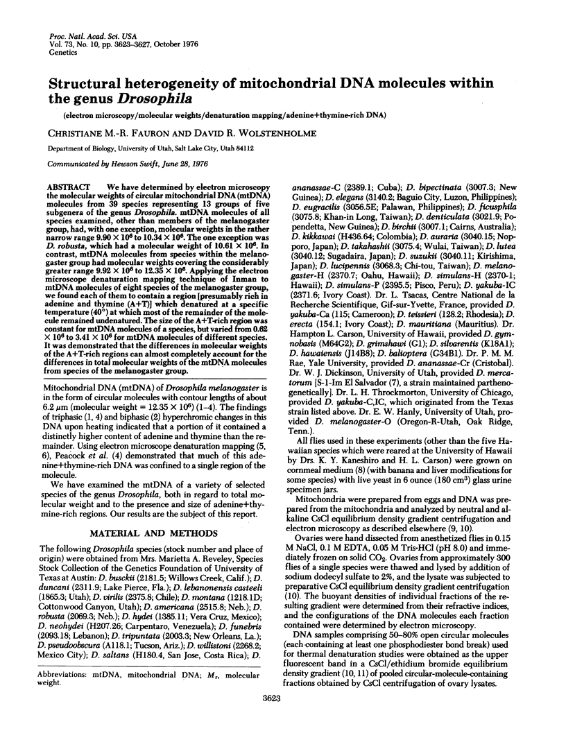
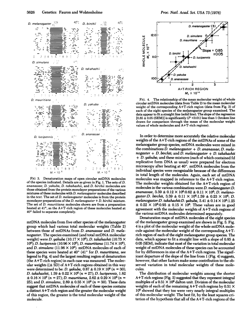
We have determined by electron microscopy the molecular weight of circular mitochondrial DNA (mtDNA) molecules from 39 species representing 13 groups of five subgenera of the genus Drosophila. mtDNA molecules of all species examined, other than members of the melanogaster group, had, with one exception, molecular weights in the rather narrow range 9.90 X 10(6). The one exception was D. robusta, which had a molecular weight of 10.61 X 10(6). In contrast, mtDNA molecules from species within the melanogaster group had molecular weights covering the considerably greater range 9.92 X 10(6) to 12.35 X 10(6). Applying the electron microscope denaturation mapping technique of Inman to mtDNA molecules of eight species of the melanogaster group, we found each of them to contain a region [presumably rich in adenine and thymine (A+T)] which denatured at a specific temperature (40 degrees) at which most of the remainder of the molecule remained undenatured. The size of the A+T-rich region was constant for mtDNA molecules of a species, but varied from 0.62 X 10(6) to 3.41 X 10(6) for mtDNA molecules of different species. It was demonstrated that the differences in molecular weights of the A+T-rich regions can almost completely account for the differences in total molecular weights of the mtDNA molecules from species of the melanogaster group.
Full text
PDF


Images in this article
Selected References
These references are in PubMed. This may not be the complete list of references from this article.
- Blumenfled M., Forrest H. S. Is Drosophila dAT on the Y chromosome? Proc Natl Acad Sci U S A. 1971 Dec;68(12):3145–3149. doi: 10.1073/pnas.68.12.3145. [DOI] [PMC free article] [PubMed] [Google Scholar]
- Bultmann H., Laird C. D. Mitochondrial DNA from Drosophila melanogaster. Biochim Biophys Acta. 1973 Mar 19;299(2):196–209. doi: 10.1016/0005-2787(73)90342-0. [DOI] [PubMed] [Google Scholar]
- Carson H. L. Selection for parthenogenesis in Drosophila mercatorum. Genetics. 1967 Jan;55(1):157–171. doi: 10.1093/genetics/55.1.157. [DOI] [PMC free article] [PubMed] [Google Scholar]
- Fansler B. S., Travaglini E. C., Loeb L. A., Schultz J. Structure of Drosophila melanogaster dAT replicated in an in vitro system. Biochem Biophys Res Commun. 1970 Sep 30;40(6):1266–1272. doi: 10.1016/0006-291x(70)90003-3. [DOI] [PubMed] [Google Scholar]
- Follett E. A., Crawford L. V. Electron microscope study of the denaturation of Polyoma virus DNA. J Mol Biol. 1968 Jun 28;34(3):565–573. doi: 10.1016/0022-2836(68)90181-2. [DOI] [PubMed] [Google Scholar]
- Fouts D. L., Manning J. E., Wolstenholme D. R. Physicochemical properties of kinetoplast DNA from Crithidia acanthocephali. Crithidia luciliae, and Trypanosoma lewisi. J Cell Biol. 1975 Nov;67(2PT1):378–399. doi: 10.1083/jcb.67.2.378. [DOI] [PMC free article] [PubMed] [Google Scholar]
- Inman R. B. A denaturation map of the lambda phage DNA molecule determined by electron microscopy. J Mol Biol. 1966 Jul;18(3):464–476. doi: 10.1016/s0022-2836(66)80037-2. [DOI] [PubMed] [Google Scholar]
- Inman R. B. Denaturation maps of the left and right sides of the lambda DNA molecule determined by electron microscopy. J Mol Biol. 1967 Aug 28;28(1):103–116. doi: 10.1016/s0022-2836(67)80081-0. [DOI] [PubMed] [Google Scholar]
- Peacock W. J., Brutlag D., Goldring E., Appels R., Hinton C. W., Lindsley D. L. The organization of highly repeated DNA sequences in Drosophila melanogaster chromosomes. Cold Spring Harb Symp Quant Biol. 1974;38:405–416. doi: 10.1101/sqb.1974.038.01.043. [DOI] [PubMed] [Google Scholar]
- Polan M. L., Friedman S., Gall J. G., Gehring W. Isolation and characterization of mitochondrial DNA from Drosophila melanogaster. J Cell Biol. 1973 Feb;56(2):580–589. doi: 10.1083/jcb.56.2.580. [DOI] [PMC free article] [PubMed] [Google Scholar]
- Radloff R., Bauer W., Vinograd J. A dye-buoyant-density method for the detection and isolation of closed circular duplex DNA: the closed circular DNA in HeLa cells. Proc Natl Acad Sci U S A. 1967 May;57(5):1514–1521. doi: 10.1073/pnas.57.5.1514. [DOI] [PMC free article] [PubMed] [Google Scholar]
- Renger H. C., Wolstenholme D. R. The form and structure of kinetoplast DNA of Crithidia. J Cell Biol. 1972 Aug;54(2):346–364. doi: 10.1083/jcb.54.2.346. [DOI] [PMC free article] [PubMed] [Google Scholar]
- Wolstenholme D. R. Replicating DNA molecules from eggs of Drosophila melanogaster. Chromosoma. 1973 Jul 23;43(1):1–18. doi: 10.1007/BF01256731. [DOI] [PubMed] [Google Scholar]
- Wolstonholme D. R., Kirschner R. G., Gross N. J. Heart denaturation studies of rat liver mitrochondrial DNA. A denaturation map and changes in molecular configurations. J Cell Biol. 1972 May;53(2):393–406. doi: 10.1083/jcb.53.2.393. [DOI] [PMC free article] [PubMed] [Google Scholar]


