Persoonia : Molecular Phylogeny and Evolution of Fungi logoLink to Persoonia : Molecular Phylogeny and Evolution of Fungi
. 2014 Nov 24;33:212–289. doi: 10.3767/003158514X685680

Fungal Planet description sheets: 281–319

PW Crous 1,, MJ Wingfield 2, RK Schumacher 3, BA Summerell 4, A Giraldo 5, J Gené 5, J Guarro 5, DN Wanasinghe 6,7,8, KD Hyde 6,7,8, E Camporesi 9,10, EB Gareth Jones 11, KM Thambugala 8,12, EF Malysheva 13, VF Malysheva 13, K Acharya 14, J Álvarez 15, P Alvarado 16, A Assefa 17, CW Barnes 18, JS Bartlett 19, RA Blanchette 20, TI Burgess 21, JR Carlavilla 15, MPA Coetzee 22, U Damm 23, CA Decock 24, A den Breeÿen 25, B de Vries 26, AK Dutta 14, DG Holdom 19, S Rooney-Latham 27, JL Manjón 15, S Marincowitz 22, M Mirabolfathy 28, G Moreno 15, C Nakashima 29, M Papizadeh 30, SA Shahzadeh Fazeli 30, MA Amoozegar 30, MK Romberg 31, RG Shivas 19, JA Stalpers 1, B Stielow 1, MJC Stukely 32, WJ Swart 33, YP Tan 19, M van der Bank 34, AR Wood 25, Y Zhang 35, JZ Groenewald 1
PMCID: PMC4312934  PMID: 25737601

Abstract

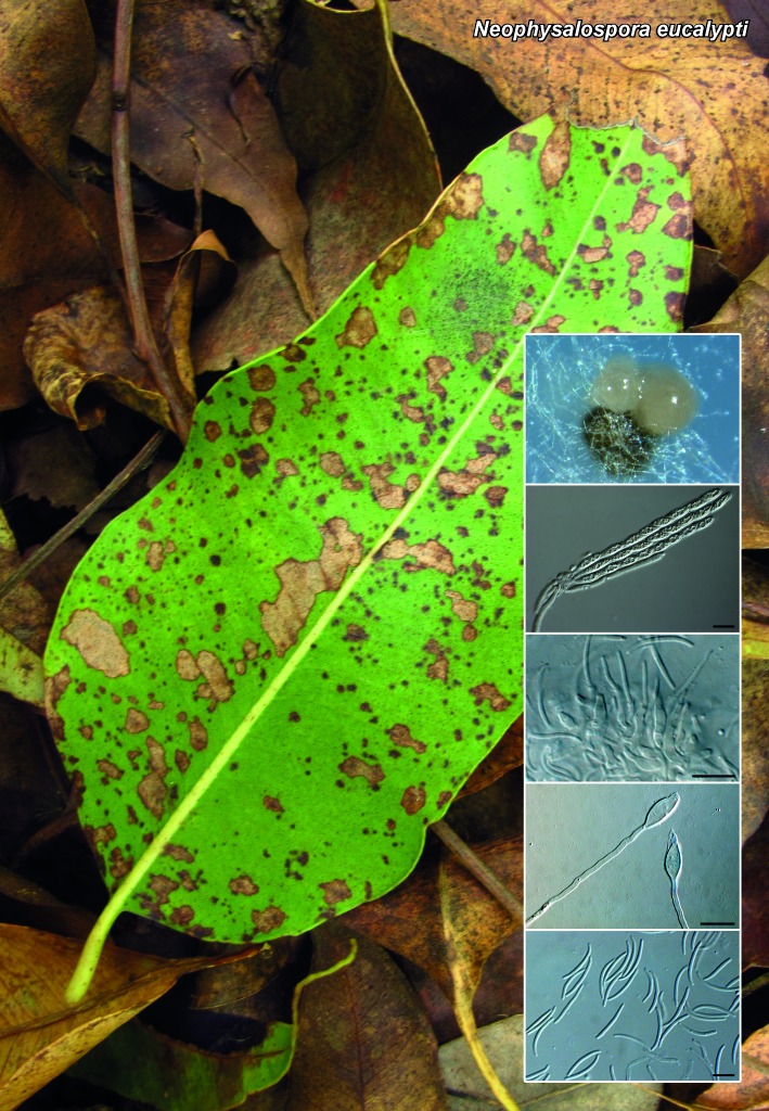
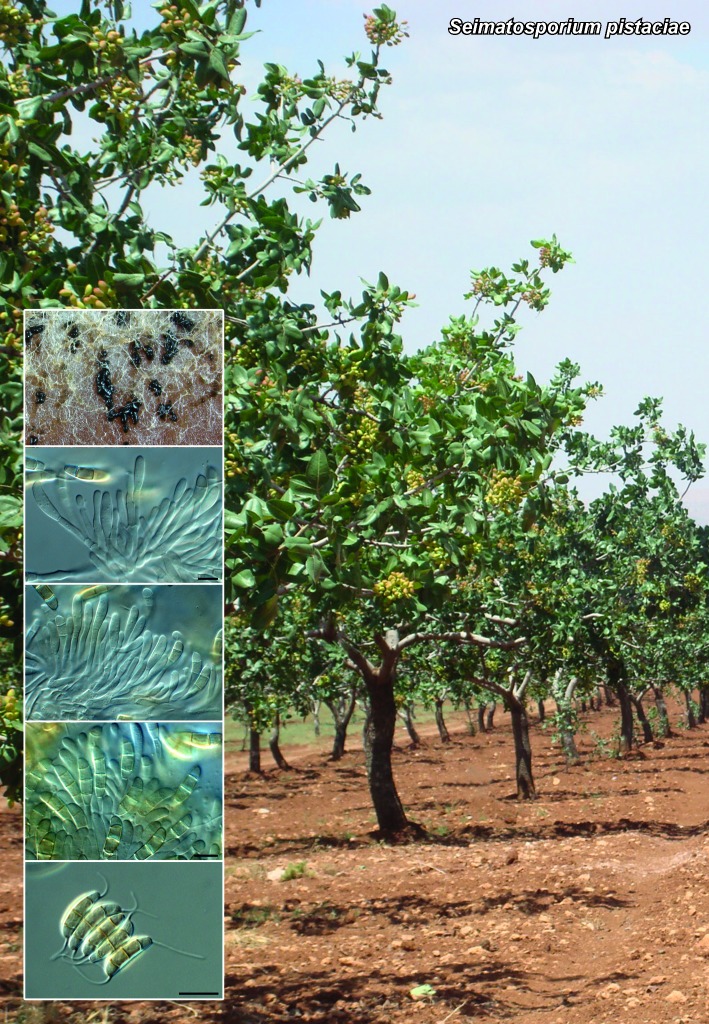
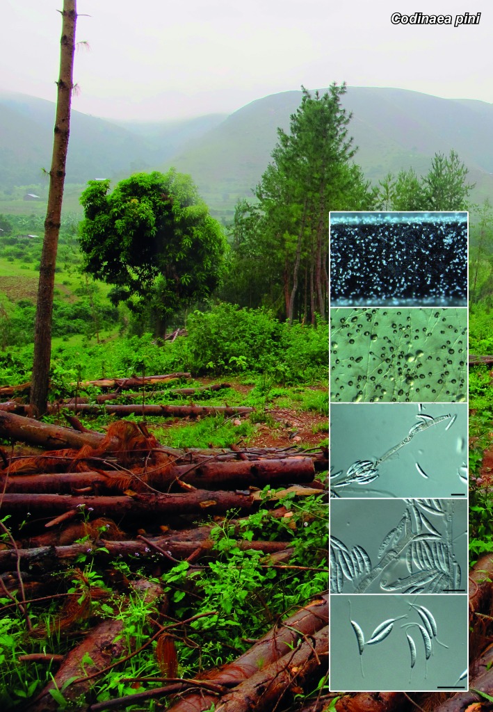
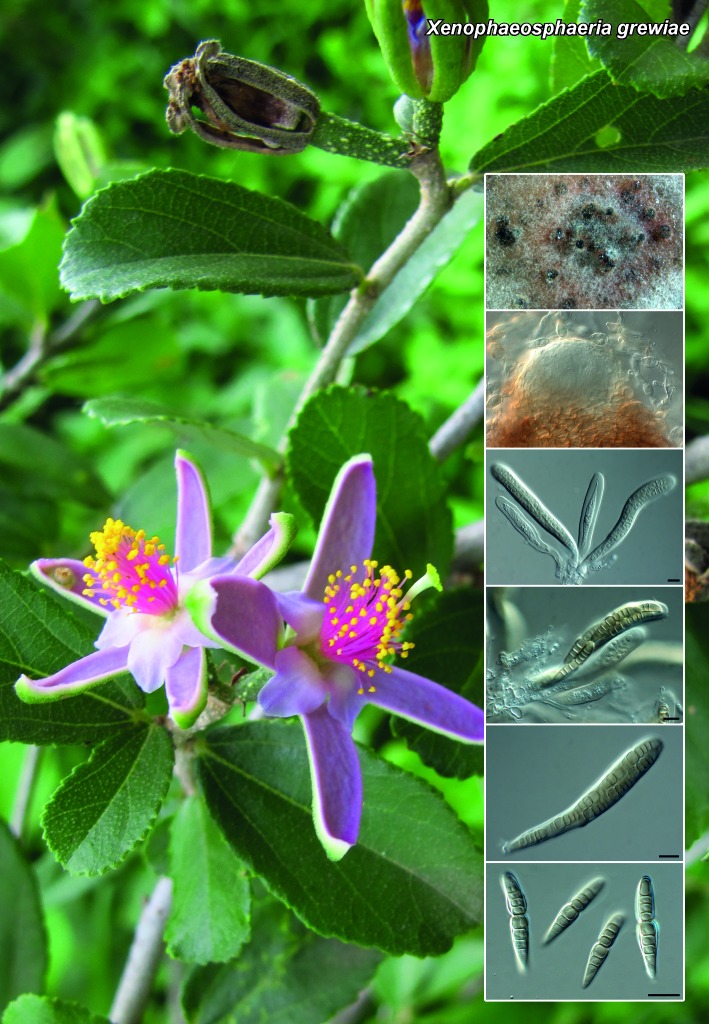
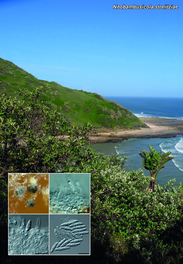
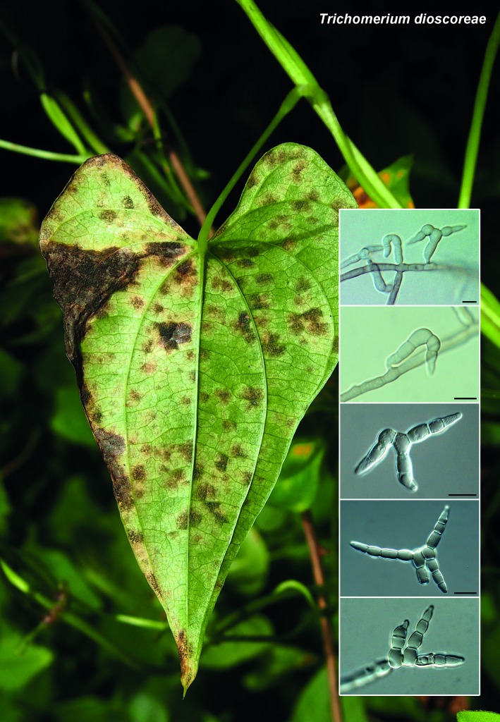
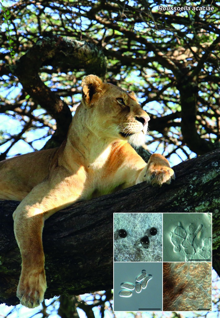
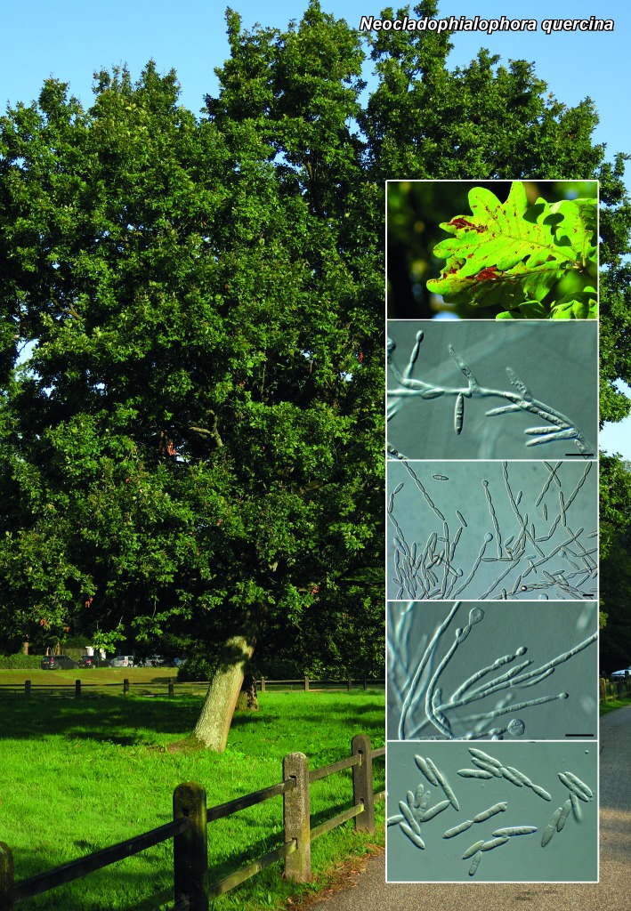
Novel species of fungi described in the present study include the following from South Africa: Alanphillipsia aloeicola from Aloe sp., Arxiella dolichandrae from Dolichandra unguiscati, Ganoderma austroafricanum from Jacaranda mimosifolia, Phacidiella podocarpi and Phaeosphaeria podocarpi from Podocarpus latifolius, Phyllosticta mimusopisicola from Mimusops zeyheri and Sphaerulina pelargonii from Pelargonium sp. Furthermore, Barssia maroccana is described from Cedrus atlantica (Morocco), Codinaea pini from Pinus patula (Uganda), Crucellisporiopsis marquesiae from Marquesia acuminata (Zambia), Dinemasporium ipomoeae from Ipomoea pes-caprae (Vietnam), Diaporthe phragmitis from Phragmites australis (China), Marasmius vladimirii from leaf litter (India), Melanconium hedericola from Hedera helix (Spain), Pluteus albotomentosus and Pluteus extremiorientalis from a mixed forest (Russia), Rachicladosporium eucalypti from Eucalyptus globulus (Ethiopia), Sistotrema epiphyllum from dead leaves of Fagus sylvatica in a forest (The Netherlands), Stagonospora chrysopyla from Scirpus microcarpus (USA) and Trichomerium dioscoreae from Dioscorea sp. (Japan). Novel species from Australia include: Corynespora endiandrae from Endiandra introrsa, Gonatophragmium triuniae from Triunia youngiana, Penicillium coccotrypicola from Archontophoenix cunninghamiana and Phytophthora moyootj from soil. Novelties from Iran include Neocamarosporium chichastianum from soil and Seimatosporium pistaciae from Pistacia vera. Xenosonderhenia eucalypti and Zasmidium eucalyptigenum are newly described from Eucalyptus urophylla in Indonesia. Diaporthe acaciarum and Roussoella acacia are newly described from Acacia tortilis in Tanzania. New species from Italy include Comoclathris spartii from Spartium junceum and Phoma tamaricicola from Tamarix gallica. Novel genera include (Ascomycetes): Acremoniopsis from forest soil and Collarina from water sediments (Spain), Phellinocrescentia from a Phellinus sp. (French Guiana), Neobambusicola from Strelitzia nicolai (South Africa), Neocladophialophora from Quercus robur (Germany), Neophysalospora from Corymbia henryi (Mozambique) and Xenophaeosphaeria from Grewia sp. (Tanzania). Morphological and culture characteristics along with ITS DNA barcodes are provided for all taxa.

Keywords: ITS DNA barcodes, LSU, novel fungal species, systematics

HIGHER ORDER CLASSIFICATION OF TAXONOMIC NOVELTIES.

ASCOMYCOTA
Dothideomycetes
    Acrospermales, Acrospermaceae
        Gonatophragmium triuniae
    Botryosphaeriales, Botryosphaeriaceae
        Alanphillipsia aloeicola
    Botryosphaeriales, Phyllostictaceae
        Phyllosticta mangiferae-indica
        Phyllosticta mimusopisicola
        Phyllosticta rubella
    Capnodiales, Cladosporiaceae
        Rachicladosporium eucalypti
    Capnodiales, Mycosphaerellaceae
        Sphaerulina pelargonii
        Xenosonderhenia eucalypti
        Zasmidium eucalyptigenum
    Incertae sedis
        Arxiella dolichandrae
    Pleosporomycetidae, Pleosporales, Incertae
    sedis
        Phellinocrescentia guianensis
    Pleosporomycetidae, Pleosporales, Incertae
    sedis, Corynesporascaceae
        Corynespora endiandrae
    Pleosporomycetidae, Pleosporales, Incertae
    sedis, Roussoellaceae
        Roussoella acaciae
    Pleosporomycetidae, Pleosporales,
    Massarineae, Bambusicolaceae
        Neobambusicola strelitziae
    Pleosporomycetidae, Pleosporales,
    Massarineae, Massarinaceae
        Stagonospora chrysopyla
    Pleosporomycetidae, Pleosporales,
    Pleosporineae, Didymellaceae
        Phoma tamaricicola
    Pleosporomycetidae, Pleosporales,
    Pleosporineae, Phaeosphaeriaceae
        Phaeosphaeria podocarpi
        Xenophaeosphaeria grewiae
    Pleosporomycetidae, Pleosporales,
    Pleosporineae, Pleosporaceae
        Comoclathris spartii
        Neocamarosporium chichastianum
Eurotiomycetes
    Chaetothyriomycetidae, Chaetothyriales,
    Trichomeriaceae
        Trichomerium dioscoreae
    Eurotiomycetidae, Eurotiales, Trichocomaceae
        Penicillium coccotrypicola
    Incertae sedis
        Neocladophialophora quercina
Lecanoromycetes
    Ostropomycetidae, Ostropales, Incertae sedis
        Phacidiella podocarpi
Leotiomycetes
    Helotiales, Hyaloscyphaceae
        Crucellisporiopsis marquesiae
Pezizomycetes
    Pezizomycetidae, Pezizales, Helvellaceae
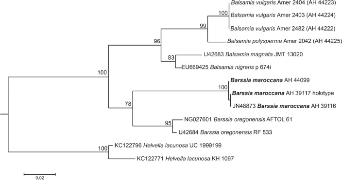
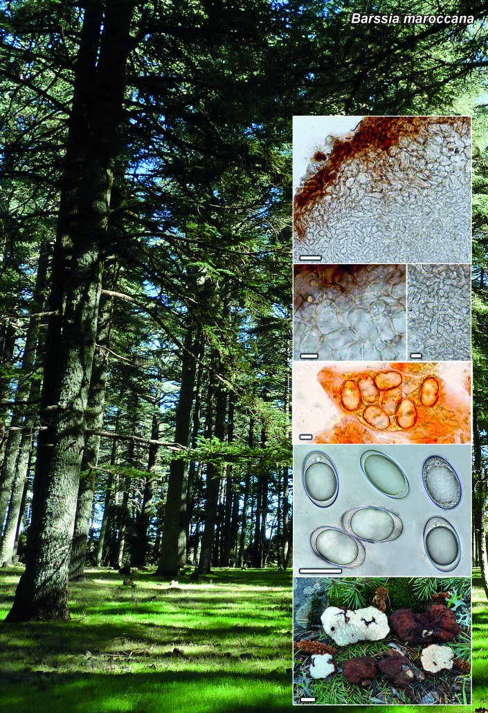
        Barssia maroccana
Sordariomycetes
    Hypocreomycetidae, Hypocreales,
    Clavicipitaceae
        Collarina aurantiaca
    Hypocreomycetidae, Hypocreales, Incertae
    sedis
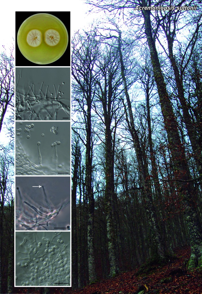
        Acremoniopsis suttonii
    Hypocreomycetidae, Incertae sedis
        Neophysalospora eucalypti
    Sordariomycetidae, Chaetosphaeriales,
    Chaetosphaeriaceae
        Codinaea pini
        Dinemasporium ipomoeae
    Sordariomycetidae, Diaporthales,
    Diaporthaceae
        Diaporthe acaciarum
        Diaporthe phragmitis
    Sordariomycetidae, Diaporthales,
    Melanconidaceae
        Melanconium hedericola
    Xylariomycetidae, Xylariales,
    Amphisphaeriaceae
        Seimatosporium pistaciae
BASIDIOMYCOTA
    Agaricomycetes, Agaricomycetidae, Agaricales,
    Marasmiaceae
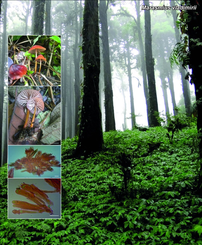
        Marasmius vladimirii
    Agaricomycetes, Agaricomycetidae, Agaricales,
    Pluteaceae
        Pluteus albotomentosus
        Pluteus extremiorientalis
    Agaricomycetes, Agaricomycetidae,
    Polyporales, Ganodermataceae
        Ganoderma austroafricanum
    Agaricomycetes, Incertae sedis, Cantharellales,
    Hydnaceae
        Sistotrema epiphyllum
CHROMISTA
    Oomycota, Oomycetes, Pythiales, Pythiaceae
        Phytophthora moyootj

Acknowledgments

Alejandra Giraldo acknowledges support from the Spanish Ministerio de Economía y Competitividad, grant CGL 2011-27185. Ekaterina F. Malysheva & Vera F. Malysheva thank Drs A. Kovalenko and N. Psurtseva (Komarov Botanical Institute) for help in collecting specimens. Financial support was provided by the Russian Foundation for Basic Research (projects 13-04-00838a and 12-04-33018). Gabriel H. Moreno acknowledges financial support from the Spanish grant (project RTA2012-00007-00-00) and to Dr L. Monje and Mr A. Pueblas of the Department of Drawing and Scientific Photography at the Alcalá University for his help in the digital preparation of the photographs, and to Dr J. Rejos, curator of the AH herbarium for his assistance with the specimens examined in the present study. We also thank the technical staff, A. van Iperen (cultures), M. Vermaas (photographic plates), and M. Starink-Willemse (DNA isolation, amplification and sequencing) for their invaluable assistance. Cony Decock acknowledges the financial support received from the Belgian State – Belgian Federal Science Policy through the BCCM research programme and the FNRS / FRFC (convention FRFC 2.4544.10).

REFERENCES

  1. Aa HA van der, Vanev S. 2002. A revision of the species described in Phyllosticta. Centraalbureau voor Schimmelcultures, Utrecht, The Netherlands. [Google Scholar]
  2. Alvarado P, Moreno G, Manjón JL, et al. 2011. First molecular data on Delastria rosea, Fischerula macrospora and Hydnocystis piligera. Boletín de la Sociedad Micológica de Madrid 35: 31–37. [Google Scholar]
  3. Antonín V, Ryoo R, Shin HD. 2012. Marasmioid and gymnopoid fungi of the Republic of Korea. 4. Marasmius sect. Sicci. Mycological Progress 11: 615–638. [Google Scholar]
  4. Ariyawansa HA, Phookamsak R, Tibpromma S, et al. 2014. A molecular and morphological reassessment of Diademaceae. The Scientific World Journal: doi.org/10.1155/2014/675348. [DOI] [PMC free article] [PubMed] [Google Scholar]
  5. Arzanlou M, Groenewald JZ, Gams W, et al. 2007. Phylogenetic and morphotaxonomic revision of Ramichloridium and allied genera. Studies in Mycology 58: 57–93. [DOI] [PMC free article] [PubMed] [Google Scholar]
  6. Aveskamp MM, Gruyter J de, Woudenberg JHC, et al. 2010. Highlights of the Didymellaceae: A polyphasic approach to characterize Phoma and related pleosporalean genera. Studies in Mycology 65: 1–60. [DOI] [PMC free article] [PubMed] [Google Scholar]
  7. Bensch K, Braun U, Groenewald JZ, et al. 2012. The genus Cladosporium. Studies in Mycology 72: 1–401. [DOI] [PMC free article] [PubMed] [Google Scholar]
  8. Braun U, Crous PW, Schubert K, et al. 2010. Some reallocations of Stenella species to Zasmidium. Schlechtendalia 20: 99–104. [Google Scholar]
  9. Braun U, Hill CF. 2008. New species and new records of foliicolous hyphomycetes from New Zealand. Australasian Mycologist 27: 45–56. [Google Scholar]
  10. Castellani E, Germano G. 1977. Le Stagonosporae graminicole. Annali della Facoltà di Scienze Agrarie della Universitá degli Studi di Torino 10: 1–135. [Google Scholar]
  11. Chomnunti P, Bhat DJ, Gareth Jones EB, et al. 2012. Trichomeriaceae, a new sooty mould family of Chaetothyriales. Fungal Diversity 56: 63–76. [Google Scholar]
  12. Corner EJH. 1996. The agaric genera Marasmius, Chaetocalathus, Crinipellis, Heimiomyces, Resupinatus, Xerula and Xerulina in Malesia. Nova Hedwigia Beihefte 111: 1–175. [Google Scholar]
  13. Crous PW, Braun U, Groenewald JZ. 2007a. Mycosphaerella is polyphyletic. Studies in Mycology 58: 1–32. [DOI] [PMC free article] [PubMed] [Google Scholar]
  14. Crous PW, Braun U, Schubert K, et al. 2007b. Delimiting Cladosporium from morphologically similar genera. Studies in Mycology 58: 33–56. [DOI] [PMC free article] [PubMed] [Google Scholar]
  15. Crous PW, Groenewald JZ, Shivas RG, et al. 2011. Fungal Planet description sheets: 69–91. Persoonia 26: 108–156. [DOI] [PMC free article] [PubMed] [Google Scholar]
  16. Crous PW, Schoch CL, Hyde KD, et al. 2009a. Phylogenetic lineages in the Capnodiales. Studies in Mycology 64: 17–47. [DOI] [PMC free article] [PubMed] [Google Scholar]
  17. Crous PW, Schubert K, Braun U, et al. 2007c. Opportunistic, human-pathogenic species in the Herpotrichiellaceae are phenotypically similar to saprobic or phytopathogenic species in the Venturiaceae. Studies in Mycology 58: 185–217. [DOI] [PMC free article] [PubMed] [Google Scholar]
  18. Crous PW, Shivas RG, Quaedvlieg W, et al. 2014. Fungal Planet description sheets: 214–280. Persoonia 32: 184–306. [DOI] [PMC free article] [PubMed] [Google Scholar]
  19. Crous PW, Summerell BA, Carnegie AJ, et al. 2009b. Unravelling Mycosphaerella: do you believe in genera? Persoonia 23: 99–118. [DOI] [PMC free article] [PubMed] [Google Scholar]
  20. Crous PW, Summerell BA, Shivas RG, et al. 2012a. Fungal Planet description sheets: 107–127. Persoonia 28: 138–182. [DOI] [PMC free article] [PubMed] [Google Scholar]
  21. Crous PW, Verkley GJM, Christensen M, et al. 2012b. How important are conidial appendages? Persoonia 28: 126–137. [DOI] [PMC free article] [PubMed] [Google Scholar]
  22. Crous PW, Wingfield MJ, Guarro J, et al. 2013. Fungal Planet description sheets: 154–213. Persoonia 31: 188–296. [DOI] [PMC free article] [PubMed] [Google Scholar]
  23. Crous PW, Wingfield MJ, Koch SH. 1990. New and interesting records of South African fungi. X. New records of Eucalyptus leaf fungi. South African Journal of Botany 56: 583–586. [Google Scholar]
  24. Cunnell GJ. 1957. Stagonospora spp. on Phragmites communis Trin. Transactions of the British Mycological Society 40: 443–445. [Google Scholar]
  25. Dai DQ, Jayarama Bhat D, Liu JK, et al. 2012. Bambusicola, a new genus from bamboo with asexual and sexual morphs. Cryptogamie, Mycologie 33: 363–379. [Google Scholar]
  26. Doidge EM. 1942. Revised descriptions of South African species of Phyllachora and related genera. Bothalia 4: 421–463. [Google Scholar]
  27. Ellis MB. 1971. Dematiaceous Hyphomycetes. CMI, Kew, England. [Google Scholar]
  28. Ellis MB. 1976. More Dematiaceous Hyphomycetes. CMI, Kew, England. [Google Scholar]
  29. Gams W. 1971. Cephalosporium-artige Schimmelpilze (Hyphomycetes). Stuttgart, Gustav Fischer Verlag. [Google Scholar]
  30. Gams W, Turham G. 1996. An extreme modification of Monocillium: Monocillium curvisetosum n. sp. Mycotaxon 59: 343–348. [Google Scholar]
  31. Gilkey HM. 1925. Five new hypogaeous fungi. Mycologia 17: 250–255. [Google Scholar]
  32. Gilkey HM. 1961. New species and revisions in the order Tuberales. Mycologia 53: 215–220. [Google Scholar]
  33. Giordano L, Garbelotto M, Nicolotti G, et al. 2013. Characterization of fungal communities associated with the bark beetle Ips typographus varies depending on detection method, location, and beetle population levels. Mycological Progress 12: 127–140. [Google Scholar]
  34. Giraldo A, Gené J, Cano J, et al. 2012. Two new species of Acremonium from Spanish soils. Mycologia 104: 1456–1465. [DOI] [PubMed] [Google Scholar]
  35. Giraldo A, Gené J, Cano J, et al. 2014. Acremonium with catenate elongate conidia: phylogeny of Acremonium fusidioides and related species. Mycologia 106: 328–338. [DOI] [PubMed] [Google Scholar]
  36. Girlanda M, Luppi-Mosca AM. 1998. Monocillium ligusticum, a new species from mycorrhizal roots in Mediterranean north Italy. Mycotaxon 67: 265–274. [Google Scholar]
  37. Glenn A, Bacon CW, Price R, et al. 1996. Molecular phylogeny of Acremonium and its taxonomic implications. Mycologia 88: 369–383. [Google Scholar]
  38. Gomes RR, Glienke C, Videira SIR, et al. 2013. Diaporthe: a genus of endophytic, saprobic and plant pathogenic fungi. Persoonia 31: 1–41. [DOI] [PMC free article] [PubMed] [Google Scholar]
  39. Gruyter J de,, Aveskamp MM, Woudenberg JHC, et al. 2009. Molecular phylogeny of Phoma and allied anamorph genera: Towards a re-classification of the Phoma complex. Mycological Research 113: 508–519. [DOI] [PubMed] [Google Scholar]
  40. Gruyter J de, Woudenberg JHC, Aveskamp MM, et al. 2010. Systematic reappraisal of species in Phoma section Paraphoma, Pyrenochaeta and Pleurophoma. Mycologia 102: 1066–1081. [DOI] [PubMed] [Google Scholar]
  41. Gruyter J de,, Woudenberg JHC, Aveskamp MM, et al. 2013. Redisposition of Phoma-like anamorphs in Pleosporales. Studies in Mycology 75: 1–36. [DOI] [PMC free article] [PubMed] [Google Scholar]
  42. Hashimoto A, Sato G, Matsuda T, et al. 2014. Molecular taxonomy of Dinemasporium and its allied genera. Mycoscience (in press) doi.org/10.1016/j.myc.2014.04.001. [Google Scholar]
  43. Hirooka Y, Rossman AY, Samuels GJ, et al. 2012. A monograph of Allantonectria, Nectria, and Pleonectria (Nectriaceae, Hypocreales, Ascomycota) and their pycnidial, sporodochial, and synnematous anamorphs. Studies in Mycology 71: 1–210. [DOI] [PMC free article] [PubMed] [Google Scholar]
  44. Houbraken J, Samson RA. 2011. Phylogeny of Penicillium and the segregation of Trichocomaceae into three families. Studies in Mycology 70: 1–51. [DOI] [PMC free article] [PubMed] [Google Scholar]
  45. Hughes SJ, Kendrick WB. 1968. New Zealand fungi 12. Menispora, Codinaea, Menisporopsis. New Zealand Journal of Botany 6: 323–375. [Google Scholar]
  46. Hyde KD, Jones EBG, Lui J-K, et al. 2013. Families of Dothideomycetes. Fungal Diversity 63: 1–313. [Google Scholar]
  47. Johnston P, Seifert KA, Stone J, et al. 2014. Recommendations on generic names competing for use in Leotiomycetes (Ascomycota). IMA Fungus 5: 91–120. [DOI] [PMC free article] [PubMed] [Google Scholar]
  48. Jung T, Stukely MJC, Hardy GEStJ, et al. 2011. Multiple new Phytophthora species from ITS Clade 6 associated with natural ecosystems in Australia: evolutionary and ecological implications. Persoonia 26: 13–39. [DOI] [PMC free article] [PubMed] [Google Scholar]
  49. Kimbrough JW, Li LTL, Wu CG. 1996. Ultrastructural evidence for the placement of the truffle Barssia in the Helvellaceae (Pezizales). Mycologia 88: 38–46. [Google Scholar]
  50. Kornerup A, Wanscher JH. 1978. Methuen handbook of colour. 3rd ed London, Eyre Methuen. [Google Scholar]
  51. Kuthubutheen AJ, Nawawi A. l991. A key to Dictyochaeta and Codinaea species. Mycological Research 95: 1224–1229. [Google Scholar]
  52. Ławrynowicz M, Skirgiello A. 1984. Barssia, a new genus in Europe. Acta Mycologia 20: 277–280. [Google Scholar]
  53. Liu J-K, Phooksamsak R, Dai D-Q, et al. 2014. Roussoellaceae, a new pleosporalean family to accommodate the genera Neoroussoella gen. nov., Roussoella and Roussoellopsis. Phytotaxa 181: 1–33. [Google Scholar]
  54. Lombard L, Leeuwen GCM van, Guarnaccia V, et al. 2014. Diaporthe species associated with Vaccinium, with specific reference to Europe. Phytopathologia Mediterranea 53: 85–97. [Google Scholar]
  55. Mel’nik V, Lee S, Groenewald JZE, et al. 2004. New hyphomycetes from Restionaceae in fynbos: Parasarcopodium ceratocaryi gen. et sp. nov., and Rhexodenticula elegiae sp. nov. Mycological Progress 3: 19–28. [Google Scholar]
  56. Minnis AM, Sundberg WJ. 2010. Pluteus section Celluloderma in the U.S.A. North American Fungi 5: 1–107. [Google Scholar]
  57. Montecchi A, Sarasini M. 2000. Funghi ipogei d’Europa. Associazione Micologica Bresadola. Fondazione Centro Studi Micologici; Trento. [Google Scholar]
  58. Nag Raj TR. 1993. Coelomycetous anamorphs with appendage-bearing conidia. Mycologue Publications, Waterloo,Ontario. [Google Scholar]
  59. Papendorf MC. 1967. Two new genera of soil fungi from South Africa. Transactions of the British Mycological Society 50: 69–75. [Google Scholar]
  60. Park RF, Keane PJ, Wingfield MJ, et al. 2000. Fungal diseases of eucalypt foliage. In: Keane PJ, Kile GA, Podger FD, et al. (eds), Diseases and pathogens of eucalypts: 153–239 CSIRO publishing, Australia. [Google Scholar]
  61. Pascoe IG. 1990. Observations on ascus structure of Plectosphaera eucalypti (Phyllachoraceae). Mycological Research 94: 675–684. [Google Scholar]
  62. Percudani R, Trevisi A, Zambonelli A, et al. 1999. Molecular phylogeny of truffles (Pezizales: Terfeziaceae, Tuberaceae) derived from nuclear rDNA sequence analysis. Molecular Phylogenetics and Evolution 13: 169–180. [DOI] [PubMed] [Google Scholar]
  63. Perdomo H, Sutton DA, García D, et al. 2011. Spectrum of clinically relevant Acremonium species in the United States. Journal of Clinical Microbiology 49: 243–256. [DOI] [PMC free article] [PubMed] [Google Scholar]
  64. Peterson SW, Pérez J, Vega FE, et al. 2003. Penicillium brocae, a new species associated with the coffee berry borer in Chipas, Mexico. Mycologia 95: 141–147. [PubMed] [Google Scholar]
  65. Phillips AJL, Alves A, Abdollahzadeh J, et al. 2013. The Botryosphaeriaceae: genera and species known from culture. Studies in Mycology 76: 51–167. [DOI] [PMC free article] [PubMed] [Google Scholar]
  66. Pitt JI. 1979. The genus Penicillium and its teleomorphic states Eupenicillium and Talaromyces. Academic Press, London. [Google Scholar]
  67. Quaedvlieg W, Verkley GJM, Shin H-D, et al. 2013. Sizing up Septoria. Studies in Mycology 75: 307–390. [DOI] [PMC free article] [PubMed] [Google Scholar]
  68. Ramaley AW. 2001. Hyaloseta nolinae, its anamorph Monocillium nolinae and Niesslia agavacearum, new members of the Niessliaceae, Hypocreales, from leaves of Agavaceae. Mycotaxon 79: 267–274. [Google Scholar]
  69. Rayner RW. 1970. A mycological colour chart. Commonwealth Mycological Institute, Kew, Surrey, UK. [Google Scholar]
  70. Reblová M, Winka K. 2000. Phylogeny of Chaetosphaeria and its anamorphs based on morphological and molecular data. Mycologia 92: 939–954. [Google Scholar]
  71. Ruscoe QW. 1970. Arxiella lunata sp. nov. Transactions of the British Mycological Society 55: 506–509. [Google Scholar]
  72. Seifert KA, Hoekstra ES, Frisvad JC, et al. 2004. Penicillium cecidicola, a new species on cynipid insect galls on Quercus pacifica in the western United States. Studies in Mycology 50: 517–523. [Google Scholar]
  73. Siboe GM, Kirk PM, Cannon PF. 1999. New dematiaceous hyphomycetes from Kenyan rare plants. Mycotaxon 73: 283–302. [Google Scholar]
  74. Sigler L, Gibas CFC, Kokotovic B, et al. 2010. Disseminated mycosis in veiled Chameleons (Chamaeleo calyptratus) caused by Chamaeleomyces granulomatis, a new fungus related to Paecilomyces viridis. Journal of Clinical Microbiology 48: 3182–3192. [DOI] [PMC free article] [PubMed] [Google Scholar]
  75. Summerbell RC, Gueidan C, Schroers HJ, et al. 2011. Acremonium phylogenetic overview and revision of Gliomastix, Trichothecium and Sarocladium. Studies in Mycology 68: 139–162. [DOI] [PMC free article] [PubMed] [Google Scholar]
  76. Sung GH, Hywel-Jones NL, Sung JM, et al. 2007. Phylogenetic classification of Cordyceps and the clavicipitaceous fungi. Studies in Mycology 57: 5–63. [DOI] [PMC free article] [PubMed] [Google Scholar]
  77. Sutton BC. 1980. The Coelomycetes: Fungi imperfecti with pycnidia, acervuli and stromata. Commonwealth Mycological Institute, Kew, Surrey, England. [Google Scholar]
  78. Takahashi H. 2000. Three new species of Marasmius section Sicci from eastern Honshu, Japan. Mycoscience 41: 313–321. [Google Scholar]
  79. Tehon LR. 1933. Notes on the parasitic fungi of Illinois. Mycologia 4: 237–257. [Google Scholar]
  80. Trappe JM. 1979. The orders, families, and genera of hypogenous Ascomycotina (truffles and their relatives). Mycotaxon 9: 297–340. [Google Scholar]
  81. Uecker FA. 1988. A world list of Phomopsis names with notes on nomenclature, morphology, and biology. Mycologia Memoirs 13: 1–231. [Google Scholar]
  82. Vellinga EC. 1990. Pluteus. In: Bas C, Kuyper ThW, Noordeloos ME, et al. (eds), Flora Agaricina Neerlandica, vol. 2: 31–55 Balkema, Rotterdam. [Google Scholar]
  83. Verkley GJM, Crous PW, Groenewald JZ, et al. 2004. Mycosphaerella punctiformis revisited: morphology, phylogeny, and epitypification of the type species of the genus Mycosphaerella (Dothideales, Ascomycota). Mycological Research 108: 1271–1282. [DOI] [PubMed] [Google Scholar]
  84. Verkley GJM, Quaedvlieg W, Shin HD, et al. 2013. A new approach to species delimitation in Septoria. Studies in Mycology 75: 213–305. [DOI] [PMC free article] [PubMed] [Google Scholar]
  85. Wannathes N, Desjardin DE, Hyde KD, et al. 2009. A monograph of Marasmius(Basidiomycota) from Northern Thailand based on morphological and molecular (ITS sequences) data. Fungal Diversity 37: 209–306. [Google Scholar]
  86. Warcup JH, Talbot PHB. 1962. Ecology and identity of mycelia isolated from soil. Transactions of the British Mycological Society 45: 495–518. [Google Scholar]
  87. Weresub LK, LeClair PM. 1971. On Papulaspora and bulbilliferous basidiomycetes Burgoa and Minimedusa. Canadian Journal of Botany 49: 2203–2213. [Google Scholar]
  88. Whitton SR, McKenzie EHC, Hyde KD. 2000. Dictyochaeta and Dictyochaetopsis species from the Pandanaceae. Fungal Diversity 4: 133–158. [Google Scholar]
  89. Wijayawardene NN, Crous PW, Kirk PM, et al. 2014. Naming and outline of Dothideomycetes – 2014. Fungal Diversity (in press). [DOI] [PMC free article] [PubMed] [Google Scholar]
  90. Wikee S, Lombard L, Nakashima C, et al. 2013. A phylogenetic re-evaluation of Phyllosticta (Botryosphaeriales). Studies in Mycology 76: 1–29. [DOI] [PMC free article] [PubMed] [Google Scholar]
  91. Woudenberg JHC, Groenewald JZ, Binder M, et al. 2013. Alternaria redefined. Studies in Mycology 75: 171–212. [DOI] [PMC free article] [PubMed] [Google Scholar]
  92. Zare R, Gams W, Evans HC. 2001. A revision of Verticillium section Prostrata. V. The genus Pochonia, with notes on Rotiferophthora. Nova Hedwigia 73: 51–86. [Google Scholar]
  93. Zhang Y, Crous PW, Schoch CL, et al. 2012. Pleosporales. Fungal Diversity 53: 1–221. [DOI] [PMC free article] [PubMed] [Google Scholar]
  94. Zhang Y, Schoch CL, Fournier J, et al. 2009. Multi-locus phylogeny of Pleosporales: a taxonomic, ecological and evolutionary re-evaluation. Studies in Mycology 64: 85–102. [DOI] [PMC free article] [PubMed] [Google Scholar]
Persoonia. 2014 Nov 24;33:214–215.

Fungal Planet 281 – 24 November 2014

Crucellisporiopsis marquesiae Crous, sp. nov.

Pedro W Crous 1, Johannes Z Groenewald 1, Michelle van der Bank 2

Etymology. Name reflects the host genus Marquesia, from which the species was isolated.

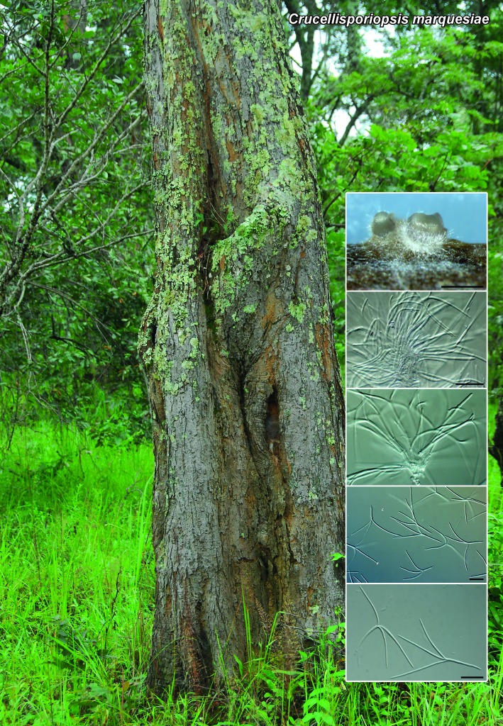
Foliicolous. Conidiomata stromatic, scattered to gregarious, erumpent, erect, acervuloid to cup-shaped, up to 400 μm diam; basal stroma up to 100 μm deep, consisting of textura angularis, hyaline, thick-walled; excipulum of textura prismatica and textura intricata; cavity surrounded by sterile hyphae, hyaline, 3–6-septate, with obtuse ends, up to 150 μm long, 2–2.5 μm diam. Conidiophores arising from conidiomatal cavity, septate, branched, hyaline, thin- and smooth-walled, branches fertile or ending in obtusely rounded, sterile setae, 10–50 × 2–2.5 μm. Conidiogenous cells integrated or discrete, subcylindrical, hyaline, smooth, 8–15 × 2–2.5 μm, with mucoid layer; proliferating inconspicuous percurrently at apex. Conidia tetra-radiate, main axis cylindrical, 0–1-septate, cells unequal, base narrow, truncate with marginal frill, hyaline, smooth, 15–20 × 2–2.5 μm, with tubular, unbranched central appendage, 1–3.5 μm long; arms 3(–4), at different apical loci on main axis, separated by septa, attenuated, septate, hyaline, smooth, not constricted at septa, (15–)30–40(–55) × 1.5 μm.

Culture characteristics — Colonies reaching 12 mm diam after 2 wk at 25 °C in the dark, erumpent, with moderate aerial mycelium and even, lobed margin. On MEA, PDA and OA surface dirty white to buff, reverse luteous with patches of buff.

Typus. ZAMBIA, OM 4142, -11.81730 24.36443, on twigs of Marquesia acuminata (Dipterocarpaceae), 24 Feb. 2013, M. van der Bank (holotype CBS H-21977, culture ex-type CPC 22539 = CBS 138895; ITS sequence GenBank KP004443, LSU sequence GenBank KP004471, MycoBank MB810587).

Notes — The genus Crucellisporiopsis was treated by Nag Raj (1993), who accepted three species. The genus is characterised by having stromatic, acervuloid conidiomata, hyaline structures with conidiogenous cells giving rise to conidia via inconspicuous percurrent proliferation, and conidia with a sub-cylindrical central axis with basal appendage, and 4–5 radiate, septate arms. Crucellisporiopsis marquesiae can be distinguished from all three species based on its conidia having a basal appendage, and the dimensions of its central axis, and lateral, 3(–4) radiating arms.

ITS. Based on a megablast search of NCBIs GenBank nucleotide database, the closest hits using the ITS sequence are Crucellisporium umtamvunae (GenBank GU291797; Identities = 546/560 (98 %), Gaps = 1/560 (0 %)), Lachnum varians (GenBank AB481267; Identities = 465/511 (91 %), Gaps = 8/511 (1 %)) and Lachnellula tricolor (GenBank KC464643; Identities = 488/541 (90 %), Gaps = 8/541 (1 %)).

LSU. Based on a megablast search of NCBIs GenBank nucleotide database, the closest hits using the LSU sequence are Lachnellula suecica (GenBank KC492980; Identities = 788/809 (97 %), no gaps), Lachnellula flavovirens (GenBank KC492975; Identities = 788/809 (97 %), no gaps) and Lachnum cf. bicolor (GenBank AY544674; Identities = 788/809 (97 %), no gaps).

Colour illustrations. Marquesia acuminata in Zambia; conidiomata, conidiogenous cells and conidia. Scale bars: conidiomata = 400 μm, all others = 10 μm.

graphic file with name per-33-212-g001.jpg

Crucellisporiopsis marquesiae

Persoonia. 2014 Nov 24;33:216–217.

Fungal Planet 282 – 24 November 2014

Alanphillipsia aloeicola Crous, sp. nov.

Pedro W Crous 1, Johannes Z Groenewald 1, Michael J Wingfield 2

Etymology. Name reflects the host genus Aloe, from which the species was isolated.

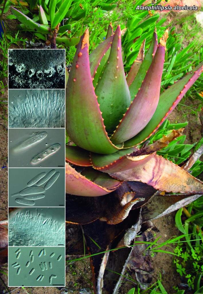
Conidiomata pycnidial, erumpent, brown, subglobose, up to 350 μm diam with central ostiole; wall of 6–8 layers of thick-walled, brown textura angularis. Conidiophores reduced to conidiogenous cells. Conidiogenous cells lining the inner cavity, hyaline, smooth, ampulliform, 15–25 × 4–6 μm, proliferating several times percurrently at apex. Paraphyses intermingled among conidiogenous cells, hyaline, smooth, subcylindrical, unbranched, septate, up to 80 μm long, 4–6 μm diam. Conidia solitary, thick-walled, guttulate, initially hyaline, becoming pale brown, finely verruculose, with longitudinal striations (when mature) along the length of its body, (25–)30–35(–42) × (10–)12–14(–17) μm, clavate to subcylindrical, apex obtuse, base truncate, 4–6 μm diam, with marginal frill up to 2 μm long. Spermatial state developing in same conidioma. Spermatophores tightly aggregated, hyaline, smooth, branched, subcylindrical, 15–25 × 3–4 μm. Spermatogenous cells terminal, subcylindrical, 8–12 × 2–3 μm. Spermatia hyaline, smooth, subcylindrical, 3–6 × 2 μm.

Culture characteristics — Colonies reaching 40 mm diam after 2 wk at 25 °C in the dark. On MEA flat, spreading with sparse aerial mycelium and lobed, feathery margins; surface olivaceous-grey in centre, outer region dirty white. On OA and PDA olivaceous-grey with a dirty white outer region.

Typus. SOUTH AFRICA, Western Cape province, Clanwilliam, Ramskop, on Aloe sp. (Aloaceae), Sept. 2013, M.J. Wingfield (holotype CBS H-21978, culture ex-type CPC 23674 = CBS 138896; ITS sequence GenBank KP004444, LSU sequence GenBank KP004472, MycoBank MB810590).

Notes — The genus Alanphillipsia (Botryosphaeriaceae, see Phillips et al. 2013) was recently introduced to accommodate four species that are aplosporella-like in morphology, but have conidia with a hyaline outer layer. Of the three species known from Aloe, A. aloeicola is most similar to A. aloetica in morphology, but distinct in that conidia of A. aloeicola (25–)30–35(–42) × (10–)12–14(–17) μm are wider than those of A. aloetica (20–)30–33(–35) × (5–)6(–7) μm (Crous et al. 2013).

ITS. Based on a megablast search of NCBIs GenBank nucleotide database, the closest hits using the ITS sequence are Alanphillipsia aloetica (GenBank KF777139; Identities = 568/571 (99 %), Gaps = 2/571 (0 %)), Alanphillipsia aloeigena (GenBank KF777137; Identities = 564/571 (99 %), Gaps = 4/571 (0 %)) and Alanphillipsia aloes (GenBank KF777138; Identities = 547/566 (97 %), Gaps = 9/566 (1 %)).

LSU. Based on a megablast search of NCBIs GenBank nucleotide database, the closest hits using the LSU sequence are Alanphillipsia aloetica (GenBank KF777195; Identities = 811/812 (99 %), no gaps), Alanphillipsia aloeigena (GenBank KF777193; Identities = 783/784 (99 %), no gaps) and Alanphillipsia aloes (GenBank KF777194; Identities = 810/812 (99 %), no gaps).

Colour illustrations. Aloe sp. in Clanwilliam; conidiogenous cells with conidia, spermatophores and spermatia. Scale bars = 10 μm.

graphic file with name per-33-212-g002.jpg

Alanphillipsia aloeicola

Persoonia. 2014 Nov 24;33:218–219.

Fungal Planet 283 – 24 November 2014

Diaporthe phragmitis Crous, sp. nov.

Pedro W Crous 1, Johannes Z Groenewald 1, Ying Zhang 2

Etymology. Name reflects the host genus Phragmites, from which the species was isolated.

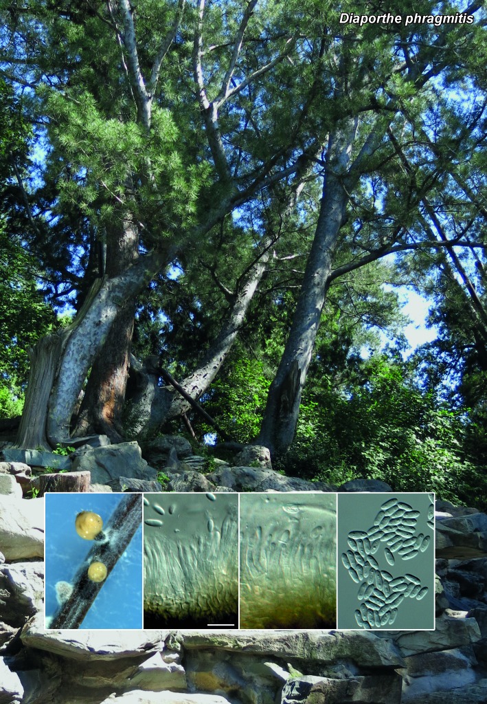
Sporulating on PNA. Conidiomata pycnidial, globose, up to 250 μm diam, black, erumpent, exuding creamy conidial droplets from central ostioles; walls consisting of 3–6 layers of medium brown textura angularis. Conidiophores hyaline, smooth, 1–3-septate, rarely branched, densely aggregated, cylindrical, straight to sinuous, 20–30 × 3–4 μm. Conidiogenous cells 10–17 × 2–2.5 μm, phialidic, cylindrical, terminal and intercalary, with slight apical taper, 1–1.5 μm diam, with visible periclinal thickening; collarette prominently flared, up to 3 μm long. Paraphyses not observed. Alpha conidia aseptate, hyaline, smooth, multi- or bi-guttulate, fusoid to ellipsoid, tapering towards both ends, straight apex subobtuse, base subtruncate, (6–)7–8(–8.5) × (2–)2.5(–3) μm. Gamma and beta conidia not observed.

Culture characteristics — Colonies covering dish after 2 wk at 25 °C in the dark. On MEA flat, spreading with moderate aerial mycelium and lobed, feathery margins; surface dirty white, reverse apricot. On OA and PDA dirty white.

Typus. CHINA, Beijing, Fragrant Hill, N39°59’18.4" E116°11’25", on Phragmites australis (Poaceae), 31 Aug. 2013, P.W. Crous & Y. Zhang (holotype CBS H-21979, culture ex-type CPC 23607 = CBS 138897; ITS sequence GenBank KP004445, LSU sequence GenBank KP004473, HIS sequence GenBank KP004503, TUB sequence GenBank KP004507, MycoBank MB810588).

Notes — Diaporthe phragmitis was isolated as endophyte from leaves of Phragmites australis. Phylogenetically, it is similar to species such as P. cotoneastri, P. juglandica and P. vaccinii based on DNA sequence data of the ITS gene, but can be distinguished from these taxa based on other loci (Lombard et al. 2014).

ITS. Based on a megablast search of NCBIs GenBank nucleotide database, the closest hits using the ITS sequence are Phomopsis vaccinii (GenBank KJ739481; Identities = 561/567 (99 %), no gaps), Phomopsis juglandina (GenBank KC242236; Identities = 530/536 (99 %), Gaps = 1/536 (0 %)) and Diaporthe cotoneastri (GenBank KJ609015; Identities = 564/572 (99 %), Gaps = 2/572 (0 %)).

LSU. Based on a megablast search of NCBIs GenBank nucleotide database, the closest hits using the LSU sequence are Diaporthe eres (GenBank AF362565; Identities = 794/794 (100 %), no gaps), Diaporthe maytenicola (GenBank KF777210; Identities = 793/794 (99 %), no gaps) and Phomopsis vaccinii (GenBank AF439630; Identities = 793/794 (99 %), no gaps).

HIS. Closest hits using the HIS sequence had highest similarity to numerous sequences of Diaporthe eres (e.g. GenBank KJ420886; Identities = 319/319 (100 %), no gaps), as well as hits with Diaporthe cf. nobilis (GenBank KC343635; Identities = 319/319 (100 %), no gaps) and Diaporthe nitschkei (GenBank KJ420875; Identities = 317/319 (99 %), no gaps).

TUB. Based on a megablast search of NCBIs GenBank nucleotide database, the closest hits using the TUB sequence are Diaporthe ‘sp. YY-2013’ (an unpublished species from jujube in China; GenBank KF600610; Identities = 773/785 (98 %), Gaps = 1/785 (0 %)), Diaporthe cf. nobilis (GenBank KC344115; Identities = 692/697 (99 %), no gaps) and Diaporthe bicincta (GenBank KC344102; Identities = 688/697 (99 %), no gaps).

Colour illustrations. Fragrant Hill, Beijing; conidiomata, conidiophores and conidia. Scale bar = 10 μm.

graphic file with name per-33-212-g003.jpg

Diaporthe phragmitis

Persoonia. 2014 Nov 24;33:220–221.

Fungal Planet 284 – 24 November 2014

Dinemasporium ipomoeae Crous, sp. nov.

Pedro W Crous 1, Johannes Z Groenewald 1, Ulrike Damm 2

Etymology. Name reflects the host genus Ipomoea, from which the species was isolated.

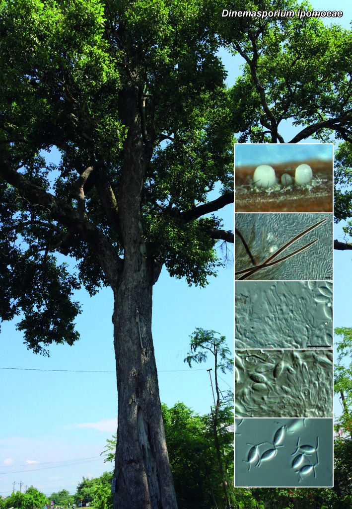
Conidiomata stromatic, scattered or aggregated, superficial, pale brown, cupulate, unilocular, globose, up to 250 μm diam, setose with a central crystalline conidial mass on PNA; basal stroma of textura angularis, layer 20–30 μm thick. Setae of two types. Type A brown to black, simple, subulate with acute apex, unbranched, smooth, thick-walled, up to 6-septate, 50–200 × 5–8 μm, 1 μm wide at acute apex, arising from basal stroma or lateral from excipulum. Type B setate pale brown, flexuous, septate, up to 100 μm long, 1.5–2 μm diam. Conidiophores lining the basal stroma, 1–2-septate, sparingly branched, cylindrical, thin-walled, smooth, base pale brown, apex hyaline, 15–20 × 2–3 μm. Conidiogenous cells determinate, phialidic with periclinal thickening, hyaline, smooth, subcylindrical, 8–12 × 2–2.5 μm. Conidia hyaline, aseptate, thin-walled, smooth, fusoid-ellipsoid, straight, ends acutely rounded, guttulate, (7–)8(–9) × (2.5–)3(–3.5) μm, with three, unbranched, flexuous, centric, tubular appendages at each end, 3–5 μm.

Culture characteristics — Colonies after 2 wk at 25 °C in the dark spreading, flat, with sparse to moderate aerial mycelium and feathery margins. On MEA surface white, reverse white to ochreous. On OA buff. On PDA surface dirty white, reverse buff.

Typus. VIETNAM, Can Dao Islands, Con Son, sea shore, on leaves of Ipomoea pes-caprae (Convolvulaceae), 12 Dec. 2012, U. Damm (holotype CBS H-21980, culture ex-type CPC 21885 = CBS 138898; ITS sequence GenBank KP004446, LSU sequence GenBank KP004474, MycoBank MB810589).

Notes — The genus Dinemasporium and allied genera were recently treated in separate studies (Crous et al. 2012b, 2014, Hashimoto et al. 2014), in which Diarimella and Stauronema were reduced to synonymy under Dinemasporium. In conidiomata of Dinemasporium ipomoeae, dehiscence by a longitudinal raphe was not seen, but the conidial appendages and two types of setae suggest that this is a member of the genus Diarimella sensu Sutton (1980). This adds further support to reduce Diarimella to synonymy with Dinemasporium (Hashimoto et al. 2014). Dinemasporium ipomoeae is phylogenetically distinct from other members.

ITS. Based on a megablast search of NCBIs GenBank nucleotide database, the closest hits using the ITS sequence are Dinemasporium polygonum (GenBank JQ889276; Identities = 428/445 (96 %), Gaps = 8/445 (1 %)), Dinemasporium americana (GenBank JQ889274; Identities = 474/509 (93 %), Gaps = 13/509 (2 %)) and Dinemasporium strigosum (GenBank JQ889283; Identities = 521/560 (93 %), Gaps = 16/560 (2 %)).

LSU. Based on a megablast search of NCBIs GenBank nucleotide database, the closest hits using the LSU sequence are Dinemasporium polygonum (GenBank JQ889292; Identities = 788/793 (99 %), no gaps), Dinemasporium morbidum (GenBank JQ889297; Identities = 786/793 (99 %), no gaps) and Dinemasporium pseudostrigosum (GenBank JQ889295; Identities = 786/793 (99 %), no gaps).

Colour illustrations. Scenic tree from Can Dao Islands, Vietnam; conidiomata on CLA, setae, conidiogenous cells and conidia. Scale bars = 10 μm.

graphic file with name per-33-212-g004.jpg

Dinemasporium ipomoeae

Persoonia. 2014 Nov 24;33:222–223.

Fungal Planet 285 – 24 November 2014

Phyllosticta mimusopisicola Crous & W.J. Swart, sp. nov.

Pedro W Crous 1, Johannes Z Groenewald 1, Wijnand J Swart 2

Etymology. Name reflects the host genus Mimusops, from which the species was isolated.

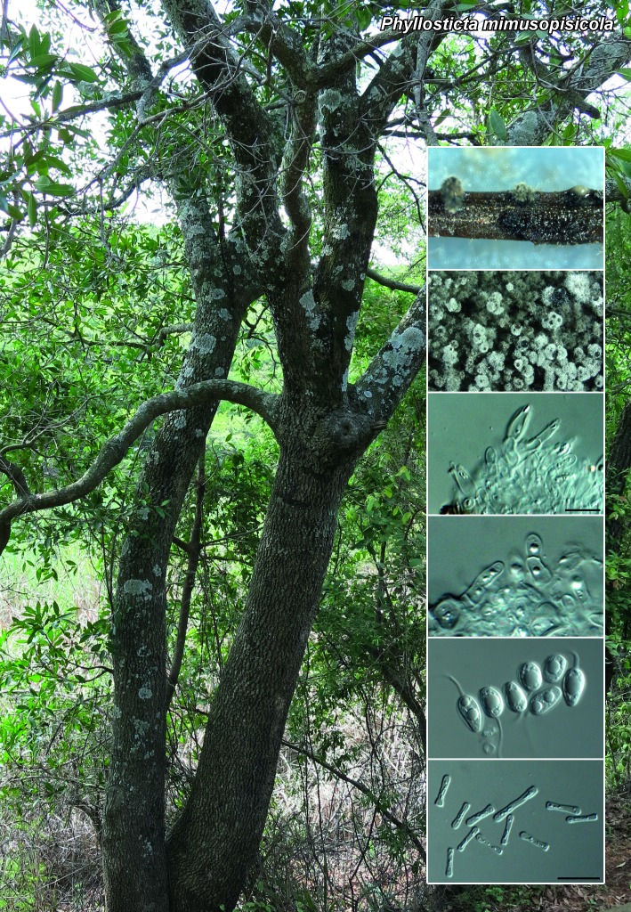
Leaf spots brown, amphigenous, subcircular, associated with leaf margins, up to 2 cm diam. Conidiomata pycnidial, solitary, black, erumpent, globose, exuding colourless to opaque conidial masses; pycnidia up to 150 μm diam; pycnidial wall of several layers of textura angularis, up to 30 μm thick; inner wall of hyaline textura angularis. Ostiole central, up to 10–20 μm diam. Conidiophores subcylindrical to ampulliform, reduced to conidiogenous cells, or with 1–2 supporting cells, at times branched at base, 20–30 × 5–7 μm. Conidiogenous cells terminal, subcylindrical, hyaline, smooth, coated in a mucoid layer, 7–15 × 2.5–3 μm; proliferating several times percurrently near apex. Conidia 10–11(–12) × (5.5–)6–6.5(–7) μm, solitary, hyaline, aseptate, thin- and smooth-walled, coarsely guttulate, or with a single large central guttule, ellipsoid to obovoid, tapering towards a narrow truncate base, 2.5–3 μm diam, enclosed in a thin, persistent mucoid sheath, 1–2 μm thick and bearing a hyaline, apical mucoid appendage, (8–)17–25(–35) × 1.5(–2) μm, flexible, unbranched, tapering towards an acute tip. Spermatogonia resembling conidiomata. Spermatia hyaline, smooth, subcylindrical with obtuse apex and truncate base, 7–15 × 1.5–2 μm.

Culture characteristics — Colonies flat, spreading with sparse aerial mycelium, and feathery, lobate margins. On PDA surface greenish black, reverse iron-grey. On OA surface iron-grey. On MEA surface olivaceous-grey in centre, pale olivaceous-grey in outer region, olivaceous-grey underneath.

Typus. SOUTH AFRICA, Limpopo province, Klein Kariba ATKV resort, S24°50’11.6" E28°19’55.6", on leaves of Mimusops zeyheri (Sapotaceae), 22 Jan. 2013, P.W. Crous & W.J. Swart (holotype CBS H-21981, culture ex-type CPC 22063 = CBS 138899; ITS sequence GenBank KP004447, LSU sequence GenBank KP004475, MycoBank MB810591).

Notes — Several species of Phyllosticta have been described from Mimusops, namely P. mimusopsidis Henn., which turned out to be a species of Phomopsis, P. mimusopsidis Cufino, which appears to be a species of Phoma, along with P. mimusopsidis-elengi (van der Aa & Vanev 2002). As far as we are aware, Phyllosticta mimusopisicola is thus the first true species of Phyllosticta reported from Mimusops.

In a recent phylogenetic re-evaluation of the genus Phyllosticta (Wikee et al. 2013), two nomenclatural errors were made that need to be corrected, namely P. rubra Berl. & Voglino (1886) was added to the MycoBank repository after the deposit of P. rubra Wikee & Crous (2013), rendering the latter invalid, while the name P. mangifera-indica Wikee, Crous, K.D. Hyde & McKenzie was never deposited in MycoBank.

Phyllosticta rubella Wikee & Crous, nom. nov. — MycoBank MB810592

Phyllosticta rubra Wikee & Crous, Stud. Mycol. 76: 25. 2013 (nom. illegit., Art. 53.1), non P. rubra Berl. & Voglino (1886).

Description and illustration: Wikee et al. (2013).

Specimen examined. USA, Missouri, on Acer rubrum, July 1999, G. Carroll (holotype CBS H-21398, culture ex-type CBS 111635).

Phyllosticta mangiferae-indicae Wikee, Crous, K.D. Hyde & McKenzie, sp. nov. –– MycoBank MB810593

Phyllosticta mangifera-indica Wikee, Crous, K.D. Hyde & McKenzie, Stud. Mycol. 76: 18. 2013 (nom. illegit., Art. 42.1).

Description and illustration: Wikee et al. (2013).

Specimen examined. THAILAND, Chiangrai, Nanglae, on healthy leaf of Mangifera indica, July 2011, S. Wikee (holotype MFU13-0108; ex-type culture CPC 20274 = MFLUCC10-0029 = CBS 136061).

ITS. Based on a megablast search of NCBIs GenBank nucleotide database, the closest hits using the ITS sequence are Phyllosticta podocarpicola (GenBank KF206173; Identities = 389/409 (95 %), Gaps = 7/409 (1 %)), Phyllosticta cornicola (GenBank KF170307; Identities = 384/409 (94 %), Gaps = 9/409 (2 %)) and Phyllosticta minima (GenBank KF766216; Identities = 384/409 (94 %), Gaps = 6/409 (1 %)).

LSU. Based on a megablast search of NCBIs GenBank nucleotide database, the closest hits using the LSU sequence are Phyllosticta philoprina (GenBank KF766342; Identities = 762/773 (99 %), no gaps), Guignardia rhodorae (GenBank KF206292; Identities = 745/756 (99 %), no gaps) and Phyllosticta foliorum (GenBank KF206287; Identities = 745/756 (99 %), no gaps).

Colour illustrations. Mimusops zeyheri at the Klein Kariba ATKV resort; conidiomata, conidiophores, conidia and spermatia. Scale bars = 10 μm.

graphic file with name per-33-212-g005.jpg

Phyllosticta mimusopisicola

Persoonia. 2014 Nov 24;33:224–225.

Fungal Planet 286 – 24 November 2014

Rachicladosporium eucalypti Crous, sp. nov.

Pedro W Crous 1, Johannes Z Groenewald 1, Addisu Assefa 1

Etymology. Name reflects the host genus Eucalyptus, from which the species was isolated.

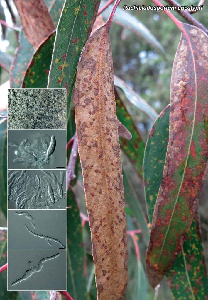
Leaf spots brown, amphigenous, subcircular to irregular, up to 15 mm diam. Colonies homothallic, sporulating on OA. Ascomata pseudothecial, erumpent, up to 90 μm diam, with central ostiole; wall of 3–6 layers of brown textura angularis. Asci fasciculate, bitunicate, subsessile, hyaline, smooth, 8-spored, narrowly obovoid, with minute apical chamber, 1 μm diam, 23–40 × 7–12 μm. Pseudoparaphyses absent. Ascospores hyaline, smooth, guttulate, fusoid-ellipsoid, widest in middle of apical cell, tapering towards both ends, constricted at median septum, (10–)11–12 × 3(–3.5) μm; ascospores germinating from both ends, frequently with lateral branches, ascospores becoming distorted, 6–8 μm diam, brown and verruculose.

Culture characteristics — Colonies reaching 12 mm diam after 2 wk at 25 °C in the dark, spreading with moderate aerial mycelium, and even, smooth margins. On MEA surface olivaceous-grey, reverse iron-grey. On PDA surface smoke grey, reverse olivaceous-grey. On OA surface olivaceous-grey.

Typus. ETHIOPIA, Addis Ababa, Addis Ababa Botanical Garden, N09°05’ 16.2" E38°43’4.7", on leaves of Eucalyptus globulus (Myrtaceae), 24 June 2013, P.W. Crous & A. Assefa (holotype CBS H-21982, culture ex-type CPC 23241 = CBS 138900; ITS sequence GenBank KP004448, LSU sequence GenBank KP004476, MycoBank MB810594).

Notes — The genus Rachicladosporium was established for taxa associated with leaf spots that are cladosporium-like in morphology, but distinct in that they have conidiophores with an apical rachis, and conidia that are pigmented, occur in chains and have slightly thickened hila (Crous et al. 2007b). Rachicladosporium eucalypti is the first species in the genus with a known sexual morph, which is mycosphaerella-like in morphology.

ITS. Based on a megablast search of NCBIs GenBank nucleotide database, the closest hits using the ITS sequence are Rachicladosporium alpinum (GenBank KF309941; Identities = 451/464 (97 %), Gaps = 4/464 (0 %)), Rachicladosporium inconspicuum (GenBank KF309939; Identities = 451/464 (97 %), Gaps = 4/464 (0 %)) and Rachicladosporium pini (GenBank JF951145; Identities = 564/584 (97 %), Gaps = 3/584 (0 %)).

LSU. Based on a megablast search of NCBIs GenBank nucleotide database, the closest hits using the LSU sequence are Rachicladosporium alpinum (GenBank KF309988; Identities = 705/707 (99 %), no gaps), Rachicladosporium pini (GenBank JF951165; Identities = 756/759 (99 %), no gaps) and Rachicladosporium luculiae (GenBank EU040237; Identities = 756/759 (99 %), no gaps).

Colour illustrations. Eucalyptus globules leaves at the Addis Ababa Botanical Garden, Ethiopia; ascomata, asci and germinating ascospores. Scale bars = 10 μm.

graphic file with name per-33-212-g006.jpg

Rachicladosporium eucalypti

Persoonia. 2014 Nov 24;33:226–227.

Fungal Planet 287 – 24 November 2014

Arxiella dolichandrae Crous, sp. nov.

Pedro W Crous 1, Johannes Z Groenewald 1, Alana den Breeÿen 1

Etymology. Name reflects the host genus Dolichandra, from which the species was isolated.

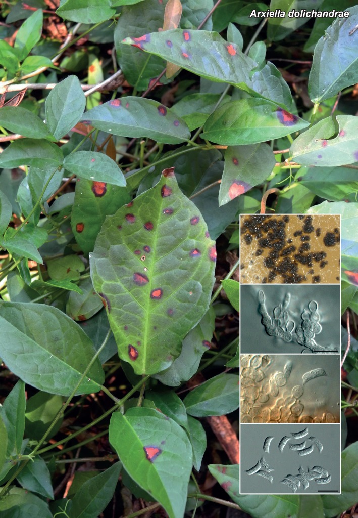
Conidiomata sporodochial, forming loose, brown, superficial sporodochia on agar surface, up to 300 μm diam, consisting of brown textura angularis to textura globulosa, that become fertile at the edges. Conidiogenous cells smooth, brown, globose to somewhat elongated, 4–6 μm diam, phialidic, with minute periclinal thickening. Conidia solitary, hyaline, smooth, guttulate, reniform, medianly 1-septate, inner plane with apical and basal horn-like appendages following curvature of inner plane, 2–3 μm long; conidia (incl. appendages) 10–11 × 2.5–3 μm, with a slightly raised hilum (0.5 μm diam) at the base where the basal appendage joins the conidium body.

Culture characteristics — Colonies flat, appressed, spreading with sparse aerial mycelium, surface folded with smooth, lobate margin, reaching 3 cm diam after 2 wk at 25 °C in the dark. On MEA surface dirty white, reverse ochreous. On PDA surface dirty white, reverse pale luteous. On OA surface pale luteous.

Typus. SOUTH AFRICA, KwaZulu-Natal, Pietermaritzburg, S29°37’50.95" E30°25’51.67", on leaves of Dolichandra unguiscati (Bignoniaceae), 24 May 2013, A. King (holotype CBS H-21983, culture ex-type CPC 22951 = CBS 138853; ITS sequence GenBank KP004449, LSU sequence GenBank KP004477, MycoBank MB810595).

Notes — The genus Arxiella was established for a fungus collected from leaf litter and soil under Acacia karroo in South Africa (Papendorf 1967) and presently includes two species. Arxiella dolichandrae is distinct from these species by its conidial dimensions (A. terrestris, 6–16 × 3–4.5 μm; A. lunata, 10–17 × 3–4 μm) (Papendorf 1967, Ruscoe 1970).

ITS. Based on a megablast search of NCBIs GenBank nucleotide database, the closest hits using the ITS sequence are Mycoleptodiscus terrestris (GenBank JN711860; Identities = 363/420 (86 %), Gaps = 17/420 (4 %)), Polychaeton citri (GenBank GU214649; Identities = 445/538 (83 %), Gaps = 22/538 (4 %)) and Leptoxyphium madagascariense (GenBank GQ303277; Identities = 409/501 (82 %), Gaps = 28/501 (5 %)).

LSU. Based on a megablast search of NCBIs GenBank nucleotide database, the closest hits using the LSU sequence are Chlamydotubeufia huaikangplaensis (GenBank JN865198; Identities = 722/809 (89 %), Gaps = 5/809 (0 %)), Hysterium vermiforme (GenBank GQ221897; Identities = 719/810 (89 %), Gaps = 6/810 (0 %)) and Chlamydotubeufia khunkornensis (GenBank JN865190; Identities = 720/813 (89 %), Gaps = 11/813 (1 %)).

Colour illustrations. Symptomatic leaves of Dolichandra unguiscati; colonies on OA, conidiogenous cells and conidia. Scale bars = 10 μm.

graphic file with name per-33-212-g007.jpg

Arxiella dolichandrae

Persoonia. 2014 Nov 24;33:228–229.

Fungal Planet 288 – 24 November 2014

Corynespora endiandrae Crous & Summerell, sp. nov.

Pedro W Crous 1, Johannes Z Groenewald 1, Brett A Summerell 2

Etymology. Name reflects the host genus Endiandra, from which the species was isolated.

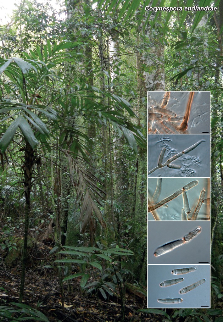
Mycelium consisting of hyaline, smooth, branched, septate, 3–4 μm diam hyphae. Conidiophores solitary, erect, straight to flexuous, subcylindrical, unbranched, brown, thick-walled, finely roughened, base bulbous, lacking rhizoids, 10–12 μm diam, stipe 200–300 × 5–7 μm, 8–16-septate. Conidiogenous cells integrated, terminal and lateral, monotretic, subcylindrical, brown, finely roughened, slightly darkened, 2 μm diam. Conidia obclavate, solitary or in short chains (2–3), thick-walled, brown, finely roughened, 3(–4) distoseptate, (35–)37–45(–57) × (7–)8(–9) μm; hila darkened, thickened, 2.5–3.5 μm diam.

Culture characteristics — Colonies reaching 20 mm diam after 2 wk at 25 °C in the dark, with moderate aerial mycelium and smooth, even margins. On MEA, PDA and OA surface and reverse dirty white.

Typus. AUSTRALIA, New South Wales, Nightcap National Park, S28.33.918 E153.20.228, on leaves of Endiandra introrsa (Lauraceae), 9 Mar. 2013, B.A. Summerell (holotype CBS H-21984, culture ex-type CPC 22194 = CBS 138902; ITS sequence GenBank KP004450, LSU sequence GenBank KP004478, MycoBank MB810596).

Notes — Species of Corynespora are commonly associated with leaf spots as necrotrophic pathogens. Species have mainly been described based on host association, and the genus is in need of revision. No species have thus far been recorded on Endiandra, and based on the key provided by Siboe et al. (1999), C. endiandrae appears to represent a novel taxon.

ITS. Based on a megablast search of NCBIs GenBank nucleotide database, the closest hits using the ITS sequence are Helminthosporium velutinum (GenBank JN198435; Identities = 453/505 (90 %), Gaps = 9/505 (1 %)), Helminthosporium solani (GenBank KC106739; Identities = 501/560 (89 %), Gaps = 13/560 (2 %)) and Helminthosporium chlorophorae (GenBank AF120259; Identities = 422/475 (89 %), Gaps = 16/475 (3 %)).

LSU. Based on a megablast search of NCBIs GenBank nucleotide database, the closest hits using the LSU sequence are Corynespora leucadendri (GenBank KF251654; Identities = 806/819 (98 %), no gaps), Corynespora olivacea (GenBank JQ044448; Identities = 806/820 (98 %), Gaps = 1/820 (0 %)) and Byssothecium circinans (GenBank GU205217; Identities = 802/819 (98 %), no gaps).

Colour illustrations. Nightcap National Park, Australia; conidiophores and conidia. Scale bars = 10 μm.

graphic file with name per-33-212-g008.jpg

Corynespora endiandrae

Persoonia. 2014 Nov 24;33:230–231.

Fungal Planet 289 – 24 November 2014

Gonatophragmium triuniae Crous & Summerell, sp. nov.

Pedro W Crous 1, Johannes Z Groenewald 1, Brett A Summerell 2

Etymology. Name reflects the host genus Triunia, from which the species was isolated.

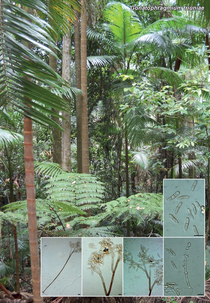
Mycelium consisting of hyaline, septate, branched, 2–3 μm diam hyphae. Conidiophores solitary, macronematous, erect, arising from superficial hyphae, straight to flexuous, T-cell at base slightly swollen (up to 7 μm diam) or not, stipe 200–280 μm long, 4–5 μm diam at the base, 4–7-septate, brown, smooth, thin-walled, branched in upper part. Primary branches pale brown, verruculose, subcylindrical, aseptate, 25–35 × 3–4 μm, giving rise to 1–2 secondary branches, pale brown, subcylindrical, aseptate, 15–20 × 3–4 μm. Secondary branches giving rise to a conidiogenous region consisting of 3–4 subcylindrical cells, pale brown, finely verruculose to smooth, each cell with an upper fertile region consisting of aggregated denticulate loci, 0.5 μm long, 1 μm diam, darkened and thickened; at times cells also have a fertile lateral branch, 13–20 × 3–3.5 μm. Conidia solitary, clavate, pale brown, guttulate, roughened, apex obtuse, lower part attenuating towards truncate base, 1 μm diam; conidia 1-septate, slightly constricted at septum, straight to slightly curved, apical cell 5–6 μm long, basal cell 7–8 μm long, conidia (10–)12–14(–15) × (3.5–)4(–4.5) μm (apical cell rarely developing a second septum); hila 0.5–1 μm diam, somewhat darkened and thickened.

Culture characteristics — Colonies reaching 15 mm diam after 2 wk at 25 °C in the dark, with moderate aerial mycelium and smooth, even margins. On MEA surface ochreous, reverse umber. On PDA surface luteous to buff, with diffuse, luteous pigment, but umber in reverse. On OA surface dirty white with diffuse buff pigment.

Typus. AUSTRALIA, New South Wales, Nightcap National Park, S28.38.413 E153.20.179, on leaves of Triunia youngiana (Proteaceae), 9 Mar. 2013, B.A. Summerell (holotype CBS H-21985, culture ex-type CPC 22191, 22192 = CBS 138901; ITS sequence of CPC 22191, GenBank KP004451, LSU sequence GenBank KP004479, MycoBank MB810597).

Notes — Species of Gonatophragmium are commonly associated with leaf spots on a wide range of hosts (Ellis 1971, 1976, Braun & Hill 2008). Of the approximately 15 species presently known to occur in the genus, G. triuniae is easily distinguished based on its small, 1-septate conidia. It is also the only species thus far reported from Triunia.

ITS. Based on a megablast search of NCBIs GenBank nucleotide database, the closest hits using the ITS sequence are Arthothelium spectabile (GenBank AF138814; Identities = 446/469 (95 %), Gaps = 9/469 (1 %)), Phaeodactylium stadleri (GenBank HF678526; Identities = 317/369 (86 %), Gaps = 8/369 (2 %)) and Radulidium subulatum (GenBank EU041790; Identities = 436/544 (80 %), Gaps = 36/544 (6 %)).

LSU. Based on a megablast search of NCBIs GenBank nucleotide database, the closest hits using the LSU sequence are Acrospermum adeanum (GenBank EU940104; Identities = 758/800 (95 %), no gaps), Pseudovirgaria grisea (GenBank JF957610; Identities = 780/827 (94 %), Gaps = 2/827 (0 %)) and Pseudovirgaria hyperparasitica (GenBank EU041822; Identities = 780/827 (94 %), Gaps = 2/827 (0 %)).

Colour illustrations. Nightcap National Park, Australia; conidiophores, conidiogenous cells and conidia. Scale bars = 10 μm.

graphic file with name per-33-212-g009.jpg

Gonatophragmium triuniae

Persoonia. 2014 Nov 24;33:232–233.

Fungal Planet 290 & 291 – 24 November 2014

Phaeosphaeria podocarpi Crous & A.R. Wood, sp. nov.

Pedro W Crous 1, Johannes Z Groenewald 1, Alan R Wood 2

Etymology. Name reflects the host genus Podocarpus, from which the species was isolated.

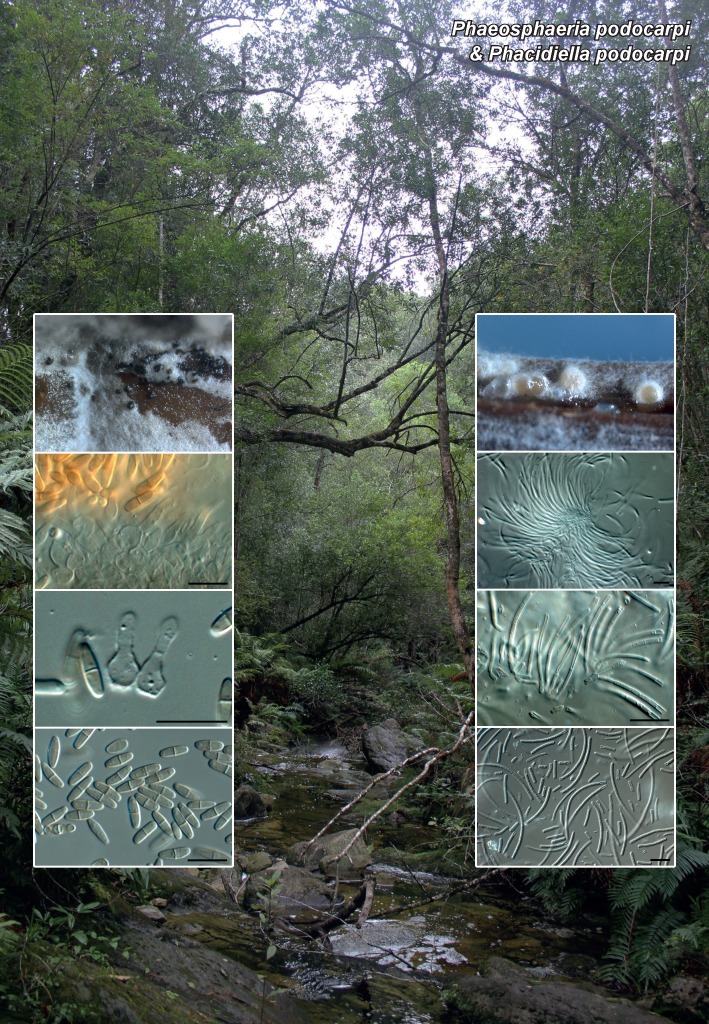
Conidiomata pycnidial, erumpent, brown, globose, solitary, up to 300 μm diam, with central ostiole; wall of 4–8 layers of brown textura angularis. Conidiophores reduced to conidiogenous cells. Conidiogenous cells lining the inner cavity, hyaline, smooth, ampulliform, 4–7 × 3–4 μm, phialidic with inconspicuous periclinal thickening at apex. Paraphyses intermingled among conidiogenous cells, subcylindrical, hyaline, 1–2-septate, up to 25 μm long, 2–3 μm diam. Conidia solitary, red-brown in mass, smooth, fusoid-ellipsoidal, apex obtuse, base truncate, 1–1.5 μm diam, medianly 1-septate, mostly straight, (7–)8–10(–12) × (2–)2.5(–3) μm.

Culture characteristics — Colonies reaching 30 mm diam after 2 wk at 25 °C in the dark, surface folded, with moderate aerial mycelium and even, lobate margins. On MEA surface dirty white, reverse apricot. On OA surface pale olivaceous-grey. On PDA surface pale olivaceous-grey, reverse olivaceous-grey.

Typus. SOUTH AFRICA, Western Cape Province, Knysna, Garden Route National Park, Velbroeksdraai picnic site, Diepwalle Forest, S33°56’ E23°09’, on leaves of Podocarpus latifolius (Podocarpaceae), 1 July 2013, A.R. Wood (holotype CBS H-21986, culture ex-type CPC 23433 = CBS 138903; ITS sequence GenBank KP004452, LSU sequence GenBank KP004480, ACT sequence GenBank KP004502, TUB sequence GenBank KP004508, MycoBank MB810598).

Notes — The genus Phaeoseptoria was shown to be synonymous with Phaeosphaeria by Quaedvlieg et al. (2013). Although most of the asexual morphs of Phaeosphaeria species have multiseptate conidia, the general morphology of P. podocarpi corresponds with other members of the genus. However, there are several unresolved lineages that are phaeosphaeria-like awaiting study. It is thus possible that P. podocarpi could still be segregated in a distinct genus based on additional collections and DNA sequence data.

ITS. Based on a megablast search of NCBIs GenBank nucleotide database, the closest hits using the ITS sequence are Ascochyta manawaorae (GenBank GU230751; Identities = 461/478 (96 %), Gaps = 3/478 (0 %)), Phaeosphaeria poagena (GenBank KJ869114; Identities = 522/542 (96 %), Gaps = 3/542 (0 %)) and Parastagonospora nodorum (GenBank KF512822; Identities = 510/532 (96 %), Gaps = 9/532 (1 %)).

LSU. Based on a megablast search of NCBIs GenBank nucleotide database, the closest hits using the LSU sequence are Phaeosphaeria oryzae (GenBank KF251689; Identities = 816/816 (100 %), no gaps), Phaeosphaeriopsis musae (GenBank DQ885894; Identities = 816/816 (100 %), no gaps) and Phaeosphaeria papayae (GenBank KF251690; Identities = 815/815 (100 %), no gaps).

Persoonia. 2014 Nov 24;33:232–233.

Fungal Planet 290 & 291 – 24 November 2014

Phacidiella podocarpi Crous & A.R. Wood, sp. nov.

Pedro W Crous 1, Johannes Z Groenewald 1, Alan R Wood 2

Etymology. Name reflects the host genus Podocarpus, from which the species was isolated.

Conidiomata pycnidial, hyaline to subhyaline on SNA, aggregated in clusters, globose, up to 350 μm diam; wall of 3–6 layers of hyaline textura intricata. Conidiophores lining the inner cavity, subcylindrical, smooth, hyaline, 1-septate, giving rise to 1–2 conidiogenous cells, 5–8 × 2–2.5 μm. Conidiogenous cells terminal, hyaline, smooth, subcylindrical to doliiform, proliferating sympodially at apex, 3–6 × 2–2.5 μm. Conidia solitary, hyaline, smooth, subcylindrical, flexuous, apex obtuse, base truncate, (45–)50–70(–90) × 2(–2.5) μm, 9–18-septate, disarticulating into phragmospores, cylindrical with truncate ends, 5–6 μm long.

Culture characteristics — Colonies reaching 10 mm diam after 2 wk at 25 °C in the dark, spreading, with fluffy aerial mycelium and feathery margin. On MEA surface and reverse dirty white, with diffuse apricot zone in agar. On OA surface apricot. On PDA surface salmon, reverse dirty white.

Typus. SOUTH AFRICA, Western Cape Province, Knysna, Garden Route National Park, Velbroeksdraai picnic site, Diepwalle Forest, S33°56’ E23°09’, on leaves of Podocarpus latifolius (Podocarpaceae), 1 July 2013, A.R. Wood (holotype CBS H-21987, culture ex-type CPC 23447 = CBS 138904; ITS sequence GenBank KP004453, LSU sequence GenBank KP004481, MycoBank MB810599).

Notes — The genus Phacidiella (1884) has been linked to Pyrenopeziza (1870) sexual morphs (Sutton 1980). However, Pyrenopeziza is also linked to Cylindrosporium (1823), while some species of Cylindrosporium are linked to Blumeriella (1961) (Johnston et al. 1914). Sutton (1980) stated that Phacidiella and its generic synonyms are in need of revision, as some have 1-septate conidia (e.g. Ramulariospora), and others are aseptate, suggesting that they may not all be congeneric. Phacidiella podocarpi is thus best described in the genus Phacidiella based on its hyaline, aseptate conidia.

ITS. Based on a megablast search of NCBIs GenBank nucleotide database, the closest hits using the ITS sequence are Glomerobolus gelineus (GenBank DQ247782; Identities = 232/248 (94 %), Gaps = 6/248 (2 %)), Caloplaca albopruinosa (GenBank EF093566; Identities = 240/260 (92 %), Gaps = 3/260 (1 %)) and Umbilicaria mammulata (GenBank DQ782851; Identities = 243/265 (92 %), Gaps = 5/265 (1 %)).

LSU. Based on a megablast search of NCBIs GenBank nucleotide database, the closest hits using the LSU sequence are Stictis radiata (GenBank AY300864; Identities = 751/783 (96 %), no gaps), Carestiella socia (GenBank AY661682; Identities = 790/827 (96 %), Gaps = 3/827 (0 %)) and Conotrema populorum (GenBank AY300833; Identities = 780/826 (94 %), Gaps = 4/826 (0 %)).

Colour illustrations. Velbroeksdraai picnic site, Diepwalle Forest, South Africa; Phaeosphaeria podocarpi (left column): colonies on OA, conidiogenous cells and conidia; Phacidiella podocarpi (right column): conidiomata on PNA, conidiogenous cells and conidia. Scale bars = 10 μm.

graphic file with name per-33-212-g010.jpg

Phaeosphaeria podocarpi & Phacidiella podocarpi

Persoonia. 2014 Nov 24;33:235.

Fungal Planet 292 – 24 November 2014

Phellinocrescentia Crous & Decock, gen. nov.

Pedro W Crous 1, Johannes Z Groenewald 1, Cony A Decock 2

Etymology. L. = crescit, growing on, referring to its ecological habit, growing on Phellinus.

Conidiomata pycnidial, globose, solitary or aggregated, uni- to multilocular, with central ostiole; wall consisting of 3–6 layers of brown textura angularis; outer surface covered in brown, warty hyphae. Conidiophores reduced to conidiogenous cells or a supporting cell. Conidiogenous cells lining the inner cavity, hyaline, smooth, tightly aggregated, subcylindrical, straight to curved, phialidic with periclinal thickening. Conidia aseptate, solitary, hyaline, smooth, guttulate, thin-walled, ellipsoid to tear-drop-shaped.

Type species. Phellinocrescentia guianensis.

MycoBank MB810600.

Persoonia. 2014 Nov 24;33:234–235.

Fungal Planet 292 – 24 November 2014

Phellinocrescentia guianensis Crous & Decock, sp. nov.

Pedro W Crous 1, Johannes Z Groenewald 1, Cony A Decock 2

Etymology. Name reflects the locality, French Guiana, where this species was collected.

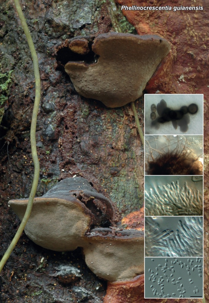
Conidiomata pycnidial, globose, solitary or aggregated, uni- to multilocular, up to 220 μm diam, with central ostiole; wall consisting of 3–6 layers of brown textura angularis, becoming hyaline towards inner centrum; outer surface covered in brown, warty hyphae, 3–4 μm diam. Conidiophores reduced to conidiogenous cells or a supporting cell. Conidiogenous cells lining the inner cavity, hyaline, smooth, tightly aggregated, subcylindrical, straight to curved, phialidic with periclinal thickening, 5–12 × 1.5 μm. Conidia aseptate, solitary, hyaline, smooth, guttulate, thin-walled, ellipsoid to tear-drop-shaped, (2.5–)3(–4) × 1.5 μm.

Culture characteristics — Colonies reaching 7 mm diam after 2 wk at 25 °C in the dark, surface folded, with sparse aerial mycelium and even, lobed margins. On MEA surface a mixture of dirty white and olivaceous-grey, reverse olivaceous-grey. On OA and PDA olivaceous-grey.

Typus. FRENCH GUIANA, on polypore No. 742 (Phellinus sp.), 12 July 2013, C. Decock (holotype CBS H-21988, culture ex-type CPC 23600 = CBS 138913; ITS sequence GenBank KP004454, LSU sequence GenBank KP004482, MycoBank MB810601).

Notes — The strain was found growing on the sporocarp of a Phellinus sp. and isolated by plating it on malt agar. Phellinocrescentia guianensis is phoma-like in morphology, but distinct in having solitary or aggregated, uni- to multilocular conidiomata, and ellipsoid to tear-drop-shaped conidia. It also has a unique ecological habit, growing on basidiocarps of a Phellinus sp. It was not possible to assign a genus to this fungus based on phylogenetic inference and the new genus, Phellinocrescentia, is introduced to accommodate it.

ITS. Based on a megablast search of NCBIs GenBank nucleotide database, the closest hits using the ITS sequence are Didymosphaeria futilis (GenBank EU552123; Identities = 470/574 (82 %), Gaps = 38/574 (6 %)), Funbolia dimorpha (GenBank JF951136; Identities = 362/424 (85 %), Gaps = 15/424 (3 %)) and Geomyces pannorum var. asperulatus (GenBank AJ938166; Identities = 329/395 (83 %), Gaps = 25/395 (6 %)).

LSU. Based on a megablast search of NCBIs GenBank nucleotide database, the closest hits using the LSU sequence are Pseudopassalora gouriqua (GenBank JN712565; Identities = 743/790 (94 %), no gaps), Heleiosa barbatula (GenBank GU479787; Identities = 735/793 (93 %), Gaps = 5/793 (0 %)) and Funbolia dimorpha (GenBank JF951156; Identities = 729/790 (92 %), Gaps = 1/790 (0 %)).

Colour illustrations. Basidiomata of Phellinus sp. on a dead standing tree in French Guiana; conidiomata in agar, warty hyphae, conidiogenous cells and conidia. Scale bars = 10 μm.

graphic file with name per-33-212-g011.jpg

Phellinocrescentia guianensis

Persoonia. 2014 Nov 24;33:236–237.

Fungal Planet 293 – 24 November 2014

Neocamarosporium chichastianum Papizadeh, Crous, Shahzadeh Fazeli & Amoozegar, sp. nov.

Pedro W Crous 1, Johannes Z Groenewald 1, Moslem Papizadeh 2, Seyed Abolhassan Shahzadeh Fazeli 2, Mohammad Ali Amoozegar 2

Etymology. Name reflects the location, Lake Urmia (formerly known as Chichast), from which the species was isolated.

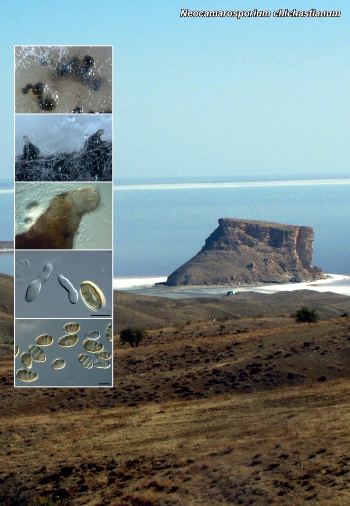
Conidiomata pycnidial, solitary, uniloculate, black, up to 200 μm diam, with 1–3 papillate necks (up to 150 μm diam), with central ostioles 5–10 μm diam. Conidiophores reduced to conidiogenous cells. Conidiogenous cells lining the inner cavity, hyaline, smooth, subcylindrical, 7–15 × 4–5 μm, proliferating percurrently at apex. Paraphyses intermingled between conidiogenous cells, hyaline, smooth, subcylindrical, 1–2-septate with obtuse ends, up to 35 μm long, 4–5 μm diam. Conidia solitary, brown, finely roughened, ellipsoid, widest in middle, apex obtuse, muriformly septate, thick-walled, base truncate, 2–3 μm diam, (11–)15–19(–22) × (6–)8–9(–11) μm; 3 transverse septa, 1–2 oblique or vertical septa.

Culture characteristics — Colonies flat, spreading with sparse aerial mycelia. On OA surface olivaceous-grey. On MEA surface pale olivaceous-grey to olivaceous-grey. Optimum growth occurred at 25 °C, but the fungus grew at 15 °C up to 35 °C. Furthermore, optimum growth was recorded at pH values between 5.5 and 6.5, although it could grow at a broad range of pH values (4–10).

Typus. IRAN, Lake Urmia, soil, 2011, M. Papizadeh & M.R. Soudi (holo-type CBS H-21989, culture ex-type IBRC-M 30126 = CBS 137502; ITS sequence GenBank KP004455, LSU sequence GenBank KP004483, MycoBank MB810602).

Notes — Neocamarosporium chichastianum clusters with N. goegapense, the type species of the genus Neocamarosporium, which is morphologically similar to the genus Camarosporium based on its pycnidial conidiomata, hyaline, percurrently proliferating conidiogenous cells, and brown, muriformly septate conidia (Crous et al. 2014). Neocamarosporium chichastianum is the second species described in this genus, and interestingly has paraphyses, which were not observed in N. goegapense.

ITS. Based on a megablast search of NCBIs GenBank nucleotide database, the closest hits using the ITS sequence are Neocamarosporium goegapense (GenBank KJ869163; Identities = 550/579 (95 %), Gaps = 5/579 (0 %)), Phoma betae (GenBank KC460811; Identities = 463/493 (94 %), Gaps = 7/493 (1 %)) and Ascochyta obiones (GenBank GU230752; Identities = 463/498 (93 %), Gaps = 4/498 (0 %)). Our ITS sequence is 98 % (517/529) identical to the sequence of Chaetosphaeronema hispidulum CBS 826.88 in Q-bank (www.q-bank.eu).

LSU. Based on a megablast search of NCBIs GenBank nucleotide database, the closest hits using the LSU sequence are Neocamarosporium goegapense (GenBank KJ869220; Identities = 804/806 (99 %), no gaps), Chaetosphaeronema hispidulum (GenBank EU754145; Identities = 848/851 (99 %), no gaps) and Coniothyrium obiones (GenBank DQ678054; Identities = 849/853 (99 %), no gaps).

Colour illustrations. Lake Urmia in Iran; conidiomata on OA and PNA, conidiomatal neck, conidiogenous cells and conidia. Scale bars = 10 μm.

graphic file with name per-33-212-g012.jpg

Neocamarosporium chichastianum

Persoonia. 2014 Nov 24;33:238–239.

Fungal Planet 294 – 24 November 2014

Sphaerulina pelargoniiCrous & M.J. Wingf., sp. nov.

Pedro W Crous 1, Johannes Z Groenewald 1, Michael J Wingfield 2

Etymology. Name reflects the host genus Pelargonium, from which this species was isolated.

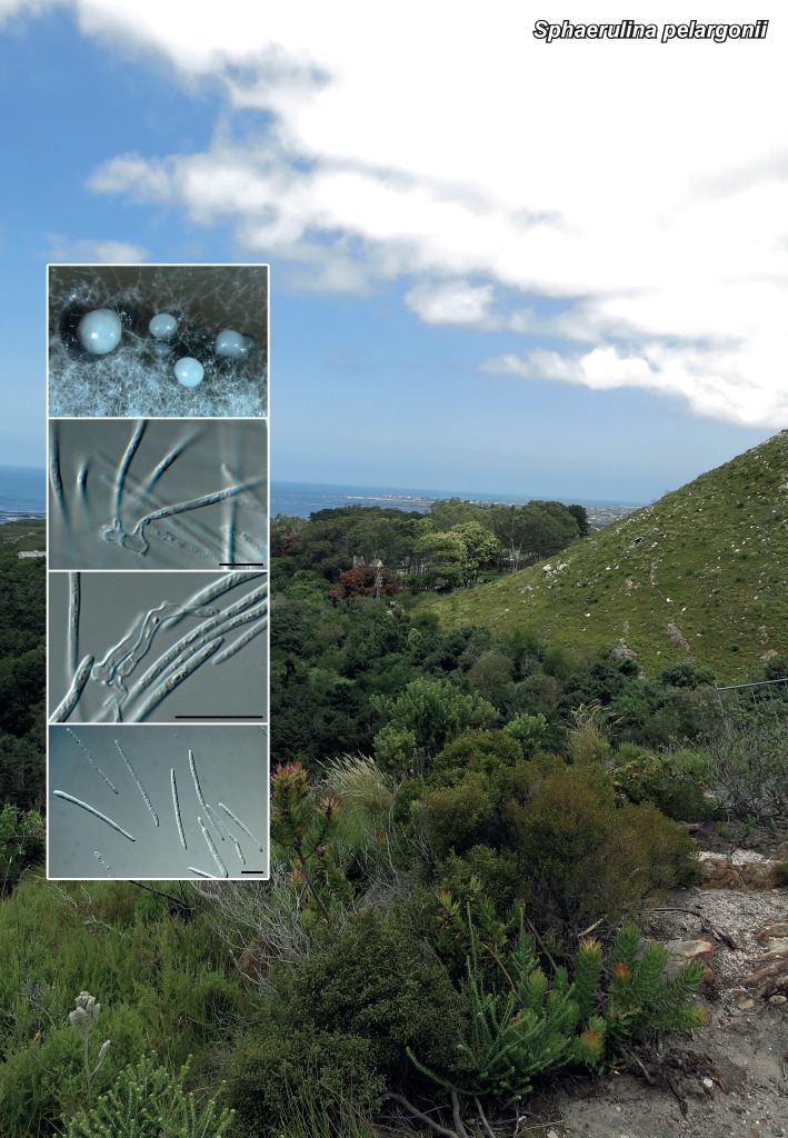
Sporulating on SNA. Conidiomata pycnidial, brown, separate, immersed to erumpent, globose, up to 150 μm diam, exuding a creamy crystalline conidial mass via a central ostiole; wall of 3–4 layers of brown textura angularis. Conidiophores reduced to conidiogenous cells. Conidiogenous cells hyaline, at times pale brown, smooth, subcylindrical, straight to geniculous-sinuous, 7–15 × 3–5 μm, proliferating sympodially. Conidia hyaline, smooth, guttulate, filiform, narrowly obclavate, apex subobtusely rounded, base long obconically truncate, (1–)3–4-septate, (15–)28–45(–60) × 1.5–2(–2.5) μm.

Culture characteristics — Colonies on PDA, MEA and OA spreading with sparse to moderate aerial mycelium, and smooth, lobate margins, reaching 20 mm diam after 2 wk at 25 °C in the dark. On MEA surface olivaceous-grey with apricot sporulation, iron-grey underneath. On PDA surface olivaceous-grey with strands of dirty white mycelium, reverse iron-grey. On OA surface dirty white.

Typus. SOUTH AFRICA, Western Cape Province, Betty’s Bay, Harold Porter National Botanical Garden, on leaves of Pelargonium sp. (Geraniaceae), 15 Jan. 2014, P.W. Crous & M.J. Wingfield (holotype CBS H-21990, culture ex-type CPC 24151 = CBS 138857; ITS sequence GenBank KP004456, LSU sequence GenBank KP004484, TEF sequence GenBank KP004506, MycoBank MB810603).

Notes — The genus Sphaerulina was shown to have septoria-like asexual morphs by Quaedvlieg et al. (2013), several of which were either endophytes or important plant pathogens. Although Sphaerulina pelargonii was associated with small, brown leaf spots on Pelargonium, inoculation studies have not been conducted to confirm its pathogenicity. This is the first species of Sphaerulina reported on this host. Similar taxa reported from Pelargonium include Septoria pelargonii (conidia 3–5-septate, 40–60 × 2–2.5 μm), Septoria canberrica (conidia 1–3-septate, 12–30(–36) × 1.5–2 μm), Septoria geranii (conidia 35–50 × 1 μm) and Septoria geranii-nodosi (conidia 50–65 × 2 μm). Species of Septoria that are known from culture were recently treated in detail by Verkley et al. (2013).

ITS. Based on a megablast search of NCBIs GenBank nucleotide database, the closest hits using the ITS sequence are Sphaerulina rhododendricola (GenBank KF777187; Identities = 600/614 (98 %), Gaps = 8/614 (1 %)), Mycosphaerella ribis (GenBank EU167588; Identities = 634/649 (98 %), Gaps = 5/649 (0 %)) and Pseudocercosporella chaenomelis (GenBank JQ793663; Identities = 573/587 (98 %), Gaps = 8/587 (1 %)).

LSU. Based on a megablast search of NCBIs GenBank nucleotide database, the closest hits using the LSU sequence are Sphaerulina rhododendricola (GenBank KF779493; Identities = 834/836 (99 %), no gaps), Pseudocercosporella chaenomelis (GenBank GU253834; Identities = 826/828 (99 %), no gaps) and Sphaerulina azaleae (GenBank KF252105; Identities = 823/825 (99 %), no gaps).

TEF. Based on a megablast search of NCBIs GenBank nucleotide database, the closest hits using the TEF sequence are Sphaerulina rhabdoclinis (GenBank KF253578; Identities = 344/382 (90 %), Gaps = 9/382 (2 %)), Sphaerulina amelanchier (GenBank KF253545; Identities = 344/382 (90 %), Gaps = 9/382 (2 %)) and Sphaerulina menispermi (GenBank KF253565; Identities = 343/381 (90 %), Gaps = 10/381 (2 %)).

Colour illustrations. Harold Porter National Botanical Garden, Betty’s Bay, South Africa; conidiomata on OA, conidiogenous cells and conidia. Scale bars = 10 μm.

graphic file with name per-33-212-g013.jpg

Sphaerulina pelargonii

Persoonia. 2014 Nov 24;33:240–241.

Fungal Planet 295 & 296 – 24 November 2014

Xenosonderhenia eucalypti Crous & M.J. Wingf., sp. nov.

Pedro W Crous 1, Johannes Z Groenewald 1, Michael J Wingfield 2

Etymology. Name reflects the host genus Eucalyptus, from which this species was isolated.

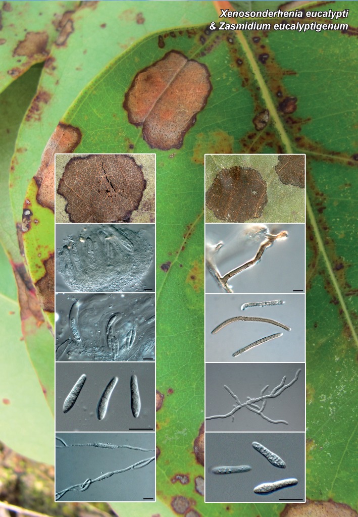
Leaf spots amphigenous, dark brown, 10–20 mm diam, with dark brown border. Co-occurring on leaf spots with Zasmidium eucalyptigenum. Ascomata hypophyllous, black erumpent, globose, solitary, up to 110 μm diam, with central ostiole; wall of 2–3 layers of brown textura angularis. Asci fasciculate, bitunicate, subsessile, hyaline, smooth, 8-spored, obovoid to ellipsoid, aparaphysate, straight to slightly curved, 35–45 × 10–12 μm. Pseudoparaphyses absent. Ascospores tri- to multi-seriate, hyaline, smooth, fusoid-ellipsoid, widest in apical cell, one third from apex, tapering towards both ends, not constricted at median septum, (17–)18–20(–22) × (3–)4 μm; ascospores germinating from both ends, not constricting or distorting, remaining hyaline, 4–5 μm diam; germ tubes developing numerous lateral branches.

Culture characteristics — Colonies spreading, erumpent with moderate aerial mycelium and smooth, lobate margins, reaching 20 mm diam on PDA, MEA and OA after 2 wk at 25 °C in the dark. On MEA surface pale luteus with patches of dirty white, reverse sienna. On OA surface saffron. On PDA surface saffron, reverse pale luteus.

Typus. MOZAMBIQUE, Forestas de Niassa, leaf spots of Eucalyptus urophylla (Myrtaceae), 2 Feb. 2014, M.J. Wingfield (holotype CBS H-21991, culture ex-type CPC 24247 = CBS 138858; ITS sequence GenBank KP004457, LSU sequence GenBank KP004485, MycoBank MB810604).

Notes — Xenosonderhenia eucalypti appears to represent an undescribed genus in the Mycosphaerellaceae. It clusters with ‘Mycosphaerellaelaeocarpi that lacks an asexual morph and Xenosonderhenia syzygii, which lacks a sexual morph (Crous et al. 2012a). Because this taxon is clearly not a species of Mycosphaerella s.str., which has Ramularia asexual morphs (Verkley et al. 2004, Crous et al. 2009b), we tentatively place it in the genus Xenosonderhenia, pending further collections.

ITS. Based on a megablast search of NCBIs GenBank nucleotide database, the closest hits using the ITS sequence are Xenosonderhenia syzygii (GenBank JX069872; Identities = 506/525 (96 %), Gaps = 3/525 (0 %)), Mycosphaerella elongata (GenBank EF394833; Identities = 492/520 (95 %), Gaps = 4/520 (0 %)) and Mycosphaerella elaeocarpi (GenBank EU040212; Identities = 514/547 (94 %), Gaps = 7/547 (1 %)).

Persoonia. 2014 Nov 24;33:240–241.

Fungal Planet 295 & 296 – 24 November 2014

Zasmidium eucalyptigenumCrous & M.J. Wingf., sp. nov.

Pedro W Crous 1, Johannes Z Groenewald 1, Michael J Wingfield 2

Etymology. Name reflects the host genus Eucalyptus, from which this species was isolated.

Co-occurring on leaf spots with Xenosonderhenia eucalypti. Ascomata hypophyllous, black erumpent, globose, solitary, up to 100 μm diam, with central ostiole; wall of 2–3 layers of brown textura angularis. Asci fasciculate, bitunicate, subsessile, hyaline, smooth, 8-spored, obovoid to ellipsoid, aparaphysate, straight to slightly curved, 25–40 × 8–10 μm. Pseudoparaphyses absent. Ascospores hyaline, smooth, fusoid-ellipsoid, widest in middle of apical cell, tapering towards both ends, constricted at median septum, 13–16 × (2.5–)3.5–4 μm; ascospores germinating from both ends, becoming constricted, but remaining hyaline and smooth, 4–6 μm diam, developing lateral branches. Mycelium brown, verruculose, typical of Zasmidium asexual morph. Conidiophores brown, verruculose, solitary on superficial hyphae, erect, branched or not, up to 50 μm tall, 3–4 μm diam, 1–3-septate. Conidiogenous cells terminal or intercalary, with several thickened, darkened, refractive scars, 1 μm diam. Conidia brown, verruculose, straight to curved, solitary or in branched chains, subcylindrical, apex obtuse, base tapering to a truncate hilum, 1–1.5 μm diam, 1–9-septate, 30–120 × (2.5–)3 μm.

Culture characteristics — Colonies erumpent, spreading, folded, with mucoid exudate and sparse to moderate aerial mycelium, and smooth, lobed margins, reaching 3 cm diam after 2 wk at 25 °C in the dark. Culture is sterile.. On OA surface olivaceous-grey. On MEA surface olivaceous-grey with patches of pale olivaceous-grey. On PDA surface olivaceous-grey with patches of pale olivaceous-grey, iron-grey in reverse.

Typus. MOZAMBIQUE, Forestas de Niassa, leaf spots of Eucalyptus urophylla (Myrtaceae), 2 Feb. 2014, M.J. Wingfield (holotype CBS H-21992, culture ex-type CPC 24251 = CBS 138860; ITS sequence GenBank KP004458, LSU sequence GenBank KP004486, MycoBank MB810605).

Notes — The genus Mycosphaerella is polyphyletic (Crous et al. 2007a), and Zasmidium is the oldest name to accommodate stenella-like taxa clustering in the Mycosphaerellaceae (Arzanlou et al. 2007). Several species have thus far been described from Eucalyptus (Crous et al. 2009a, b, Braun et al. 2010), all of which are phylogenetically distinct from Z. eucalyptigenum.

ITS. Based on a megablast search of NCBIs GenBank nucleotide database, the closest hits using the ITS sequence are Zasmidium rothmanniae (GenBank KJ869135; Identities = 461/480 (96 %), Gaps = 7/480 (1 %)), Periconiella arcuata (GenBank EU041779; Identities = 427/449 (95 %), Gaps = 6/449 (1 %)) and Mycosphaerella pseudovespa (GenBank DQ530216; Identities = 432/457 (95 %), Gaps = 6/457 (1 %)).

Colour illustrations. Leaf spots on Eucalyptus urophylla. Xenosonderhenia eucalypti (left column): leaf spot, asci with ascospores and germinating ascospores; Zasmidium eucalyptigenum (right column): leaf spots, conidiophore and conidia, ascospores and germinating ascospores. Scale bars =10 μm.

graphic file with name per-33-212-g014.jpg

Xensonderhenia eucalypti & Zasmidium eucalyptigenum

Persoonia. 2014 Nov 24;33:242–243.

Fungal Planet 297 – 24 November 2014

Diaporthe acaciarum Crous & M.J. Wingf., sp. nov.

Pedro W Crous 1, Johannes Z Groenewald 1, Michael J Wingfield 2

Etymology. Name reflects the host genus Acacia, from which this species was isolated.

Sporulating on PNA. Conidiomata pycnidial, globose, up to 300 μm diam, black, erumpent, exuding creamy conidial droplets from central ostioles; walls consisting of 3–6 layers of medium brown textura angularis. Conidiophores hyaline, smooth, 2–3-septate, branched, densely aggregated, cylindrical, straight to sinuous, 20–30 × 2.5–4 μm. Conidiogenous cells 15–25 × 2–3 μm, phialidic, cylindrical, terminal and intercalary, with slight apical taper, 1–1.5 μm diam, with visible periclinal thickenening; collarette not flared, up to 2 μm long. Paraphyses not observed. Alpha conidia (6–)6.5–7(–7.5) × (2–)2.5(–3) μm, aseptate, hyaline, smooth, bi-guttulate, fusoid-ellipsoid, tapering towards both ends, straight apex subobtuse, base subtruncate, 1 μm diam. Beta conidia spindle-shaped, aseptate, smooth, hyaline, apex acutely rounded, base truncate, tapering from the lower third towards the base, (20–)25–35(–40) × 1.5(–2) μm.

Culture characteristics — Colonies covering the dish after 2 wk at 25 °C in the dark, with sparse to moderate aerial mycelium. On MEA, PDA and OA surface dirty white with patches of grey-olivaceous, reverse dirty white with patches of sienna.

Typus. TANZANIA, Serengeti, on thorns of Acacia tortilis (Fabaceae), Feb. 2014, M.J. Wingfield (holotype CBS H-21994, culture ex-type CPC 24324 = CBS 138862; ITS sequence GenBank KP004460, LSU sequence GenBank KP004488, HIS sequence GenBank KP004504, TUB sequence GenBank KP004509, MycoBank MB810606).

Notes — No Diaporthe (incl. Phomopsis) species have been described from Acacia tortilis (Uecker 1988, Gomes et al. 2013). Phylogenetically, D. acaciarum is closely related to several species based on ITS (see below), but it can be distinguished from them based on TUB sequence data.

ITS. Based on a megablast search of NCBIs GenBank nucleotide database, the closest hits using the ITS sequence are Phomopsis chimonanthi (GenBank KF746059; Identities = 505/513 (98 %), Gaps = 5/513 (0 %)), Diaporthe helianthi (GenBank JQ936257; Identities = 546/556 (98 %), Gaps = 1/556 (0 %)) and Diaporthe infecunda (GenBank KF939614; Identities = 525/536 (98 %), Gaps = 4/536 (0 %)).

LSU. Based on a megablast search of NCBIs GenBank nucleotide database, the closest hits using the LSU sequence are Diaporthe leucospermi (GenBank JN712524; Identities = 836/839 (99 %), no gaps), Taeniolella alta (GenBank DQ377938; Identities = 834/839 (99 %), no gaps) and Diaporthe arctii (GenBank AF362562; Identities = 831/836 (99 %), no gaps).

HIS. Closest hits using the HIS sequence had highest similarity to Diaporthe infecunda (GenBank KC343613; Identities = 359/369 (97 %), Gaps = 1/369 (0 %)), Diaporthe terebinthifolii (GenBank KC343702; Identities = 353/371 (95 %), Gaps = 6/371 (1 %)) and Diaporthe melonis (GenBank KC343626; Identities = 351/373 (94 %), Gaps = 5/373 (1 %)).

TUB. Closest hits using the TUB sequence had highest similarity to Diaporthe infecunda (GenBank KF939619; Identities = 753/783 (96 %), Gaps = 4/783 (0 %)), Diaporthe beilharziae (GenBank KF170921; Identities = 743/777 (96 %), Gaps = 1/777 (0 %)) and Diaporthe terebinthifolii (GenBank KC344186; Identities = 662/708 (94 %), Gaps = 1/708 (0 %)).

Colour illustrations. Leopard in Acacia tortilis tree in the Serengeti, Tanzania; conidiomata on PNA, conidiogenous cells, alpha and beta conidia. Scale bars = 10 μm.

graphic file with name per-33-212-g015.jpg

Diaporthe acaciarum

Persoonia. 2014 Nov 24;33:244–245.

Fungal Planet 298 – 24 November 2014

Melanconium hedericola Crous & R.K. Schumach., sp. nov.

Pedro W Crous 1, Johannes Z Groenewald 1, René K Schumacher 2

Etymology. Name reflects the host genus Hedera, from which this species was isolated.

Conidiomata pycnidial single or in densely crowded groups under loosened bark, superficial on a black stromatic layer which continues as a black stromatic line deep in the wood, ± pyroid with flattened base, black, rough, soft, distinctly thick, ostiolum central and indistinct; periphyses not seen, with a few setae on the outer site of the peridium. Setae 1-celled, stiff, pointed, basally enlarged and flattened, brown, thick-walled, smooth, eguttulate, up to 26 μm long. Peridium multi-layered, consisting of a red-brown textura epidermoidea (outer layer) and hyaline textura angularis-prismatica (inner layer), cells thick-walled, smooth and eguttulate. Sporulating on PNA. Conidiomata pycnidial, globose, up to 600 μm diam, black, immersed, exuding creamy conidial droplets from central ostioles, or developing 1–4 black necks on OA (not on PNA); walls consisting of 3–6 layers of medium brown textura angularis. Conidiophores hyaline, smooth, 1–2-septate, branched below, densely aggregated, cylindrical, straight to sinuous, 20–40 × 3–4 μm. Conidiogenous cells 10–17 × 2–3 μm, phialidic, cylindrical, terminal and intercalary, with slight apical taper, 2 μm diam, with visible periclinal thickening; collarette flared, up to 5 μm long. Paraphyses not observed. Alpha conidia (6–)7(–8) × (3.5–)4(–4.5) μm, aseptate, hyaline, smooth, ellipsoid with large central guttule, becoming brown with age. Beta conidia spindle-shaped, aseptate, smooth, hyaline, guttulate, apex bluntly rounded, base truncate, tapering from the middle towards the base, (17–)18–20(–25) × 2.5(–3) μm.

Culture characteristics — Colonies flat, spreading, covering the dish after 2 wk at 25 °C in the dark, with sparse aerial mycelium. On MEA, PDA and OA surface umber with patches of dirty white and iron-grey; reverse umber with patches of dirty white and iron-grey.

Typus. SPAIN, Sarasibar (Navarra), on branch of Hedera helix (Araliaceae), 26 Jan. 2014, S. Garcia (holotype CBS H-21995, culture ex-type CPC 24278 = CBS 138863; ITS sequence GenBank KP004461, LSU sequence GenBank KP004489, HIS sequence GenBank KP004505, TUB sequence GenBank KP004510, MycoBank MB810607).

Notes — Although Melanconium hedericola clusters within the genus Diaporthe, the LSU region lacks resolution within the Diaporthales. We have thus chosen to describe it in Menanconium based on the ellipsoid alpha conidia that turn brown with age, and their characteristic large, central guttules (Sutton 1980). However, Melanconium is known to not have species with beta conidia, which suggests that M. hedericola might represent an unknown genus within this complex. Further studies and collections are required before this question can be resolved. Previously published taxa on Hedera include Coniothyrium hederae and its possible synonym, Melanconium hederae, which have similar alpha conidia (6–8 × 4.5–6 μm), but that lack beta conidia.

ITS. Based on a megablast search of NCBIs GenBank nucleotide database, the closest hits using the ITS sequence are Phomopsis columnaris (GenBank FN394688; Identities = 528/541 (98 %), Gaps = 2/541 (0 %)), Diaporthe endophytica (GenBank AB899789; Identities = 566/583 (97 %), Gaps = 7/583 (1 %)) and Diaporthe phaseolorum (GenBank JQ936148; Identities = 565/583 (97 %), Gaps = 7/583 (1 %)).

LSU. Based on a megablast search of NCBIs GenBank nucleotide database, the closest hits using the LSU sequence are Phomopsis columnaris (GenBank AF439627; Identities = 834/840 (99 %), no gaps), Diaporthe ambigua (GenBank JQ862833; Identities = 821/828 (99 %), no gaps) and Phomopsis sclerotioides (GenBank AF439628; Identities = 831/840 (99 %), no gaps).

HIS. Based on a megablast search of NCBIs GenBank nucleotide database, the closest hits using the HIS sequence are Diaporthe longispora (GenBank KC343619; Identities = 360/378 (95 %), Gaps = 5/378 (1 %)), Diaporthe sclerotioides (GenBank KC343678; Identities = 359/381 (94 %), Gaps = 4/381 (1 %)) and Diaporthe ‘sp. 4’ (GenBank KC343690; Identities = 356/378 (94 %), Gaps = 5/378 (1 %)).

TUB. Based on a megablast search of NCBIs GenBank nucleotide database, the closest hits using the TUB sequence are Diaporthe longispora (GenBank KC344103; Identities = 398/420 (95 %), Gaps = 2/420 (0 %)), Diaporthe sclerotioides (GenBank KC344161; Identities = 391/416 (94 %), Gaps = 2/416 (0 %)) and Diaporthe scabra (GenBank HQ450372; Identities = 418/450 (93 %), Gaps = 2/450 (0 %)).

Colour illustrations. Hedera helix growing along a tree trunk; conidiomata on PNA and OA, conidiogenous cells, alpha and beta conidia. Scale bars =10 μm.

graphic file with name per-33-212-g016.jpg

Melanconium hedericola

Persoonia. 2014 Nov 24;33:247.

Fungal Planet 299 – 24 November 2014

NeophysalosporaCrous & M.J. Wingf., gen. nov.

Pedro W Crous 1, Johannes Z Groenewald 1, Michael J Wingfield 2

Etymology. Name reflects the fact that the genus is morphologically similar to the genus Physalospora.

Endophytic and plant pathogenic. Ascomata globose, solitary, brown, immersed, with central ostiole; wall of 2–3 layers of brown textura angularis. Asci cylindrical, hyaline, stipitate, unitunicate with apical mechanism staining in Melzer’s reagent, ascospores uniseriate but overlapping, with 8 ascospores per ascus. Paraphyses intermingled among asci, hyaline, septate, branched. Ascospores hyaline, smooth, guttulate, fusoid-ellipsoid, with acutely rounded ends. Conidiomata globose, solitary to aggregated, brown, with central ostiole; wall of 2–3 layers of brown textura angularis. Conidiophores lining the inner conidiomatal wall, subcylindrical, straight to curved, branched, septate, or reduced to conidiogenous cells, hyaline to pale brown, smooth or finely verruculose. Conidiogenous cells ampulliform to subcylindrical, hyaline, smooth, terminal or intercalary, phialidic with flared collarette at the apex. Conidia solitary, hyaline, smooth, subcylindrical, curved, with obtuse apex and truncate base, thick-walled.

Type species. Neophysalospora eucalypti.

MycoBank MB810608.

Persoonia. 2014 Nov 24;33:246–247.

Fungal Planet 299 – 24 November 2014

Neophysalospora eucalyptiCrous & M.J. Wingf., sp. nov.

Pedro W Crous 1, Johannes Z Groenewald 1, Michael J Wingfield 2

Etymology. Name reflects the host genus Eucalyptus, from which this species was isolated.

Associated with brown leaf spots in plantations, and cutting rot in nurseries. Ascomata globose, solitary, brown, up to 250 μm diam, with central ostiole, up to 80 μm diam; wall of 2–3 layers of brown textura angularis. Asci cylindrical, hyaline, stipitate, unitunicate with apical mechanism staining in Melzer’s reagent, ascospores uniseriate but overlapping, with 8 ascospores per ascus, 70–100 × 6–8 μm. Paraphyses intermingled among asci, hyaline, septate, branched, 2–3 μm diam. Ascospores hyaline, smooth, guttulate, fusoid-ellipsoid, widest in middle, tapering to acutely rounded ends, (13–)15–17(–19) × (4–)4.5(–5) μm. Ascospores germinate from one end, remain hyaline, fusoid-ellipsoid, 15–17 × 7 μm. Conidiomata globose, solitary to aggregated, brown, up to 200 μm diam, with central ostiole; wall of 2–3 layers of brown textura angularis. Conidiophores lining the inner conidiomatal wall, subcylindrical, straight to curved, branched, 1–3-septate, or reduced to conidiogenous cells, hyaline to pale brown, smooth or finely verruculose, 10–30 × 2.5–4 μm. Conidiogenous cells ampulliform to subcylindrical, hyaline, smooth, terminal or intercalary, 7–12 × 1.5–3 μm, phialidic with flared collarette at the apex, 1.5–2 μm diam. Conidia solitary, hyaline, smooth, subcylindrical, curved, with obtuse apex and truncate base, thick-walled, (13–)14–15(–16) × 1.5 μm.

Culture characteristics — Colonies spreading with sparse, appressed aerial mycelium, reaching 6 mm diam after 2 wk at 25 °C in the dark; surface folded with feathery, lobate margin. On MEA surface flesh with patches of saffron, reverse red with patches of coral. On OA surface red with patches of peach and saffron. On PDA surface white, reverse saffron.

Typus. MOZAMBIQUE, on leaves of Corymbia henryi (Myrtaceae), 1 Feb. 2014, M.J. Wingfield (holotype CBS H-21996, culture ex-type CPC 24209 = CBS 138864; ITS sequence GenBank KP004462, LSU sequence GenBank KP004490, MycoBank MB810609). – SOUTH AFRICA, Mpumalanga province, Piet Retief, forestry nursery, cutting rot of Eucalyptus grandis × camaldulensis, 9 Jan. 1989, P.W. Crous, CPC 123 = CBS 110740 (ITS, LSU sequence GenBank KP031106, KP031108), CPC 124 = CBS 111123 (ITS, LSU sequence GenBank KP031107, KP031109) (specimen also deposited in IMI, now Kew).

Notes — The genus Physalospora (Hyponectriaceae) is polyphyletic, and in need of taxonomic revision. Neophysalospora eucalypti is reminiscent of Clypeophysalospora latitans and to some extent also Plectosphaera eucalypti (= Phyllachora eucalypti). Clypeophysalospora latitans is a saprobe or weak pathogen that is commonly encountered on living eucalypt leaves and leaf litter (Crous et al. 1990). It has a melanised clypeus, and unitunicate asci that strain in Melzer’s reagent, and does not produce an asexual morph in culture. Plectosphaera eucalypti has black ascomata embedded in the leaf tissue, with a melanised pseudoclypeus, forming circular to irregular colonies on the leaf surface. It has bitunicate asci and ascospores wit a prominent mucoid sheath. Infections are usually visible on only one side of the leaf surface (Pascoe 1990, Park et al. 2000).

ITS. Based on a megablast search of NCBIs GenBank nucleotide database, the closest hits using the ITS sequence are Bagadiella lunata (GenBank GQ303269; Identities = 586/625 (94 %), Gaps = 15/625 (2 %)), Bagadiella koalae (GenBank JF951142; Identities = 586/627 (93 %), Gaps = 14/627 (2 %)) and Bagadiella victoriae (GenBank JF951141; Identities = 584/627 (93 %), Gaps = 12/627 (1 %)).

LSU. Based on a megablast search of NCBIs GenBank nucleotide database, the closest hits using the LSU sequence are Plectosphaera eucalypti (GenBank DQ923538; Identities = 821/840 (98 %), Gaps = 2/840 (0 %)), Bagadiella victoriae (GenBank JF951161; Identities = 815/839 (97 %), no gaps) and Bagadiella lunata (GenBank GQ303300; Identities = 815/839 (97 %), no gaps).

Colour illustrations. Corymbia henryi leaves in Mozambique; conidiomata on SNA, asci and ascospores, conidiogenous cells, germinating ascospores and conidia. Scale bars = 10 μm.

graphic file with name per-33-212-g017.jpg

Neophysalospora eucalypti

Persoonia. 2014 Nov 24;33:248–249.

Fungal Planet 300 – 24 November 2014

Seimatosporium pistaciae Crous & Mirab., sp. nov.

Pedro W Crous 1, Johannes Z Groenewald 1, Mansoureh Mirabolfathy 2

Etymology. Name reflects the host genus Pistacia, from which this species was isolated.

Foliicolous. Conidiomata pycnidioid, separate to gregarious, becoming erumpent, oval to elongate, up to 150 μm diam. Conidiophores arising from a central stroma, hyaline, 3–4-septate, branched, subcylindrical, 20–45 × 3–4 μm. Conidiogenous cells terminal and intercalary, hyaline, smooth, subcylindrical, straight to somewhat curved, 10–15 × 2–2.5 μm, proliferating inconspicuously percurrently at apex. Conidia ellipsoid to fusoid, 3-septate, smooth, not constricted at septa, two median cells medium brown, basal and apical cell hyaline, granular, (15–)17–20(–22) × (4–)4.5(–5) μm, apical cell obtusely rounded with apical appendage single, unbranched, filiform, flexuous, 10–14 μm; basal appendage single, unbranched, filiform, flexuous, excentric, 12–20 μm.

Culture characteristics — Colonies spreading, appressed with moderate aerial mycelium and smooth margin, reaching 7 cm diam after 2 wk at 25 °C in the dark. On MEA surface peach with patches of olivaceous-grey due to sporulation, and diffuse red pigment, reverse similar. On PDA surface dirty white with patches of luteous and olivaceous-grey, reverse salmon with patches of olivaceous-grey. On OA surface salmon with patches of grey-olivaceous.

Typus. IRAN, Saveh, on buds of Pistacia vera (Anacardiaceae), 29 Apr. 2014, M. Mirabolfathy (holotype CBS H-21997, culture ex-type CPC 24455 = CBS 138865; CPC 24455 ITS sequence GenBank KP004463, CPC 24455 LSU sequence GenBank KP004491, CPC 24457 ITS sequence GenBank KP004464, CPC 24457 LSU sequence GenBank KP004492, MycoBank MB810610).

Notes — The genus Seimatosporium (1833) is linked to sexual morphs in Discostroma (1909). Because the former genus is better established in literature, and represents the older name with many more species, it has preference over Discostroma. As far as we are aware, no species of Seimatosporium have been described from Pistacia. Of the species treated by Nag Raj (1993), S. pistaciae morphologically most closely matches S. lonicerae (conidia 9–16 × 3.5–5 μm) and S. rosae (conidia 12.5–16.5 × 3.5–4 μm), but can be distinguished based on its larger conidial dimensions.

ITS. Based on a megablast search of NCBIs GenBank nucleotide database, the closest hits using the ITS sequence are Discostroma fuscellum (GenBank JF320818; Identities = 559/566 (99 %), Gaps = 2/566 (0 %)), Seimatosporium parasiticum (GenBank AB594808; Identities = 542/551 (98 %), no gaps) and Seimatosporium discosioides (GenBank AB594800; Identities = 544/555 (98 %), Gaps = 4/555 (0 %)).

LSU. Based on a megablast search of NCBIs GenBank nucleotide database, the closest hits using the LSU sequence are Discostroma botan (GenBank DQ368629; Identities = 826/830 (99 %), no gaps), Seimatosporium parasiticum (GenBank AB593741; Identities = 795/799 (99 %), no gaps) and Discostroma fuscellum (GenBank AB593726; Identities = 795/799 (99 %), no gaps).

Colour illustrations. Pistacia vera trees; conidiomata on PDA, conidiophores and conidia. Scale bars = 10 μm.

graphic file with name per-33-212-g018.jpg

Seimatosporium pistaciae

Persoonia. 2014 Nov 24;33:250–251.

Fungal Planet 301 – 24 November 2014

Codinaea pini Crous & M.J. Wingf., sp. nov.

Pedro W Crous 1, Johannes Z Groenewald 1, Michael J Wingfield 2

Etymology. Name reflects the host genus Pinus, from which this species was isolated.

Conidiophores erect, brown, flexuous, finely verruculous, appearing somewhat granular, unbranched, arising from superficial mycelium, mononematous, macronematous, cylindrical, 1–3-septate, 30–100 × 2.5–4 μm. Conidiogenous cells terminal, pale to medium brown, mono- to rarely polyphialidic, with one lateral aperture, 25–60 × 3–3.5 μm; collarette funnel-shaped, 3–4 μm diam, 2–3 μm deep, pale brown. Conidia (12–)13–15 × (2–)2.5(–3) μm, solitary, aggregating in a globose mucoid mass, hyaline, smooth, granular, fusoid, slightly curved or straight, widest in middle, tapering towards acute apices that give rise to setulae at each end, 8–9(–10) μm.

Culture characteristics — Colonies flat, appressed, spreading, with sparse aerial mycelium and smooth, even, lobate margin, reaching 35 mm diam after 2 wk at 25 °C in the dark. On OA surface iron-grey in centre, crystalline in outer region. On MEA surface olivaceous-grey, pale olivaceous-grey in outer region, olivaceous-grey in reverse. On PDA surface olivaceous-grey in centre, pale olivaceous-grey in outer region; in reverse olivaceous-grey in centre, pale olivaceous-grey in outer region.

Typus. UGANDA, on dead needles of Pinus patula (Pinaceae), Jan. 2014, M.J. Wingfield (holotype CBS H-21998, culture ex-type CPC 24400 = CBS 138866; ITS sequence GenBank KP004465, LSU sequence GenBank KP004493, MycoBank MB810611).

Notes — The species is described in the genus Codinaea because Réblová & Winka (2000) suggested separating Codinaea (setulate conidia) from Dictyochaeta (asetulate conidia). Based on the keys provided by Kuthubutheen & Nawawi (1991) and Whitton et al. (2000), C. pini appears to be distinct from presently known taxa. Phylogenetically, it is part of the C. simplex species complex (Hughes & Kendrick 1968).

ITS. Based on a megablast search of NCBIs GenBank nucleotide database, the closest hits using the ITS sequence are Dictyochaeta simplex (GenBank EF029193; Identities = 472/515 (92 %), Gaps = 21/515 (4 %)), Pseudolachnella guaviyunis (GenBank KJ834524; Identities = 493/548 (90 %), Gaps = 27/548 (4 %)) and Dictyochaeta fertilis (GenBank AF178540; Identities = 463/507 (91 %), Gaps = 23/507 (4 %)).

LSU. Based on a megablast search of NCBIs GenBank nucleotide database, the closest hits using the LSU sequence are Dictyochaeta simplex (GenBank AF178559; Identities = 827/832 (99 %), Gaps = 1/832 (0 %)), Rattania setulifera (GenBank HM171322; Identities = 811/838 (97 %), Gaps = 1/838 (0 %)) and Pseudolachnella guaviyunis (GenBank KJ834525; Identities = 803/831 (97 %), Gaps = 1/831 (0 %)).

Colour illustrations. Pinus patula plantation in Uganda; colonies sporulating on PNA and SNA, conidiophores and conidia. Scale bars = 10 μm.

graphic file with name per-33-212-g019.jpg

Codinaea pini

Persoonia. 2014 Nov 24;33:253.

Fungal Planet 302 – 24 November 2014

Xenophaeosphaeria Crous & M.J. Wingf., gen.nov.

Pedro W Crous 1, Johannes Z Groenewald 1, Michael J Wingfield 2

Etymology. Name reflects a morphological similarity to Phaeosphaeria.

Caulicolous. Ascomata solitary to gregarious, immersed, opening via a central ostiole, somewhat papillate, globose, brown; wall of 3–4 layers of brown textura angularis. Pseudoparaphyses intermingled among asci, hyaline, smooth, septate, hyphal-like, anastomosing. Asci clavate to fusoid-ellipsoid, fasciculate, short-stipitate with apical chamber, bitunicate, ascospores uni- to biseriate, overlapping, 8-spored. Ascospores brown, guttulate, smooth, fusoid-ellipsoid, straight, apex obtusely rounded, base subobtusely rounded, medianly euseptate, but each cell contains 2–4 distosepta.

Type species. Xenophaeosphaeria grewiae.

MycoBank MB810612.

Persoonia. 2014 Nov 24;33:252–253.

Fungal Planet 302 – 24 November 2014

Xenophaeosphaeria grewiae Crous & M.J. Wingf., sp. nov.

Pedro W Crous 1, Johannes Z Groenewald 1, Michael J Wingfield 2

Etymology. Name reflects the host genus Grewia, from which this species was isolated.

Caulicolous. Ascomata solitary to gregarious, immersed, opening via a central ostiole, up to 100 μm diam, somewhat papillate; ascomata up to 350 μm diam, globose, brown; wall of 3–4 layers of brown textura angularis. Pseudoparaphyses intermingled among asci, hyaline, smooth, septate, hyphal-like, anastomosing, 3–5 μm diam, frequently constricted at septa. Asci clavate to fusoid-ellipsoid, fasciculate, short-stipitate with apical chamber, 3–4 μm diam, bitunicate, ascospores uni- to biseriate, overlapping, 8-spored, 80–120 × 12–15 μm. Ascospores brown, guttulate, smooth, fusoid-ellipsoid, straight, apex obtusely rounded, base subobtusely rounded, 1-euseptate, apical cell (12–15 μm) shorter than basal cell (15–20 μm), widest at top of basal cell, but each cell contains 2–4 distosepta, (28–)32–37(–40) × (6–)7(–8) μm.

Culture characteristics — Colonies appressed, spreading with sparse aerial mycelium and smooth, even, lobate margins, reaching 20 mm diam after 2 wk at 25 °C in the dark. On PDA surface greyish sepia with patches of mouse grey, reverse greyish sepia. On MEA surface greyish sepia with patches of dirty white, reverse greyish sepia. On OA surface mouse grey with patches of red in outer zone.

Typus. TANZANIA, Masek Lake, on twigs of Grewia sp. (Malvaceae), Feb. 2014, M.J. Wingfield (holotype CBS H-21999, culture ex-type CPC 24398 = CBS 138867; ITS sequence GenBank KP004466, LSU sequence GenBank KP004494, MycoBank MB810613).

Notes — Xenophaeosphaeria represents a novel genus in the Phaeosphaeriaceae (Zhang et al. 2009, 2012). This resembles genera such as Neomassariosphaeria and Neophaeosphaeria, but is distinct with regards to the development of its ascospore septation. Xenophaeosphaeria grewiae sporulates readily in culture, and is not associated with an asexual morph.

ITS. Based on a megablast search of NCBIs GenBank nucleotide database, the closest hits using the ITS sequence are Setophoma vernoniae (GenBank KJ869141; Identities = 423/481 (88 %), Gaps = 15/481 (3 %)), Ophiobolus disseminans (GenBank KM014664; Identities = 424/483 (88 %), Gaps = 15/483 (3 %)) and Chaetosphaeronema hispidulum (GenBank KF871469; Identities = 423/483 (88 %), Gaps = 16/483 (3 %)).

LSU. Based on a megablast search of NCBIs GenBank nucleotide database, the closest hits using the LSU sequence are Phaeosphaeria juncicola (GenBank KF251686; Identities = 786/810 (97 %), Gaps = 2/810 (0 %)), Coniothyrium concentricum (GenBank EU754152; Identities = 786/810 (97 %), Gaps = 2/810 (0 %)) and Leptospora rubella (GenBank DQ195792; Identities = 86/810 (97 %), Gaps = 2/810 (0 %)).

Colour illustrations. Grewia sp. in Tanzania; colony sporulating on OA, ostiolar region, asci and ascospores. Scale bars = 10 μm.

graphic file with name per-33-212-g020.jpg

Xenophaeosphaeria grewiae

Persoonia. 2014 Nov 24;33:255.

Fungal Planet 303 – 24 November 2014

Neobambusicola Crous & M.J. Wingf., gen. nov.

Pedro W Crous 1, Johannes Z Groenewald 1, Michael J Wingfield 2

Etymology. Name reflects the morphological similarity with the genus Bambusicola.

Conidiomata separate or aggregated, erumpent, globose, dark brown, opening via central ostiole; wall of 3–6 layers of brown textura angularis. Conidiophores reduced to conidiogenous cells. Conidiogenous cells hyaline, smooth, subcylindrical to ampulliform, phialidic, proliferating percurrently at apex, or with prominent periclinal thickening. Conidia solitary, hyaline, smooth (becoming olivaceous with age), prominently guttulate, medianly 1-septate, constricted at septum, fusoid-ellipsoid, apex bluntly subobtusely rounded, tapering to a distinctly truncate base, mostly straight, but at times slightly curved. Microconidial state occurring in same conidioma. Microconidiogenous cells hyaline, smooth, doliiform to subcylindrical, proliferating percurrently at apex. Microconidia solitary, hyaline, smooth, guttulate to granular, aseptate, subglobose to subcylindrical, apex obtusely rounded, base truncate.

Type species. Neobambusicola strelitziae.

MycoBank MB810614.

Persoonia. 2014 Nov 24;33:254–255.

Fungal Planet 303 – 24 November 2014

Neobambusicola strelitziae Crous & M.J. Wingf., sp. nov.

Pedro W Crous 1, Johannes Z Groenewald 1, Michael J Wingfield 2

Etymology. Name reflects the host genus Strelitzia, from which this species was isolated.

Conidiomata separate or aggregated, erumpent, globose, dark brown, up to 200 μm diam, opening via central ostiole; wall of 3–6 layers of brown textura angularis. Conidiophores reduced to conidiogenous cells. Conidiogenous cells hyaline, smooth, subcylindrical to ampulliform, 7–15 × 3–5 μm, phialidic, proliferating percurrently at apex, or with prominent periclinal thickening. Conidia solitary, hyaline, smooth (becoming olivaceous with age), prominently guttulate, medianly 1-septate, constricted at septum, fusoid-ellipsoid, apex bluntly subobtusely rounded, tapering to a distinctly truncate base, 2 μm diam, mostly straight, but at times slightly curved, (15–)17–19(–21) × (3–)3.5(–4) μm. Microconidial state occurring in same conidioma. Microconidiogenous cells hyaline, smooth, doliiform to subcylindrical, 3–6 × 3–5 μm, proliferating percurrently at apex. Microconidia solitary, hyaline, smooth, guttulate to granular, aseptate, subglobose to subcylindrical, 3–7 × 3–4 μm, apex obtusely rounded, base truncate, 2–2.5 μm diam.

Culture characteristics — Colonies erumpent with sparse aerial mycelium and smooth, even, lobate margin, reaching 15 mm diam after 2 wk at 25 °C in the dark. On MEA surface dirty white with luteous in centre, reverse rust to red. On OA surface dirty white with diffuse luteous pigment. On PDA surface pale luteous with diffuse luteous pigment, reverse orange with diffuse luteous pigment.

Typus. SOUTH AFRICA, Eastern Cape Province, Haga Haga, on leaves of Strelitzia nicolai (Strelitziaceae), Dec. 2013, M.J. Wingfield (holotype CBS H-22000, culture ex-type CPC 24182 = CBS 138869; ITS sequence GenBank KP004467, LSU sequence GenBank KP004495, MycoBank MB810615).

Notes — Neobambusicola strelitziae was isolated from necrotic leaf tissue associated with infections of Phyllachora strelitziae. The latter fungus causes well-defined subcircular leaf spots, 3–10 mm diam, with epiphyllous black ascostromata. As these leaf spots get older and enlarge, conidiomata are observed surrounding the ascomata in the dead leaf tissue. Doidge (1942) commented on 2-celled conidia of a potential hyperparasite invading old ascomata of P. strelitziae, which we suspect is Neobambusicola strelitziae. Further collections are required, however, to resolve the relationship between these two species.

Neobambusicola resembles the genus Bambusicola in having reduced conidiophores with percurent proliferation, and conidia that turn pale brown at maturity (Dai et al. 2012, Hyde et al. 2013). However, Neobambusicola is distinct in that it does not have pycnothyrial conidiomata, and its conidia are fusoid-ellipsoid, rather than cylindrical.

ITS. Based on a megablast search of NCBIs GenBank nucleotide database, the closest hits using the ITS sequence are Microdiplodia hawaiiensis (GenBank GU361956; Identities = 391/461 (85 %), Gaps = 24/461 (5 %)), Camarographium koreanum (GenBank JQ044432; Identities = 413/494 (84 %), Gaps = 28/494 (5 %)) and Paraconiothyrium hawaiiense (GenBank KF177681; Identities = 390/461 (85 %), Gaps = 24/461 (5 %)).

LSU. Based on a megablast search of NCBIs GenBank nucleotide database, the closest hits using the LSU sequence are Bambusicola irregulispora (GenBank JX442036; Identities = 783/809 (97 %), Gaps = 3/809 (0 %)), Bambusicola massarinia (GenBank JX442037; Identities = 782/808 (97 %), Gaps = 2/808 (0 %)) and Bambusicola bambusae (GenBank JX442035; Identities = 785/814 (96 %), Gaps = 3/814 (0 %)).

Colour illustrations. Strelitzia nicolai at Haga Haga, Eastern Cape Province, South Africa; colony sporulating on OA, conidiogenous cells and conidia. Scale bars = 10 μm.

graphic file with name per-33-212-g021.jpg

Neobambusicola strelitziae

Persoonia. 2014 Nov 24;33:256–257.

Fungal Planet 304 – 24 November 2014

Trichomerium dioscoreae Crous & C. Nakash., sp. nov.

Pedro W Crous 1, Johannes Z Groenewald 1, Chiharu Nakashima 2

Etymology. Name reflects the host genus Dioscorea, from which this species was isolated.

Mycelium consists of olivaceous-brown, septate, branched, smooth, 3–4 μm diam hyphae. Conidiophores reduced to conidiogenous cells; conidia arising directly from hyphae. Conidiogenous loci inconspicuous, truncate, somewhat erumpent, 1.5–2 μm diam. Conidia solitary, pale to medium brown, smooth, consisting of a subcylindrical basal cell, 1–3-septate, 10–20 × 3–5 μm, with truncate hilum, 2 μm diam, giving rise to 2–3 lateral arms from a central cell; arms 1–2-septate, subcylindrical with obtusely rounded ends, 12–25 × 3–6 μm.

Culture characteristics — Colonies erumpent, spreading, surface folded, with sparse to moderate aerial mycelium and even, lobate margins, reaching 35 mm diam after 2 wk at 25 °C in the dark. On MEA, PDA and OA surface iron-grey to fuscous-black, reverse fuscous-black.

Typus. JAPAN, Iwate, Morioka, Koma, on leaves of Dioscorea sp. (Dioscoreaceae), 10 Sept. 2013, C. Nakashima (holotype CBS H-22001, culture ex-type CPC 24259 = CBS 138870; ITS sequence GenBank KP004468, LSU sequence GenBank KP004496, MycoBank MB810616).

Notes — The genus Trichomerium (1918) was recently studied by Chomnunti et al. (2012), who established the new family, Trichomeriaceae to accommodate it. Although these authors focused on the sexual morph, they did note that it possibly had Tripospermum (1918) asexual morphs. The present collection of a Tripospermum morph that clusters in Trichomerium, thus conforms this association.

ITS. Based on a megablast search of NCBIs GenBank nucleotide database, the closest hits using the ITS sequence are Trichomerium deniqulatum (GenBank JX313654; Identities = 627/672 (93 %), Gaps = 20/672 (2 %)), Trichomerium gleosporum (GenBank JX313656; Identities = 628/684 (92 %), Gaps = 31/684 (4 %)) and Trichomerium foliicola (GenBank JX313655; Identities = 628/684 (92 %), Gaps = 31/684 (4 %)).

LSU. Based on a megablast search of NCBIs GenBank nucleotide database, the closest hits using the LSU sequence are Trichomerium foliicola (GenBank JX313659; Identities = 834/845 (99 %), no gaps), Trichomerium deniqulatum (GenBank JX313660; Identities = 828/839 (99 %), no gaps) and Trichomerium gleosporum (GenBank JX313662; Identities = 831/843 (99 %), Gaps = 1/843 (0 %)).

Colour illustrations. Leaf of Dioscorea sp. infected with Distocercospora pachyderma and co-colonised by Trichomerium dioscoreae; conidiogenous loci and conidia. Scale bars = 10 μm.

graphic file with name per-33-212-g022.jpg

Trichomerium dioscoreae

Persoonia. 2014 Nov 24;33:258–259.

Fungal Planet 305 – 24 November 2014

Roussoella acaciae Crous & M.J. Wingf. sp. nov.

Pedro W Crous 1, Johannes Z Groenewald 1, Michael J Wingfield 2

Etymology. Name reflects the host genus Acacia, from which this species was isolated.

Conidiomata eustromatic, multilocular, separate, globose, immersed, brown, up to 200 μm diam, opening via central ostiole, exuding a brown conidial mass; wall of 3–6 layers of brown textura angularis. Conidiophores reduced to conidiogenous cells. Conidiogenous cells lining the inner cavity, hyaline, smooth, ampulliform to doliiform, 3–7 × 5–7 μm, with prominent periclinal thickening at apex, or with tightly aggregated percurrent proliferations at apex. Conidia solitary, pale to medium brown, smooth, guttulate, subcylindrical, straight to slightly curved, apex obtuse, base truncate, 2–3 μm diam, mostly central, but at times also displaced laterally, (5–)6–7(–10) × (2–)2.5–3 μm.

Culture characteristics — Colonies flat, spreading, with sparse aerial mycelium and even, smooth margin, reaching 6 mm diam after 2 wk at 25 °C in the dark. On MEA surface olivaceous-grey with patches of pale luteous, reverse sienna in centre, orange in outer region. On OA olivaceous-grey in centre, with dirty white in outer region. On PDA centre olivaceous-grey on surface and reverse.

Typus. TANZANIA, Serengeti, on leaves of Acacia tortilis (Fabaceae), Feb. 2014, M.J. Wingfield (holotype CBS H-22002, culture ex-type CPC 24314 = CBS 138873; ITS sequence GenBank KP004469, LSU sequence GenBank KP004497, MycoBank MB810617).

Notes — Members of the genus Roussoella (1888) (Roussoellaceae; Liu et al. 2014) mostly occur on monocotyledons, thus the occurrence of R. acaciae on Acacia is unusual. Although we isolated only the Cytoplea (1885) asexual morph, which is unknown for most species of Roussoella, the fungus on Acacia appears to be phylogenetically distinct from other members of the genus. Both the genera Roussoella and Cytoplea are in need of revision.

ITS. Based on a megablast search of NCBIs GenBank nucleotide database, the closest hits using the ITS sequence are Roussoella chiangraina (GenBank KJ474828; Identities = 360/395 (91 %), Gaps = 17/395 (4 %)), Roussoella siamensis (GenBank KJ474837; Identities = 352/387 (91 %), Gaps = 16/387 (4 %)) and Arthopyrenia salicis (GenBank KM030296; Identities = 353/390 (91 %), Gaps = 17/390 (4 %)).

LSU. Based on a megablast search of NCBIs GenBank nucleotide database, the closest hits using the LSU sequence are Roussoella percutanea (GenBank KF366449; Identities = 822/841 (98 %), Gaps = 3/841 (0 %)), Sporidesmium australiense (GenBank DQ408554; Identities = 827/847 (98 %), Gaps = 2/847 (0 %)) and Roussoella hysterioides (GenBank AB524622; Identities = 809/829 (98 %), Gaps = 2/829 (0 %)).

Colour illustrations. Lion resting in an Acacia tortilis tree, Serengeti, Tanzania; conidiomata sporulating on OA, conidiogenous cells and conidia. Scale bars = 10 μm.

graphic file with name per-33-212-g023.jpg

Roussoella acaciae

Persoonia. 2014 Nov 24;33:261.

Fungal Planet 306 – 24 November 2014

Neocladophialophora Crous & R.K. Schumach., gen. nov.

Pedro W Crous 1, Johannes Z Groenewald 1, René K Schumacher 2

Etymology. Name reflects its morphological similarity to the genus Cladophialophora.

Mycelium consisting of hyaline, smooth, septate, branched, hyphae. Conidiophores reduced to conidiogenous cells. Conidiogenous cells erect, subcylindrical, hyaline to subhyaline, mono- to polyphialidic; loci truncate. Conidia formed in long, rarely branched basipetal chains consisting of subcylindrical conidia, apical conidium clavate to subglobose, other conidia subcylindrical, 0–1-septate, somewhat constricted at the septum, guttulate to granular, subhyaline; constricted at the truncate hila, somewhat darkened. Older conidia appear pale olivaceous in mass.

Type species. Neocladophialophora quercina.

MycoBank MB810618.

Persoonia. 2014 Nov 24;33:260–261.

Fungal Planet 306 – 24 November 2014

Neocladophialophora quercina Crous & R.K. Schumach., sp. nov.

Pedro W Crous 1, Johannes Z Groenewald 1, René K Schumacher 2

Etymology. Name reflects the host genus Quercus, from which this species was isolated.

Mycelium consisting of hyaline, smooth, septate, branched, 1.5–2 μm diam hyphae. Conidiophores reduced to conidiogenous cells. Conidiogenous cells erect, subcylindrical, hyaline to subhyaline, mono- to polyphialidic, 10–30 × 3–4 μm; loci truncate, 1 μm diam. Conidia formed in long, rarely branched basipetal chains consisting of subcylindrical conidia (up to 20 per chain), apical conidium clavate to subglobose, other conidia subcylindrical, 0–1-septate, somewhat constricted at the septum, guttulate to granular, subhyaline, (9–)13–15(–17) × (2.5–)3(–5) μm; constricted at the truncate hila, somewhat darkened, 0.5 μm diam. Older conidia appear pale olivaceous in mass.

Culture characteristics — Colonies spreading, appressed, surface folded with sparse aerial mycelium and smooth, lobate margin, reaching 18 mm diam after 2 wk at 25 °C in the dark. On MEA and PDA surface and reverse ochreous. On OA surface pale luteous.

Typus. GERMANY, on dead twig of Quercus robur (Fagaceae), 23 Feb. 2014, R.K. Schumacher (holotype CBS H-22003, culture ex-type CPC 24426 = CBS 138874; ITS sequence GenBank KP004470, LSU sequence GenBank KP004498, MycoBank MB810619).

Notes — Neocladophialophora is morphologically similar to Cladophialophora, Fusicladium and Polyscytalidium (see Crous et al. 2007c, Bench et al. 2012), but different in that conidiophores are hyaline, conidiogenous cells phialidic, and conidia are constricted at their slightly darkened hila. Furthermore, conidia are pigmented in mass, and terminal conidia are frequently clavate to subglobose.

ITS. Based on a megablast search of NCBIs GenBank nucleotide database, the closest hits using the ITS sequence are Triscelophorus cf. acuminatus (GenBank KF730836; Identities = 210/235 (89 %), Gaps = 14/235 (5 %)), Triscelophorus monosporus (GenBank KF730840; Identities = 184/193 (95 %), Gaps = 1/193 (0 %)) and Hemibeltrania mitrata (GenBank EF029228; Identities = 280/351 (80 %), Gaps = 19/351 (5 %)).

LSU. Based on a megablast search of NCBIs GenBank nucleotide database, the closest hits using the LSU sequence are Scolecobasidium tropicum (GenBank KF156102; Identities = 687/786 (87 %), Gaps = 14/786 (1 %)), Isthmolongispora ampulliformis (GenBank EU107303; Identities = 707/809 (87 %), Gaps = 8/809 (0 %)) and Dactylaria humicola (GenBank EU107304; Identities = 687/793 (87 %), Gaps = 13/793 (1 %)).

Colour illustrations. Quercus robur tree; conidiophores and polyphialidic conidiogenous cells giving rise to branched chains of conidia. Scale bars = 10 μm.

graphic file with name per-33-212-g024.jpg

Neocladophialophora quercina

Persoonia. 2014 Nov 24;33:262–263.

Fungal Planet 307 – 24 November 2014

Barssia maroccana G. Moreno, Manjón, Carlavilla & P. Alvarado, sp. nov.

Gabriel Moreno 1, Juan R Carlavilla 1, Julio Álvarez 1, José L Manjón 1, Pablo Alvarado 2

Etymology. From the Berber Mur N’Akush (‘land of God’), which is presently known as Morocco, the country where this species was collected.

Hypogenous ascomata 1.7–5.3 × 1.5–2.5 cm (measurements taken from herbarium material), irregularly globose or subglobose to elongated and flattened, more or less broadly lobed, reddish brown to dark reddish brown, sometimes with a rounded to irregular apical depression. Peridium covered with broad, roughly polygonal dark reddish brown warts; about 140–200 μm thick, formed by pseudo-parenchymatic cells, 12–50 μm diam, thick-walled. The outermost cell layers are reddish and have dark walls, while these become lighter inwards. Gleba whitish to pale pinkish, compact or frequently presenting small labyrinth-like cavities, with well-defined sinuous veins, formed by a prosenchymatic structure of interwoven hyphae 8–15 μm diam. Sinuose paraphyses not well-defined, 5–7 μm diam. Asci clavate to broadly ellipsoid, indehiscent, immersed into the gleba, forming a definite hymenium, hyaline, hardly observable in mature ascomata, 8-spored, 110–130 × 30–50 μm. Ascospores ellipsoidal, 29–36 × (16–)18–22 μm, hyaline, smooth, not amyloid or dextrinoid, with an obtuse apex and a large oil droplet (L/l = 1.6–1.7). Smell and taste not recorded.

Habitat & Distribution — So far found only under Cedrus atlantica, at Ifrane, Morocco, 1 760 m asl.

Typus. MOROCCO, Azrou, province of Ifrane, Cedrus atlantica forest, 18 Nov. 2010, M.A. Sanz, J. Álvarez, P. Alvarado & J.L. Manjón (holotype AH 39117; ITS sequence GenBank KM243649, LSU sequence GenBank KM243655, MycoBank MB809666); Ifrane, Cedrus atlantica forest, 18 Nov. 2010, M.Á. Sanz, P. Alvarado & J.L. Manjón, paratype AH 39116; ibid., AH 44221; Ifrane, Cedrus atlantica forest with some Quercus ilex species, 1760 m asl, J.L. Manjón, J. Álvarez-Jiménez & M.Á. Sanz, 21 Feb. 2014, paratype AH 44099 (ITS, LSU sequences GenBank KM243648, KM243654).

Additional specimens examined. Balsamia vulgaris: ITALY, Reggio Emilia, Regnano, Quercus and Pinus mixed forest, 450 m asl, 11 Dec. 2005, A. Montecchi, Amer 2482 = AH 44222 (ITS, LSU sequences GenBank KM243645, KM243651); Reggio Emilia, Rio delle Viole, Quercus pubescens forest, 350 m asl, 9 Dec. 2002, A. Montecchi, Amer 2404 = AH 44223 (ITS, LSU sequences GenBank KM243646, KM243652); Reggio Emilia, Montalvo, Quercus pubescens forest, 350 m asl, 21 Apr. 2003, A. Montecchi, Amer 2403 = AH 44224 (ITS, LSU sequences GenBank KM243647, KM243653). Balsamia polysperma: ITALY, Reggio Emilia, Monte Duro, Ostrya and conifers, 650 m asl, 14 Dec. 1999, A. Montecchi, Amer 2042 = AH 44225 (ITS, LSU sequences GenBank KM243650, KM243656).

Notes — Barssia maroccana is morphologically characterised by its large and broad spores with obtuse apex, growing under Cedrus atlantica. The deviant phylogenetic placement of this lineage was first reported by Alvarado et al. (2011). Barssia maroccana is very similar to Balsamia polysperma, but the latter has smaller ascomata 0.5–2(–3) cm diam, with narrower ellipsoidal spores, 18–25 × 9–16 μm, L/l = 1.6–1.7, and different ecology (Montecchi & Sarasini 2000). The monotypic genus Barssia was created by Gilkey (1925) to accommodate the American species B. oregonensis. This species was originally found in Oregon and the Pacific Northwest of the USA (Trappe 1979), but later reports cited it also in Poland (Ławrynowicz & Skirgiełło 1984). Barssia oregonensis differs from B. maroccana because of its ascomata being excavated with a deep apical depression covered by the peridium, and smaller spores about 24–29 × 14.5–17 μm, L/I = 1.6–1.7, and a different ecology (Ławrynowicz & Skirgiełło 1984). The only other species in the genus, Barssia yezomontana, with globose spores (Trappe 1979), was combined into Barssia from the monotypic genus Phymatomyces. Unfortunately, the type specimen of P. yezomontanus was lost in World War II (Gilkey 1961) and it is therefore not possible to confirm this taxonomic decision until this Japanese taxon is recollected.

Gilkey (1925) placed the genus Barssia in the family Tuberaceae, but later Trappe (1979) transferred it to the family Balsamiaceae. Kimbrough et al. (1996) performed an ultrastructural study of Barssia and concluded it should be classified within the family Helvellaceae. Later Percudani et al. (1999) put Balsamia and Barssia back into the family Balsamiaceae, which was nested within Helvellaceae. Macro- and microscopical differences between Balsamia and Barssia are very subtle. Gilkey (1925) highlights the apical depression observed in B. oregonensis, and compares it to the analogous structures present in Genea, Pseudobalsamia (currently considered a synonym of Balsamia), Pachyphloeus and Hydnotrya. The similarities between Barssia and Balsamia are also commented on by Montecchi & Sarasini (2000). Glebal chambers, smooth spores under the light microscope, and the presence of paraphyses are shared by both genera. The present molecular data confirm that both genera are monophyletic, with the new species from Morocco being better accommodated within Barssia.

graphic file with name per-33-212-i001.jpg

Phylogenetic tree of Helvellaceae species constructed with a maximum likelihood (ML) analysis of LSU sequences by running RAxML-HPC2 on XSEDE online v. 8.0.24. Helvella lacunosa (KC122796 and KC122771) is the outgroup. Bootstrap support values ≥ 75 % are given above branches. The phylogenetic position of Barssia maroccana is indicated in bold.

Colour illustrations. Morocco, Ifrane, forest of Cedrus atlantica where the holotype was collected; ascomata; peridium and gleba; detail of the outermost layer of the peridium with pseudoparenchymatic structure; prosenchymatic gleba, asci and ascospores; ascospores (holotype AH 39117). Scale bars = 1 cm (ascomata), 100 μm (cortex), 20 μm (pseudoparenchymatic and prosenchymatic cells), 10 μm (ascus and spores).

graphic file with name per-33-212-g025.jpg

Barssia maroccana

Persoonia. 2014 Nov 24;33:265.

Fungal Planet 308 – 24 November 2014

Acremoniopsis Giraldo, Gené & Guarro, gen. nov.

Alejandra Giraldo 1, Josepa Gené 1, Josep Guarro 1

Etymology. Referring to the similarity with the genus Acremonium.

Mycelium consisting of branched, septate, smooth-walled hyphae. Conidiophores erect, simple or poorly branched. Conidiogenous cells enteroblastic, monophialidic, discrete, cylindrical to subulate, subhyaline. Conidia unicellular, globose or subglobose, hyaline, arranged in slimy heads.

Type species. Acremoniopsis suttonii.

MycoBank MB809883.

Persoonia. 2014 Nov 24;33:264–265.

Fungal Planet 308 – 24 November 2014

Acremoniopsis suttonii Giraldo, Gené & Guarro, sp. nov.

Alejandra Giraldo 1, Josepa Gené 1, Josep Guarro 1

Etymology. Named in honour of the American mycologist Deanna A. Sutton.

Mycelium consisting of septate, hyaline, smooth- and thin-walled hyphae, 1–1.5 μm wide. Conidiophores consisting of single phialides arising orthotropically and directly from vegetative hyphae or ropes of hyphae. Phialides cylindrical to subulate, (12–)16–30(–40) μm long, 1.5–2 μm wide at the base, with distinct periclinal thickening at the conidiogenous locus, hyaline, thin-walled and rugose towards the base. Conidia unicellular, globose or subglobose, 2–3 × 2 μm, hyaline, smooth- and thick-walled, in slimy heads. Chlamydospores and sexual morph not observed.

Culture characteristics — Colonies on OA at 25 °C attaining 8–9 mm diam after 2 wk, yellowish white (2A2) (Kornerup & Wanscher 1978), flat, membranous; reverse pastel yellow (1A4). On PDA at 25 °C reaching 15–21 mm diam after 2 wk, white (1A1), radially folded, dusty; reverse pastel yellow (2A4); exudate and diffusible pale yellow pigment (2A5). Optimum growth temperature 25 °C, minimum 12 °C, maximum 35 °C.

Typus. SPAIN, Burgos, natural area of Sierra de la Demanda, isolated from forest soil, Nov. 2010, coll. J. Gené & M. Hernández, isol. A. Giraldo (holotype CBS H-21936, cultures ex-type CBS 138708 = FMR 11780; ITS sequence GenBank KJ807182, LSU sequence GenBank KJ807179, MycoBank MB809884).

Notes — Acremonium is a polyphyletic genus with species spread across different orders of the Sordariomycetes, but are mainly placed in the Hypocreales (Glenn et al. 1996, Perdomo et al. 2011, Summerbell et al. 2011, Giraldo et al. 2012). Acremonium alternatum, the type species of the genus, was recently epitypified with the strain CBS 407.66, which was placed in the family Bionectriaceae (Summerbell et al. 2011). In Hypocreales, there are still some traditional species of Acremonium phylogenetically distant from A. alternatum and Bionectriaceae that could represent novel genera. Although Acremoniopsis shows the typical Acremonium morphology, it is phylogenetically closer to nectriaceous species such as Pleonectria pyrrhochlora and P. virens, rather than bionectriaceous species, so we preferred to accommodate the species described here within a new genus. Acremoniopsis suttonii produces a diffusible pale yellow pigment similar to Acremonium citrinum, A. vitellinum, A. chrysogenum and A. flavum. The main difference between the first two species and Acremoniopsis suttonii is that they produce conidia arranged in chains (Gams 1971, Giraldo et al. 2014). Additionally, A. chrysogenum has colonies with a yeast-like appearance and ellipsoidal conidia, and A. flavum produces ellipsoidal conidia, abundant chlamydospores and is thermotolerant. Acremonium guillematii exhibits yellow colonies, but does not produce diffusible pigment into the agar (Gams 1971). Previous phylogenetic studies have demonstrated that Acremonium citrinum, A. chrysogenum and A. flavum are members of Bionectriaceae (Summerbell et al. 2011, Giraldo et al. 2014) while A. vitellinum and A. guillematii form a weakly supported clade near to the Clavicipitaceae (Summerbell et al. 2011). Acremonium pteridii produces similar subglobose conidia to those of A. suttonii, but shows chondroid hyphae, abundant crystals and partially branched conidiophores (Gams 1971) nested in the Gliomastix/Bionectria clade (Summerbell et al. 2011).

Based on a megablast search of NCBIs GenBank nucleotide database, the closest hits using the LSU sequence are Parasarcopodium ceratocary CBS 110664 (GenBank AY425026; Identities = 772/796 (97 %), Gaps = 1/796 (0 %)), Pleonectria pyrrhochlora CBS 125131 (GenBank HM484570; Identities = 774/799 (97 %), Gaps = 3/799 (0 %)), P. virens A.R. 4558 (GenBank JF832754; Identities = 770/795 (97 %), Gaps = 3/795 (0 %)) and ‘Acremonium persicinum’ CBS 110646 (GenBank HQ232088; Identities = 773/800 (97 %), Gaps = 4/800 (0 %)). Parasarcopodium ceratocary (incertae sedis, Hypocreales) has verruculose conidiophores with rows or whorls of phialides and cylindrical conidia with amorphous mucoid appendages at both ends (Mel’nik et al. 2004); while Pleonectria species (Nectriaceae, Hypocreales) produce a pycnidial asexual morph (zythiostroma-like) on the natural substratum, with verticillated conidiophores, intercalary phialides and ellipsoidal conidia; and sporodochial conidiophores, densely branched with cylindrical phialides and allantoid conidia in culture (Hirooka et al. 2012).

Colour illustrations. Forest from the natural area of Sierra de la Demanda (Burgos, Spain), where the soil sample was collected. Colony on PDA after 21 d at 25 °C, phialides and globose or subglobose conidia. Scale bars = 10 μm.

graphic file with name per-33-212-g026.jpg

Acremoniopsis suttonii

Persoonia. 2014 Nov 24;33:266–267.

Fungal Planet 309 – 24 November 2014

Marasmius vladimirii A.K. Dutta & K. Acharya, sp. nov.

Arun K Dutta 1, Krishnendu Acharya 1

Etymology. Named after Vladimír Antonín, for the contribution that he has made to further our understanding of the genus Marasmius.

Pileus 25–29 mm diam, convex to broadly convex, orange-scarlet towards margin, disc orange-chestnut, smooth, non-striate; flesh creamy white, thin. Lamellae adnexed, creamy white, 2 mm wide at the middle, thinner towards margin, lamellulae of two tiers, intervenose, edge concolorous; collarium absent. Stipe central, well developed, 2.6–3.8 cm long, 2 mm broad, more or less equal, yellowish apricot from the middle towards lower part, upper portion whitish, hollow, cartilaginous, curved, surface glabrous. Basidiospores 7.5–11.5(–12) × 5.5–6.5(–7) μm (Xm = 10.9 ± 1.7 × 6.5 ± 0.6, Q = 1.3–2.1, Qm = 1.7 ± 0.3, n = 30, s = 1 specimen), ellipsoid, inamyloid, uni-guttulate, guttulae more or less globose. Basidia 36–40 × 8.5–9 μm, clavate, hyaline, tetrasterigmatic, sterigmata 3–3.5 μm long. Basidioles 36–39 × 11–12 μm, clavate, hyaline. Cheilocystidia present, in the form of Siccus-type of broom cells; main body 15.5–19(–20) × 6–8 μm, more or less clavate, hyaline, thin- to thick-walled; apical setulae (5–)8–12(–17) μm long, sub-acute, thin- to thick-walled. Pleurocystidia absent. Pileipellis a hymeniderm composed of Siccus-type broom cells; main body (20–)23–24(–39) × 7–7.5(–8) μm, clavate to broadly clavate, regular to irregular in outline, hyaline, thin- to thick-walled, often branched; apical setulae (5–)7.5–8(–9) × 2–2.5 μm, obtuse to sub-acute, thick-walled, deeply coloured. Pileus trama hyphae interwoven, 4–5 μm broad, hyaline, thin-walled, dextrinoid. Lamellar trama hyphae interwoven, hyaline, thin-walled. Stipitipellis composed of 7.5–8(–11) μm broad, hyaline, smooth, non-gelatinous, thin-walled hyphae. Caulocystidia absent. Clamp connections present in all tissues.

Typus. INDIA, West Bengal, Darjeeling district, towards the way of Pandama to Lebong, upon leaf litter mixed humus, 23 July 2012, A.K. Dutta (holotype CUH AMT003; ITS sequence GenBank KF991002, MycoBank MB807384).

Notes — The absence of a collarium and the presence of Siccus-type broom cells in the pileipellis, the absence of pleurocystidia, well-developed long central stipe, and adnexed lamellae suggest that M. vladimirii belongs to sect. Sicci, ser. Leonini. Based on a megablast search of NCBIs GenBank nucleotide database, the closest hits using the 5.8S (partial)-ITS2-28S (partial) sequence had highest similarity to M. hypochroides (GenBank EU935545; Identities = 299/382 (78 %), Gaps = 17/382 (4 %)), M. araucariae var. siccipes (GenBank FJ431223; Identities = 123/135 (91 %), Gaps = 6/135 (4 %)) and M. occultatus (GenBank FJ917622; Identities = 133/150 (89 %), Gaps = 9/150 (6 %)). Marasmius vladimirii differs from M. hypochroides (characterised by a rugulose pileus, brown to dark brown coloured at the disc with a brownish orange to yellowish brown margin, a stipe apex buff with an yellowish brown to reddish brown base, basidiospores 8–13 × 5–8 μm; Wannathes et al. 2009), in having a pileus coloured orange-scarlet towards margin with an orange-chestnut disc, a stipe yellowish apricot towards base and whitish at the upper portion and smaller basidiospores (7.5–12 × 5.5–7 μm). The absence of caulocystidia also distinguishes the newly described taxon from M. araucariae var. siccipes (Wannathes et al. 2009). The macroscopically similar M. occultatus, known from eastern Honshu, Japan, differs from M. vladimirii by having a smaller-sized pileus (12–27 mm), the presence of white mycelioid bristles at the stipe base, basidiospores 14–16 × 3–4 μm, and fertile lamellar edge (Takahashi 2000). Marasmius occultatiformis, described from the Republic of Korea, differs from the newly described taxon in having a smaller pileus (12 mm broad) with inflexed margin, smaller basidiospores (7.0–8.5 × 3.5–4.5 μm, av. = 7.8 × 4.0 μm), different sized cheilocystidia (11–19 × 5–8 μm) and pileipellis cells main body (14–25 × 6–10 μm; Antonín et al. 2012). Another similar taxon, M. abundans, differs by having a paler coloured, greyish orange, golden-yellow, orange, brownish yellow or ferrugineous pileus, and even larger basidiospores (12–18(–20) × 4–5 μm; Corner 1996).

Colour illustrations. Collection site at Darjeeling hills, India; basidiomes of Marasmius vladimirii (bar = 10 mm); basidiomata showing lamellae and lamellae (10 mm); Siccus-type cells of pileipellis (10 μm); basidium (10 μm).

graphic file with name per-33-212-g027.jpg

Marasmius vladimirii

Persoonia. 2014 Nov 24;33:268–269.

Fungal Planet 310 – 24 November 2014

Ganoderma austroafricanum M.P.A. Coetzee, M.J. Wingf, Marinc., Blanchette, sp. nov.

Martin PA Coetzee 1, Seonju Marincowitz 1, Michael J Wingfield 1, Robert A Blanchette 2, Charles W Barnes 3

Etymology. Name reflects the geographical origin from which the fungus was collected.

Mature basidiomata annual, pileate, sessile, dimidiate, pileus dark to reddish brown when dry, margin rounded, thickened, red brown when dry. Pore surface smooth, pores 3–4 per mm, round to angular, radially elongated, 150–390 μm long, 85–245 μm wide, dissepiments 60–200 μm diam. Hyphal system dimitic, generative hyphae, usually collapsed, thin-walled, hyaline. Vegetative hyphae observed in the tubes. Tube trama intricate and agglutinated skeletal hyphae 4–8 μm thick, with extremities tapering to elongate apices of 2–2.5 μm diam, hyaline to pale yellowish in 5 % KOH. Basidia not observed. Basidiospores brown, subglobose with a truncate base, bitunicate, verruculose, 8–11 × 5.5–7 μm. Chlamydospores on 2 % malt extract agar medium (MEA) ellipsoidal with a hyphal extension at the base, apex occasionally papillated, terminal or intercalary, 10.5–19.5 × 6.5–9.5 μm.

Culture characteristics — Colonies on 2 % MEA fertile, showing the best growth reaching 82 mm at 25 °C in the dark in 8 d, the second best 62 mm at 30 °C, 37 mm at 20 °C, 18 mm at 15 °C, no growth at 35 °C, circular with entire edge, flat, felt-like texture, white at all temperatures, with sporadic tint of yellow at inner 20 mm circle at 30 °C; chlamydospores present.

Typus. SOUTH AFRICA, Gauteng, Pretoria, Brooklyn, Jan. 2014, on Jacaranda mimosifolia, M.J. Wingfield (holotype PREM 61074, living culture ex-type CBS 138724 = CMW 41454; ITS sequence GenBank KM507324, LSU sequence GenBank KM507325, MycoBank MB810411).

Notes — Ganoderma austroafricanum is the causal agent of root and butt rot disease on large numbers of jacaranda trees in the suburb of Brooklyn (Pretoria, South Africa). ITS sequence comparisons using Blastn searches against sequences in GenBank yielded the closest hits with G. subamboinense var. laevisporum (JQ520205; query cover: 100 %, similarity 98 %). It can be differentiated from the closest related sequences of G. subamboinense var. laevisporum based on 13 nucleotide differences in the ITS sequence. Phylogenetic trees generated from ITS sequences placed G. austroafricanum within a clade that included sequences from GenBank representing G. weberianum and G. stipitatum but with low bootstrap support (parsimony bootstrap = 52 %). The latter group formed a sister group to a monophyletic clade (bootstrap support = 92 %) that included sequences from GenBank representing G. lobatum, G. lucidum, G. neojaponicum, G. oregonense and G. resinaceum.

Colour illustrations. Jacaranda mimosifolia (Jacaranda tree) growing as street tree in the suburb of Brooklyn, Pretoria, South Africa (background); crown of infected Jacaranda tree showing branch die-back; purple flowers of Jacaranda tree; basidiocarps; basidiospores (10 μm); skeletal hyphae refractive in phase contrast (25 μm); chlamydospores on 2 % MEA (10 μm).

graphic file with name per-33-212-g028.jpg

Ganoderma austroafricanum

Persoonia. 2014 Nov 24;33:271.

Fungal Planet 311 – 24 November 2014

Collarina Giraldo, Gené & Guarro, gen. nov.

Alejandra Giraldo 1, Josepa Gené 1, Josep Guarro 1

Etymology. Referring to the presence of conspicuous collarettes.

Mycelium consisting of branched, septate, smooth-walled hyphae. Setae interspersed with conidiophores, erect, unbranched, septate at the base, swollen at the apex, hyaline. Conidiophores erect, simple or poorly branched. Conidiogenous cells enteroblastic, monophialidic, discrete, cylindrical to acicular, with conspicuous collarettes, subhyaline or pale brown. Conidia unicellular, ellipsoidal or subglobose, hyaline to brownish in mass, ar-ranged in slimy heads.

Type species. Collarina aurantiaca.

MycoBank MB809407.

Persoonia. 2014 Nov 24;33:270–271.

Fungal Planet 311 – 24 November 2014

Collarina aurantiaca Giraldo, Gené & Guarro, sp. nov.

Alejandra Giraldo 1, Josepa Gené 1, Josep Guarro 1

Etymology. Referring to the diffusible orange pigment produced on PDA medium.

Mycelum consisting of septate, hyaline, smooth- and thin-walled hyphae, 1.5–2 μm wide. Setae arising directly from vegetative hyphae and interspersed with conidiophores, erect, unbranched, with a basal septum, straight to slightly flexuose, cylindrical and thick-walled towards the base, swollen and thin-walled at the apex, up to 20 μm long, 2 μm wide at the base, 3–4 μm at the apex, hyaline, smooth-walled. Conidiophores erect, simple or poorly branched, up to 40 μm long, hyaline or pale brown, smooth-walled. Phialides arising directly from vegetative hyphae or ropes of hyphae, cylindrical, slightly tapering at the apex, straight or slightly bent, 10–40 × 1–1.5 μm, with a brownish funnel-shaped collarette, subhyaline to pale brown with age, thick- and smooth-walled. Conidia ellipsoidal or subglobose, 2.5–3(–4) × 1–2 μm, hyaline to brownish in mass, smooth- and thin-walled. Chlamydospores and sexual morph not observed.

Culture characteristics — Colonies on OA and PCA at 25 °C attaining 8–14 mm diam in 14 d, brownish grey (6E2) (Kornerup & Wanscher 1978), flat, dusty. On PDA at 25 °C reaching 14–18 mm diam in 14 d, greyish white (1B1), radially folded, felt-like or fasciculate, reverse brownish grey (6D3) with a diffusible orange pigment. Optimum temperature for growth 25 °C, minimum 12 °C, maximum 30 °C.

Typus. SPAIN, Aragón, Huesca province, Ordesa y Monte Perdido National Park, isolated from sediments of Ara River, 23 Mar. 2011, coll. A. Giraldo, M. Hernández & J. Capilla, isol. A. Giraldo (holotype CBS H-21781, cultures ex-type CBS 138274 = FMR 11784; ITS sequence GenBank KJ807180, LSU sequence GenBank KJ807177, MycoBank MB809408).

Additional specimen examined. SPAIN, Aragón, Huesca province, Torla to direction Bujaruelos, from forest soil, 19 June 2009, coll. M. Hernández, J. Mena-Portales, J. Cano, isol. A. Giraldo (CBS 138273 = FMR 11134; ITS sequence GenBank KJ807181, LSU sequence GenBank KJ807178).

Notes — The SSU sequence of Collarina aurantiaca revealed that it belongs to the Clavicipitaceae s.str. (Hypocreales, Sordariomycetes), with Chamaeleomyces viridis, C. granulomatis, Pochonia bulbillosa, P. rubescens and Nomuraea rileyi being the closest species. Chamaeleomyces differs from Collarina by pale green to greenish grey colonies, and a yeast-like growth, phialides basally swollen with narrow necks and conidia in fragile chains; Pochonia has yellowish white colonies, slender acicular phialides commonly arranged in whorls, and some species produce dictyochlamydospores and conidia in chains; N. rileyi has pale green slow-growing colonies, cylindrical phialides with short necks and greenish coloured conidia (Zare et al. 2001, Sung et al. 2007, Sigler et al. 2010). Although members of Clavicipitaceae s.str. have been reported as important entomopathogens of Lepidoptera, Homoptera and Coleoptera, they are common soil fungi.

Based on a megablast search of NCBIs GenBank nucleotide database, the closest hits using the LSU sequence are Cordyceps sp. (GenBank AB027378, Identities = 975/1029 (95 %), Gaps = 12/1029 (1 %)), Eucasphaeria capensis CBS 120027 (GenBank EF110619, Identities = 973/1029 (95 %), Gaps = 5/1029 (0 %)) and Ascopolyporus philodendrus (GenBank AY886545, Identities = 977/1037 (94 %), Gaps = 14/1037 (1 %)). The closest hits using the ITS sequence had the highest similarity to ‘Acremonium psammosporum’ H28 (GenBank GU566287, Identities = 593/593 (100 %), no gaps) and with an unidentified hypocrealean fungus (GenBank KC007264, Identities = 544/550 (99 %), no gaps). Acremonium psammosporum was described by Gams (1971) and is characterised by slow-growing colonies with an orange-ochraceous reverse; conidiophores sometimes branched, up to 50 μm long, straight phialides with short collarettes; subglobose conidia, slightly apiculated at base, hyaline, 1.8–3.3 × 1.2–1.6 μm. Collarina aurantiaca differs morphologically from A. psammosporum by the presence of setae, conidiophores simple, shorter phialides (up to 40 μm), funnel-shaped collarettes and bigger and brownish conidia. In addition, the LSU sequence of the type strain of A. psammosporum (CBS 590.63) was 6.9 % different.

Collarina resembles Monocillium (asexual morphs of Niesslia) with the presence of thickened walls at the base in both phialides and setae. However, Monocillium species produce fast-growing colonies, phialides without collarettes and hyaline conidia that can be elongated and septate in several species (Gams 1971, Gams & Turham 1996, Girlanda & Luppi-Mosca 1998, Ramaley 2001).

Colour illustrations. Forest from Ordesa y Monte Perdido National Park (Aragón, Spain), where the sample was collected (photo: Javier Capilla). Colony on PDA after 21 d at 25 °C, conidiophores simple with conidia arranged in slimy heads, phialides with brownish funnel-shaped collarettes and setae, ellipsoidal conidia. Scale bars = 10 μm.

graphic file with name per-33-212-g029.jpg

Collarina aurantiaca

Persoonia. 2014 Nov 24;33:272–273.

Fungal Planet 312 – 24 November 2014

Comoclathris spartii K.M. Thambugala, E. Camporesi & K.D. Hyde, sp. nov.

Kasun M Thambugala 1, Kevin D Hyde 2, Erio Camporesi 3

Etymology. Named after the host genus from which it was collected, Spartium.

Saprobic on Spartium junceum. Ascomata solitary, scattered or aggregated in small groups, immersed in host tissue, dark brown to black, globose to subglobose up to 200 μm diam without a distinct ostiole. Ascomatal wall of 2–4 layers of medium brown cells of textura angularis up to 12–20 μm. Asci 8-spored, 100–180 × 23–28 μm, cylindro-clavate, stipitate, bitunicate, fissitunicate, apex rounded, with a small apical chamber. Pseudoparaphyses hyaline, cellular, filamentous, septate, anastomosing, 2.5–4 μm diam, extending above the asci. Ascospores uni- to biseriate in asci, muriform, yellow to pale brown, broadly fusiform, with obtuse ends, constricted at the primary septum, surrounded by a mucilaginous sheath, 25–34 × 9–14.5 μm.

Culture characteristics — Colonies on PDA surface flat, spreading, reaching 26 mm diam after 1 wk at 25 °C, white to smoke-grey, with moderate aerial mycelium and undulate, smooth to feathery margins, reverse umber.

Typus. ITALY, Castellaccio di Corniolino - Santa Sofia (province of Forlì-Cesena (FC)) on Spartium junceum (Fabaceae), 13 Oct. 2012, E. Camporesi (holotype MFLU 14-0579, culture ex-type MFLUCC 13-0214; ITS sequence GenBank KM577159, LSU sequence GenBank KM577160, SSU sequence GenBank KM577161, MycoBank MB810274).

Notes — The genus Comoclathris (based on Comoclathris lanata) is characterised by ascomata with circular lid-like openings and applanate reddish brown to dark reddish brown, muriform ascospores, with single longitudinal septa (Zhang et al. 2012, Ariyawansa et al. 2014). Some authors (Zhang et al. 2012, Woudenberg et al. 2013, Ariyawansa et al. 2014) suggested the correct phylogenetic placement of the genus in Pleosporaceae rather than Diademaceae based on both morphology and molecular phylogeny. Comoclathris spartii is phylogenetically closely allied to C. compressa (CBS 156.53 and CBS 157.53) and presently, it seems best to place this species in Comoclathris.

ITS. Based on a megablast search of NCBIs GenBank nucleotide database, the closest hits using the ITS sequence are Fungal endophyte sp. (GenBank EU977293; Identities = 511/517 (99 %), no gaps), Fungal sp. (GenBank JN578619; Identities = 523/536 (98 %), 4/536 (0 %)) and Dendryphion penicillatum (GenBank JN578618; Identities = 459/469 (98 %), no gaps).

LSU. Based on a megablast search of NCBIs GenBank nucleotide database, the closest hits using the LSU sequence are Pleospora ambigua (GenBank KC584630; Identities = 858/872 (98 %), no gaps), Comoclathris compressa (GenBank KC584372 ; Identities = 856/872 (98 %), no gaps) and Pleospora incompta (GenBank GU238087; Identities = 835/846 (99 %), no gaps).

SSU. Based on a megablast search of NCBIs GenBank nucleotide database, the closest hits using the SSU sequence are Comoclathris compressa (GenBank AY787937; Identities = 870/886 (98 %), no gaps), Comoclathris compressa (GenBank KC584631; Identities = 870/886 (98 %), no gaps) and Pleospora typhicola (GenBank JF740105; Identities = 864/888 (97 %), Gaps = 1/888 (0 %)).

Colour illustrations. Castellaccio di Corniolino - Santa Sofia, Italy; ascomata, ascus and ascospores. Scale bars = 50 μm.

graphic file with name per-33-212-g030.jpg

Comoclathris spartii

Persoonia. 2014 Nov 24;33:274–275.

Fungal Planet 313 – 24 November 2014

Pluteus albotomentosus E.F. Malysheva & Malysheva, sp. nov.

Ekaterina F Malysheva 1, Vera F Malysheva 1

Etymology. Albotomentosus refers to the colour and character of the pileus surface.

Pileus 50‒60 mm diam, hemispherical becoming applanate with low, broad umbo, pure white when fresh, yellowish white or cream (4A2‒A3) in herbarium; fine-tomentose over the entire surface, slightly flocculose towards margin. Lamellae free, crowded, broadly ventricose, white becoming pink with concolorous even edges. Stipe 60 × 8‒10 mm, broadening downwards, with vague basal bulb up to 1‒10 mm, broadening downwards, with vague basal bulb up to 12‒15 mm wide; solid, longitudinally fibrillose, white to slightly yellowish white, with white basal tomentum. Context white. Smell weak, sourish. Basidiospores 6.0‒6.6(‒6.8) × 5.3‒6.0(‒6.2) μm, Q = 1.00‒1.21(‒1.28), Q* = 1.11 (n = 30), subglobose to broadly ellipsoid, some oviform, smooth, thin- to slightly thick-walled, hyaline, contents with guttules. Basidia 23‒32 × 6‒8 μm, 4-spored, broadly clavate. Pleurocystidia abundant, 27‒70 × 11‒26 μm, utriform to broadly lageniform with very short and wide neck, some broadly clavate, hyaline, thin-walled. Lamellae edge sterile. Cheilocystidia numerous, forming dense layer, 23‒65 × 5‒11 μm, variable in shape, predominantly cylindrical, narrowly lageniform or narrowly clavate, thin-walled, hyaline. Pileipellis a cutis, made up of undifferentiated cylindrical hyphae up to 6‒10 μm wide, thin- or slightly thick-walled, colourless. Stipitipellis a cutis of 5‒12 μm wide hyaline hyphae, at lower part of stipe with scarce hyphae-like, cylindrical or narrowly clavate caulocystidia, 10‒30 × 4‒7 μm. Clamp connections absent in all tissues.

Habitat & Distribution — Solitary on mossy decayed log of deciduous tree. Found once in the south of Primorye Territory.

Typus. RUSSIA, Primorye Territory, ‘Land of the Leopard’ National Park, watershed of Ananjevka and Gryaznaya Rivers, mixed forest (Quercus mongolica, Carpinus cordata, Ulmus japonica, Abies nephrolepis), on mossy log of deciduous tree, 1 Sept. 2011, A. Kovalenko (holotype LE 289394; ITS sequence GenBank KM658284, MycoBank MB810391).

Notes — The macroscopic description is based on fresh basidiocarps from the original collection and the photos taken at the site. Colour terms are according to Kornerup & Wanscher (1978). Microscopic observations and photos were made from dried material mounted in 5 % KOH using an AxioImagerA1 light microscope.

Pluteus albotomentosus is quite different from the other white-coloured Pluteus species because of a combination of distinctive morphological characters, a scilicet distinctly tomentose pure white pileus, thin-walled utriform pleurocystidia, cylindrical cheilocystidia and a cutis-like pileipellis. The pileipellis structure and non-metuloid cystidia place P. albotomentosus in sect. Hispidoderma. Based on this it can be compared to P. ephebeus, which is easily distinguished by its dark basidiocarp colour, larger spore size (5.5‒8.5 × 4.5‒6.0(‒7.0) μm according to Vellinga 1990) and differently shaped cystidia. The molecular data (ITS sequence) indicate that it is closely related to P. ephebeus but the percentage of similarity between sequences of the two species is only 95‒96 % or less.

Colour illustrations. Russia, Primorye Territory, ‘Land of the Leopard’ National Park, area of the East Manchurian mountains, Manchurian mixed forests, where the holotype was collected; basidiocarp, pileipellis, basidiospores, pleurocystidia, cheilocystidia (all from holotype). Scale bars = 1 cm (basidiocarp), 10 μm (microscopic structures).

graphic file with name per-33-212-g031.jpg

Pluteus albotomentosus

Persoonia. 2014 Nov 24;33:276–277.

Fungal Planet 314 – 24 November 2014

Pluteus extremiorientalis E.F. Malysheva & Malysheva, sp. nov.

Ekaterina F Malysheva 1, Vera F Malysheva 1

Etymology. Named after the geographical area where it was collected (Russian Far East).

Pileus 10‒35 mm diam, firstly hemispherical, then convex becoming applanate, commonly with obtuse low umbo; surface tomentose to squamulose – densely punctate-granulose at centre and fibrillose-squamulose towards margin, often with white context exhibited between rare fibrils or squamules; centre strongly venose-rugose; dark coloured, cocoa brown, leather brown (6E6), often with reddish tint – chestnut brown (6F7‒F8) or henna (7E8), with darker disc (7F7‒F8) and lighter margin because of scarcity of squamules; margin even or rimose, slightly striate or not, often incurved. Lamellae free, moderately crowded, ventricose, white becoming pink; edges even or slightly fimbriate, fuscous (brownish), more rarely concolorous. Stipe 20‒55 × 1.5‒5 mm, cylindrical or slightly broadening downwards (up to 7 mm), but without basal bulb, longitudinally fibrillose, whitish at upper part, covered with dark brown fibrils or squamules at lower part; basal tomentum white. Context white. Smell indistinct. Basidiospores 5.6‒7 × 4.6‒5.7 μm, Q = (1.02‒)1.04‒1.39(‒1.48), Q* = 1.24 (n = 40), subglobose to broadly ellipsoid, some oviform, smooth, slightly thick-walled, hyaline, contents with one large or numerous small guttules. Basidia 20‒30 × 6‒9 μm, 4-spored, clavate. Pleurocystidia scattered, 35‒70 × 10‒17(‒25) μm, broadly lageniform to utriform, some broadly fusiform to oviform, with pedices and obtuse apices, thin-walled, hyaline. Lamellae edge sterile. Cheilocystidia numerous, 40‒90 × 10‒33 μm, varying in shape, predominantly lageniform with inflated body and narrow long neck (up to 30 μm long and 6 μm wide), a few narrow utriform to clavate-cylindrical, thin-walled, with brown intracellular pigment (including holotype), but in some specimens hyaline. Pileipellis a cystoderm, made up of spheropedunculate, pyriform, broadly clavate cells in combination with lageniform or narrowly fusiform elements with acute apices, 20‒75(‒90) × 12‒30(‒35) μm, thin- or slightly thick-walled, with dark brown intracellular pigment in vacuoles. Stipitipellis a cutis of hyaline thin- or slightly thick-walled cylindrical hyphae, 5‒12 μm wide, in stipe base with brown content and scattered clavate or cylindrical caulocystidia, 35‒70(‒100) × 8‒15 μm, often with yellow-brown intracellular pigment. Clamp connections absent in pileipellis, but occasional in stipe context.

Habitat & Distribution — Scattered to gregarious on decaying deciduous wood or soil, in floodplain broadleaf forests. Known from two localities in the Russian Far East.

Typus. RUSSIA, Primorye Territory, Ussuriysky Nature Reserve, vicinities of Peishula field station, floodplain of Koryavaya River, broadleaved forest (with Ulmus, Populus, Acer), on soil, 12 Aug. 2011, N. Psurtseva (holotype LE 262872; ITS sequence GenBank KM658280, MycoBank MB810390).

Additional specimens examined. RUSSIA, Primorye Territory, Ussuriysky Nature Reserve, vicinities of Peishula field station, floodplain of Koryavaya and Suvorovka Rivers, broadleaved forest (with Ulmus, Populus, Acer), on wood of Ulmus, 12 Aug. 2011, A. Kovalenko, LE 262871, ITS sequence GenBank KM658279; ibid., on soil, 12 Aug. 2011, E. Malysheva, LE 303463, ITS sequence GenBank KM658282; ibid., Ulmus japonica forest, on soil, 13 Aug. 2011, E. Malysheva, LE 262865, ITS sequence GenBank KM658281; Primorye Territory, Kedrovaya Pad Nature Reserve, valley of Kedrovaya River, floodplain broadleaved forest (Quercus mongolica, Carpinus cordata, Tilia amurensis, Juglans mandshurica), on decaying deciduous wood, 5 Sept. 2011, A. Kovalenko, LE 303464, ITS sequence GenBank KM658283.

Notes — Macroscopic descriptions are based on fresh basidiocarps from the original collections and photos taken at the site. Colour terms are according to Kornerup & Wanscher (1978). Microscopic observations and photos were made from dried material mounted in 5 % KOH using an AxioImagerA1 light microscope.

Pluteus extremiorientalis is characterised by small to medium-sized, brownish coloured and tomentose-squamulose pileus, fuscous edges of lamellae and dark fibrils on lower part of stipe. Microscopically, the pileipellis consists of two types of cystidioid elements (broadly clavate to sphaeropedunculate and fusiform); cheilocystidia abundant, variable in pigmentation in different collections, but most contain brown intracellular pigment; pleurocystidia not rare, predominantly lageniform or utriform.

Based on its pileipellis structure P. extremiorientalis is placed to sect. Celluloderma and subsect. Mixtini. The ITS sequences from the five studied collections are 99‒100 % identical, but morphological characters differ slightly between collections – this mainly concerns the differences in colour of basidiocarps, density of squamules or fibrils on pileus surface, pigmentation of cheilocystidia and substrate preferences.

Pluteus extremiorientalis mostly resembles P. podospileus and P. seticeps var. cystidiosus in pileus colouration, dark brown fibrils at stipe base, shape of pleurocystidia and pileipellis structure (Minnis & Sundberg 2010). It can be distinguished from both by the strong tomentose-squamulose surface of the pileus, brownish coloured lamellae edges, cheilocystidia shape and smaller elements in the pileipellis. The molecular data (generated ITS sequences) confirmed the morphological differences between all species discussed and supported the recognition of P. extremiorientalis as a separate taxon.

Colour illustrations. Russia, Primorye Territory, Ussuriysky Nature Reserve, southern spurs of the Sikhote-Alin mountains, liana coniferous-broadleaved forest; basidiocarps, pileipellis elements, basidiospores, pleurocystidia, sterile edge of lamella (with pigmented cheilocystidia), stipitipellis (all from holotype). Scale bars = 1 cm (basidiocarps), 10 μm (microscopic structures).

graphic file with name per-33-212-g032.jpg

Pluteus extremiorientalis

Persoonia. 2014 Nov 24;33:278–279.

Fungal Planet 315 – 24 November 2014

Phytophthora moyootj T.I. Burgess, sp. nov.

Treena I Burgess 1, Michael JC Stukely 2

Etymology. Named for the swamp or wetlands from which isolates were recovered (moyootj = swamp country in the local Aboriginal Nyoongar language).

Sporangia produced abundantly in non-sterile soil extract; non-caducous, non-papillate with long unbranched sporangiophores, most commonly ovoid (63 %), with broad ovoid (11.5 %), limoniform (9 %), elongated ovoid (8.5 %), globose (5 %) and mouse shapes (3 %) also found; 39.6 ± 10.8 × 26.5 ± 4.2 μm (overall range 18–73.3 × 18.3–38.4 μm), length/breadth ratio 1.5 ± 0.4. Sporangial proliferation in chains of internally proliferating sporangia, both nested and extended. Hyphal swellings absent although rarely sporangia failed to produce septa and these then continued to grow from the apex to produce ‘sporangia shaped’ swellings. Chlamydospores not observed. Gametangia not produced in single culture or when paired with A1 and A2 tester strains of P. cinnamomi. Radial growth rates on V8 agar at optimum temperature (25–30 °C) and near the maximum temperature (32.5 °C), 5.5 ± 0.5 mm/d and 0.8 ± 0.1 mm/d, respectively.

Culture characteristics — Colonies have a stellate pattern on carrot agar and V8 agar and a fluffy irregular pattern on potato dextrose agar.

Typus. AUSTRALIA, Western Australia, Walpole, mud from vehicle, 2012, collected by Department of Parks and Wildlife (holotype MURU 469, cultures ex-type CBS 138759 = VHS27218; ITS sequence GenBank KJ372256, ß-tubulin sequence GenBank KJ372303, HSP90 sequence GenBank KJ396730, coxI sequence GenBank KJ396702, NADH sequence GenBank KJ396681, LSU sequence GenBank KP004501, MycoBank MB809152).

Additional specimens examined. AUSTRALIA, Western Australia, Fitzgerald River National Park, baited from soil in native heathland, 2006, collected by Department of Parks and Wildlife, VHS16108; Jarrahdale, isolated from water from restored pit at mine site, 2012, D. Hüberli, DH103.

Notes — Phylogenetically, P. moyootj resides in a strongly supported terminal clade and shares a common ancestor with P. fluvialis, P. litoralis and P. thermophila (Jung et al. 2011, Crous et al. 2011, 2012a). In a multigene phylogeny of the ITS, HSP90, BT, NADH and coxI gene regions, P. moyootj differs from P. fluvialis by 87 bp (1.8 %), P. litoralis by 107 bp (2.3 %) and P. thermophila by 118 bp (2.6 %). These four species have all been isolated from waterways and wetlands in the south-west of Western Australia. Phytophthora moyootj has a life strategy similar to P. litoralis and P. fluvialis, being sterile in culture and having abundant and continuous asexual multiplication chains of nested and extended internally proliferating sporangia. Phytophthora moyootj can be separated from these species because it lacks external proliferation of sporangia and hyphal swellings and it has lower optimum and maximum temperatures for growth.

graphic file with name per-33-212-i002.jpg

Bayesian inference tree based on concatenated ITS, BT, HSP90, coxI and NADH sequence alignment generated in MrBayes using the GTR substitution model. The posterior probability is shown at the nodes. The species described here is printed in bold face. The tree was rooted to P. humicola (not shown). The alignment and tree are available in TreeBASE (Submission ID 15985).

Colour illustrationsTypical niche for recovery of P. moyootj; mature sporangia, ovoid, broad ovoid, ovoid just before release of zoospores, nested proliferation, empty sporangia with trapped encysted zoospores, internal nested and extended proliferation. Scale bar = 25 μm. Stellate colony on V8 agar (T.I. Burgess).

graphic file with name per-33-212-g033.jpg

Phytophthora moyootj

Persoonia. 2014 Nov 24;33:280–281.

Fungal Planet 316 – 24 November 2014

Phoma tamaricicola Wanasinghe, Camporesi, E.B.G. Jones & K.D. Hyde, sp. nov.

Dhanushka N Wanasinghe 1, Kevin D Hyde 1, Erio Camporesi 2, EB Gareth Jones 3

Etymology. Named after the host genus from which it was collected, Tamarix.

Saprobic on dead herbaceous branches. Sexual state: Ascomata 120–150 μm high, 170–210 μm diam (x̅ = 132 × 196 μm, n = 10) slightly erumpent, solitary, scattered, hardly removed from the host substrate, dark brown to black, coriaceous. Peridium 10–20 μm wide at the base, 20–25 μm wide in sides, thick, comprising 6–8 layers, outer layer heavily pigmented, thick-walled, comprising blackish to dark brown cells of textura angularis, inner layer composed of hyaline thin-walled cells of textura angularis. Hamathecium comprising numerous, 2.3 μm (n = 30) wide, filamentous, branched, septate, pseudoparaphyses. Asci (70–110) × (10–20) μm (x̅ = 14.5 × 95 μm, n = 40), 8-spored, bitunicate, fissitunicate, cylindrical to cylindric-clavate, pedicellate, thick-walled at the apex, with a minute ocular chamber. Ascospores (15–20) × (7–10) μm (x̅ = 18 × 9 μm, n = 50), overlapping 1–2-seriate, muriform, mostly ellipsoidal, 4–6 transversely septate, with 3–4 vertical septa, constricted at the septa, initially hyaline, becoming yellowish brown at maturity, conical and narrowly rounded at the ends, without a mucilaginous sheath. Asexual state: Conidiomata superficial or immersed in the agar, dark brown to black, clothed with white hyphal projections, 0.5–1.5 mm diam, simple, or complex with several merging cavities. Conidiomatal wall composed of a 30–45 μm thick outer layer and a 35–60 μm thick inner layer of textura angularis cells. Conidiogenous cells (4–7 × 3–4 μm) discrete, assembled into protruding masses of cells, or integrated in very compact conidiophores. Conidia (4–7 × 2.5–3.5 μm) narrowly ellipsoidal or short-cylindrical, straight or slightly curved, rounded at both ends, 1-celled, with 1–2 small, polar guttules, and with thin and smooth walls that are hyaline at secession, becoming light brown.

Culture characteristics — Colonies on PDA reaching 30–35 mm diam in 21 d, with abundant, fluffy grey aerial mycelium on surface, reverse smoke-grey.

Known distribution — On dead branches of Tamarix gallica (Tamaricaceae), Italy.

Typus. ITALY, Forlì-Cesena Province, Ravaldino in Monte, dead and hanging branches of Tamarix gallica, 15 Jan. 2014, E. Camporesi (holotype MFLU 14-0333, ex-type living culture = MFLUCC 14-0602, ITS sequence GenBank KM408753, LSU sequence GenBank KM408754, SSU sequence GenBank KM408755, MycoBank MB810072).

Notes — Species belonging to the genus Phoma are important plant pathogens (de Gruyter et al. 2009, Aveskamp et al. 2010, Wijayawardene et al. 2014) and characterised by ‘hyaline, unicellular conidia that may become septate due to secondary septation, phialidic, ampulliform to doliiform conidiogenous cells and (sub)globose, glabrous to pilose or setose, pseudoparenchymatous or scleroplectenchymatous pycnidia’ (de Gruyter et al. 2010).

Phoma was shown to be highly polyphyletic, and molecular based studies have shown that species are scattered throughout the Pleosporales (de Gruyter et al. 2009, 2010, 2013, Aveskamp et al. 2010), with Phoma herbarum clustering in Didymellaceae as the type species of Phoma (de Gruyter et al. 2013).

ITS. Based on a megablast search of NCBIs GenBank nucleotide database, the closest hits using the LSU sequence had highest similarity to Ascochyta pisi (GenBank DQ678070; Identities = 888/890 (99 %), no gaps), Peyronellaea prosopidis (GenBank KF777232; Identities = 887/890 (99 %), no gaps) and Coniothyrium prosopidis (GenBank KF777205; Identities = 887/890 (99 %), no gaps).

ITS. Based on a megablast search of NCBIs GenBank nucleotide database, the closest hits using the ITS sequences are Microsphaeropsis proteae (GenBank JN712497; Identities = 517/523 (99 %), Gaps = 1/523 (0 %)), Phoma macrostoma (GenBank HM036611; Identities = 514/519 (99 %), no gaps) and Phoma herbarum (GenBank FN868459; Identities = 518/525 (99 %), Gaps = 3/525 (0 %)).

Colour illustrations. Ravaldino in Monte, Italy. Ascomata on host substrate, section of ascoma, asci, ascospores, colonies on PDA; section of conidiomata, conidiomatal wall and conidia. Scale bars = 20 μm.

graphic file with name per-33-212-g034.jpg

Phoma tamaricicola

Persoonia. 2014 Nov 24;33:282–283.

Fungal Planet 317 – 24 November 2014

Stagonospora chrysopyla Romberg & Rooney-Latham, sp. nov.

Megan K Romberg 1, Suzanne Rooney-Latham 2

Etymology. Named after the location where this fungus was collected, Golden Gate Park Conservancy, San Francisco, California, USA, chryso = gold, pyla = gate.

Leaf spots spreading longitudinally along the leaf, angular, reddish brown, up to 5 cm in length, edges indistinct. Conidiomata immersed, globose to subglobose, up to 250 μm diam, dark brown, scattered, subepidermal; wall of 3–6 layers of brown textura intricata, ostiole indistinct, only present on abaxial surface, 30–50 μm diam. Conidiophores reduced to conidiogenous cells. Conidiogenous cells hyaline, smooth, lining inner cavity, ampulliform to subglobose, 10–20 × 5–12 μm. Conidia solitary, hyaline, smooth, cylindrical, straight, apex obtuse to subobtuse, base truncate, (3–)5–6(–7)-septate, (48–)50–65(–69) × (5–)7–8(–10) μm.

Culture characteristics — Colonies flat, circular, with even margins, 50 mm diam after 2 wk at 20 °C in dark on PDA. Aerial mycelium sparse, cream-coloured at first, becoming pale olivaceous-grey to olivaceous-grey, reverse olivaceous-grey (Rayner 1970).

Typus. USA, California, Marin County, Golden Gate Park Conservancy, on leaves of Scirpus microcarpus (Cyperaceae), 15 Jan. 2014, A. Shor (holotype BPI892895, culture ex-type CBS 137792; ITS sequence GenBank KM033942, TUB sequence GenBank KM033943, MycoBank MB809083).

Notes — Of the more than 500 names in Stagonospora, fewer than 20 are represented in the NCBI GenBank. Shown in the table is a comparison of conidial measurements and number of septa for the species of Stagonospora currently described from Scirpus, as well as larger-spored Stagonospora spp. described from hosts in the Cyperaceae that are not represented in GenBank (Tehon 1933, Cunnell 1957, Castellani & Germano 1977). The conidia of Stagonospora chrysopyla are larger than those of other species of Stagonospora on Scirpus and it is also the first Stagonospora described from Scirpus microcarpus. Of the larger-spored Stagonospora on Cyperaceae, S. chrysopyla most resembles S. dolosa, but differs in having smaller and thinner conidia that are 5–6-septate rather than mainly 5-septate. The conidial size of S. chrysopyla also overlaps with that of S. foliicola; however, the examined herbarium specimens of S. foliicola show a great deal of variation and the circumscription of this species may need to be re-examined. The ITS region of S. chrysopyla has 90 % identity to S. foliicola (GenBank KF251256).

ITS. Based on a megablast search of NCBIs GenBank nucleotide database, the closest hits of the ITS sequence had highest similarity to S. paludosa (GenBank KF251257; Identities = 493/518 (95 %)) and S. pseudocaricis (GenBank KF251260; Identities = 494/517 (96 %)). Both of these species differ from S. chrysopyla in conidial size and reported host (Crous et al. 2013, Quaedvlieg et al. 2013).

TUB. Based on a megablast search of NCBIs GenBank nucleotide database, the closest hits of the TUB sequences are S. paludosa (GenBank KF252740, Identities = 392/416 (94 %)) and S. pseudocaricis (GenBank KF252741, Identities = 395/420 (94 %)).

Colour illustrations. Leaves of Scirpus microcarpus growing at the Golden Gate Park Conservancy (photo: A. Shor); leaf surface with ostiole, pycnidial surface (bar = 100 μm), pycnidium (bar = 100 μm), conidiogenous cells and conidia (bars = 10 μm).

Table.

Comparison of conidial measurements and number of septa for selected species of Stagonospora.

Species Conidia (μm)1 No. of septa Specimen(s) examined Reported host(s)
Stagonospora aquatica 25–30 × 5–6 3 Scirpus
S.aquatica var.sexseptata 32–35 × 5–6 6 Scirpus
S.chrysopyla 50–65 × 7–8 5–6 BPI 892895 Scirpus
S. cylindrica 48–78 × 8–11.5 3(–4) Phragmites
S. dolosa 60–70 × 10 5 BPI 374901 Phragmites
S. elegans 52–84 × 8.5–14 (3–)4–6 Phragmites
S. foliicola 35–90 × 5.5–9.5 6–12 BPI 374911 / BPI 374909 Various
S. gigaspora 58–84 × 10–14 6–9 BPI 374955 Carex
S. maritima 16–20 × 4–6.5 2 Various
S. scirpi 20–28 × 5–6.5 (3–)4–5 Scirpus
S. scirpicola 12–18 × 2.5–3 3 BPI375122 Scirpus
S. scirpini 20 × 3–4 1–4 BPI375126 Scirpus

1Conidial measurements as presented in species descriptions in Tehon (1933), Cunnell (1957), Castellani & Germano (1977) and/or determined upon examination of herbarium specimens.

graphic file with name per-33-212-g035.jpg

Stagonospora chrysopyla

Persoonia. 2014 Nov 24;33:284–285.

Fungal Planet 318 – 24 November 2014

Penicillium coccotrypicola Holdom, Y.P. Tan & R.G. Shivas, sp. nov.

David G Holdom 1, Yu Pei Tan 1, Justin S Bartlett 1, Roger G Shivas 1

Etymology. Derived from the generic name of the palm seed borer weevil (Coccotrypes) that formed the galleries from which the fungus was isolated.

Mycelium ramified in galleries of palm seed borer (Coccotrypes carpophagus) scolytine weevils that infest seeds of Archontophoenix cunninghamiana. Synnemata protrude from the fibrous seed husk, blue-grey, clavate, often furcated and slightly flattened, up to 1 cm high, solitary or clustered in small coralloid groups. Conidiophores in vivo borne on surface of synnemata; stipes 30–120 μm, verruculose or finely roughened to partly smooth, subhyaline to pale olivaceous; penicilli biverticillate or terverticillate, rarely monoverticillate; 2–5 metulae, one metula often larger than others, (9–)10.5–13.5(–16.5) × (2.5–)3.5 × 4.5(–5) μm, smooth or finely roughened, subhyaline to pale grey-olivaceous. Conidiogenous cells phialides, in verticils of 2–6, acerose, smooth, 9–15 × 2–3.5 μm, with a distinct neck, hyaline to subhyaline. Conidia globose to subglobose (2.5–)3 × 3.5(–4) μm, smooth, pale grey-olivaceous. Synnemata produced in vitro on Czapek yeast extract agar (CYA) after 14 d in the dark at 25 °C, with similar conidiophores borne from hyphae with stipes up to 200 μm; metulae (8.5–)9–11.5(–14.5) × 2.5–3.5(–4) μm and phialides (7–)8–10(–12) × 2–2.5(–3) μm.

Culture characteristics — (after 7 d in the dark at 25 °C). On CYA colonies 40–43 mm diam, velutinous, weakly sulcate, glaucous grey to greenish grey; tinged sienna at centre and white margins, apricot droplets exuded at centre and pale brown soluble pigment released into agar; cinnamon in reverse; no growth at 5 or 37 °C. Conidial mass becoming cinnamon on older cultures on most media; increasing Cu and Zn had no effect; rare (< 1 %) germination but no growth at 30 °C. On oatmeal agar (OA) and potato dextrose agar (PDA) colonies 40–42 mm diam, with irregular concentric rings of greenish grey sporulation, synnemata form in rings. On 25 % glycerol nitrate agar colonies 12–14 mm diam with no sporulation. On creatine sucrose agar colonies 27–31 mm diam with acid production. On Czapek agar (CzA) colonies 24–30 mm diam. On malt extract agar (Pitt 1979) colonies 29–30 mm, sporulation sparse, soluble pigment not released, and colour of culture not changing to cinnamon with age. On yeast extract sucrose agar colonies 43–47 mm diam, no sporulation, primrose around a darker centre. On CYA + 5 % NaCl colonies 26–28 mm diam. On nitrite-sucrose agar colonies 27–30 mm diam. No growth on CzA + proprionic acid. On CzA + sorbic and benzoic acids colonies 5–11 mm diam.

Typus. AUSTRALIA, Queensland, Bli Bli, Maroochy Wetlands, on seeds of Archontophoenix cunninghamiana and in galleries infested by Coccotrypes carpophagus, 6 July 2013, D. Holdom & J. Hewett (holotype BRIP 59608; ITS sequence GenBank KM605436, LSU sequence GenBank KM605437, beta-tubulin sequence GenBank KM605438, MycoBank MB810327).

Notes — Penicillium s.str. has recently been redefined as a monophyletic genus by multilocus (RPB1, RPB2, Tsr1 and Cct8) phylogenetic analysis (Houbraken & Samson 2011). The formation of synnemata by species of Penicillium is uncommon (Pitt 1979, Seifert et al. 2004). Penicillium coccotrypicola is morphologically distinct from other species by having furcated synnemata. Penicillium coccotrypicola produced ramified mycelium in the galleries of palm seed borer weevils (Coccotrypes carpophagus: Coleoptera, Curculionidae, Scolytinae) in seeds of Archontophoenix cunninghamiana (Arecaceae). The palm seed borer weevils in the galleries were not colonised by P. coccotrypicola, which provides evidence that these ambrosia insects were farming the fungus. Possible mutualistic associations between Penicillium species and various scolytine weevil genera (Crypturgus, Dendroctonus, Hypothenemus, Ips, Pityogenes and Tomicus) have been noted before (Peterson et al. 2003, Giordano et al. 2013).

ITS. Based on a megablast search of the NCBI GenBank nucleotide database, the closest hits using the ITS sequence are Penicillium mononematosum (ex-type strain CBS 172.87; GenBank JX997082; Identities = 507/512 (99 %), Gaps = 0 (0 %)), P. gladioli (ex-type strain NRRL 939; GenBank DQ339568; Identities = 658/665 (99 %), Gaps = 1/665 (0 %)), P. confertum (ex-type strain CBS 171.87; GenBank JX997081; Identities = 506/512 (99 %), Gaps 0/512 (0 %)) and P. flavigenum (ex-type strain CBS 419.89; GenBank JX997105; Identities = 506/512 (99 %), Gaps = 0/512 (0 %)).

BT. Based on a megablast search of the NCBI GenBank nucleotide database, the closest hits using the BT sequence are Penicillium expansum (ex-type strain CBS 325.48; GenBank JQ965099; Identities = 585/634 (92 %), Gaps = 8/634 (1 %)) and P. sclerotigenum (ex-type strain CBS 101033; GenBank AY674393; Identities = 429/467 (92 %), Gaps = 6/467 (1 %)).

Colour illustrations. Archontophoenix cunninghamiana at Mapleton Falls National Park Queensland; seeds of A. cunninghamia with synnemata of Penicillium coccotrypicola; 7-d-old cultures on CYA (left) and OA (right); synnemata on OA; conidia and conidiophores on CYA after 14 d. Scale bars (from top to bottom) = 1 cm, 1 cm, 1 cm, 1 mm, 10 μm.

graphic file with name per-33-212-g036.jpg

Penicillium coccotrypicola

Persoonia. 2014 Nov 24;33:286–287.

Fungal Planet 319 – 24 November 2014

Sistotrema epiphyllum Stalpers, Stielow & B. de Vries, sp. nov.

Joost A Stalpers 1, Benjamin Stielow 1, Bernard de Vries 2

Etymology. Growing on leaves: epi = on, phyllon = leaf.

Basidiome resupinate, effused, arachnoid to pellicular and separable, 70–100 μm thick. Hymenium even, smooth, becoming more or less continuous, white to slightly greyish. Margin not differentiated, hyphal strands absent. Subiculum very loose. Subicular hyphae hyaline to slightly yellowish brownish, thin-walled, 3.5–7(–8) μm wide, contents hyaline to granular, individual cells sometimes swollen. Subhymenial hyphae hyaline, thin-walled, 3.5–4.5(–5) μm wide. All septa with prominent clamps. Cystidia absent. Basidia in small clusters, originating from a sprouting clamp, typically urniform, 18–25 × 4.5–5.5 μm, with 4–6(–7) sterigmata. Sterigmata 3–4 μm long. Spores hyaline, thin-walled, ellipsoid, sometimes slightly pip-shaped, flattened at the apicular side, (5.4–)5.7–6.5(–7) × 3.3–4(–4.3) μm, not amyloid, not dextrinoid, not cyanophilous. Apiculus short but distinct.

Habitat & Distribution — On the underside of brown, fallen leaves of Fagus sylvatica, which were heaped up in a depression in the terrain. The affected leaves were not directly on the surface of the leaf stack, but lower down.

Typus. NETHERLANDS, Duivekate, park forest, 20 Apr. 2013, J.A. Stalpers (holotype CBS H-21517; leg. B. de Vries, paratype BdV 7510; ITS sequence GenBank KM401576, MycoBank MB809154).

Notes — Sistotrema epiphyllum is characterised by the combination of even and pellicular hymenium and the ellipsoid-flattened spores in the range of 5.5–7 × 3.3–4.3 μm, while also the substrate is uncommon for Sistotrema spp., which are usually found on wood, or on old Ganoderma basidiomes. The impression here is that the species is actually soil-bound and fruits on more solid material, a situation also found in Tomentella. Molecular data have proven Sistotrema to be a heterogenous genus, a situation which has only been partly resolved yet. Based on the available sequences, the closest relative of S. epiphyllum is S. hypogaeum (Warcup & Talbot 1962), repeatedly isolated from soil in Adelaide, Australia. It differs in the cylindrical to subnavicular spores, (5–)7–9 × 2–2.5 μm.

Remarkably, the closest relatives to the S. epiphyllum clade belong to the genera Minimedusa and Burgoa, athelioid species with cylindrical, 4-spored basidia and characterised by the production of bulbils or sclerotioid bodies in culture. Such bodies (yellowish irregular sclerotium-like mass) are also reported for S. hypogaeum (Warcup & Talbot 1962). Unfortunately, cultures of the present species could not yet be obtained.

Weresub & LeClair (1971) mentioned a connection of Burgoa with the Sistotrema brinkmannii complex, based on the occurrence of swollen cells strongly resembling those of Sistotrema brinkmannii. Minimedusa was considered related, but not congeneric. The type species of Sistotrema is S. confluens.

ITS. Based on a megablast search against the INSDC (GenBank) nucleotide database, the closest hits using the generated ITS sequence are several environmental (clone) sequences only (e.g. DQ309120; Identities = 581/593 (98 %), Gaps = 8/593 (1 %) and KF617443; Identities = 562/591 (95 %), Gaps = 8/591 (1 %)); the closest voucher-derived sequence is of Sistotrema coronilla AFTOL-ID 618 (DQ397337; Identities = 513/550 (93 %), Gaps = 13/550 (2 %)). Amplification of the large subunit D1/D2 (28S nrDNA) failed.

Colour illustrations. Leaves of Fagus sylvatica in beech park; basidia, spores, clamps, basidiome on leaf. Scale bars = 5 μm.

graphic file with name per-33-212-g037.jpg

Sistotrema epiphyllum


Articles from Persoonia : Molecular Phylogeny and Evolution of Fungi are provided here courtesy of Naturalis Biodiversity Center & Centraalbureau voor Schimmelcultures

RESOURCES