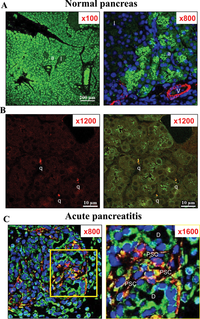
Figure 1. Differences of intracellular fractalkine receptor (CX3CR1) distribution in normal pancreas (Panel A, B) and in L-arginine induced acute pancreatitis (Panel C).
Expression of CX3CR1 (in green); glial fibrillary acidic protein (GFAP; in red) [quiescent pancreatic stellate cells (PSCs)], and alpha-smooth muscle actin (α-SMA; in red) [activated PSCs] in the pancreas of 15-week-old Wistar rats and the pancreas from L-arginine induced acute pancreatitis are examined by immunofluorescence staining. In the top two panels (A: in normal pancreas), figures are x100 and x800 each. α-SMA (in red) and CX3CR1 expression (in green) are shown. This figure demonstrates that CX3CR1 is expressed diffusely in acinar (a) and was also seen in intra-lobular duct cells but CX3CR1 is minimally expressed in the cytoplasm and the cell surface membrane of these cells in normal pancreas. Islets (I) and blood vessel cells (V) do not express CX3CR1. Blood vessel cells (V) express α-SMA, but no activated pancreatic stellate cells are seen. In the middle panels (B: in normal pancreas), figures are x1200 (left and right), and show a magnification of an area containing quiescent PSCs (q). GFAP (in red) and CX3CR1 expression (in green) are shown. Co-localization of CX3CR1 and GFAP is shown in yellow [CX3CR1 positive quiescent PSCs] In the bottom panels (C: in acute pancreatitis tissues), figures are x800 (left) and on the right x1600 (right). The area seen is shown a magnification of an area containing an increased numbers of activated PSCs (PSC) with acute pancreatitis. In the square on the left is shown at 1600× magnification on the right (Fig. 1C). Intracellular localization of CX3CR1differs from normal in that it is expressed on the cell surface membrane of acinar, duct (D) and activated PSCs (PSC). Co-localization of CX3CR1 and α-SMA shows yellow staining[CX3CR1 positive activated PSCs]. These pictures are representative immunofluorescent confocal microscopy images of four experiments.

