

Abstract
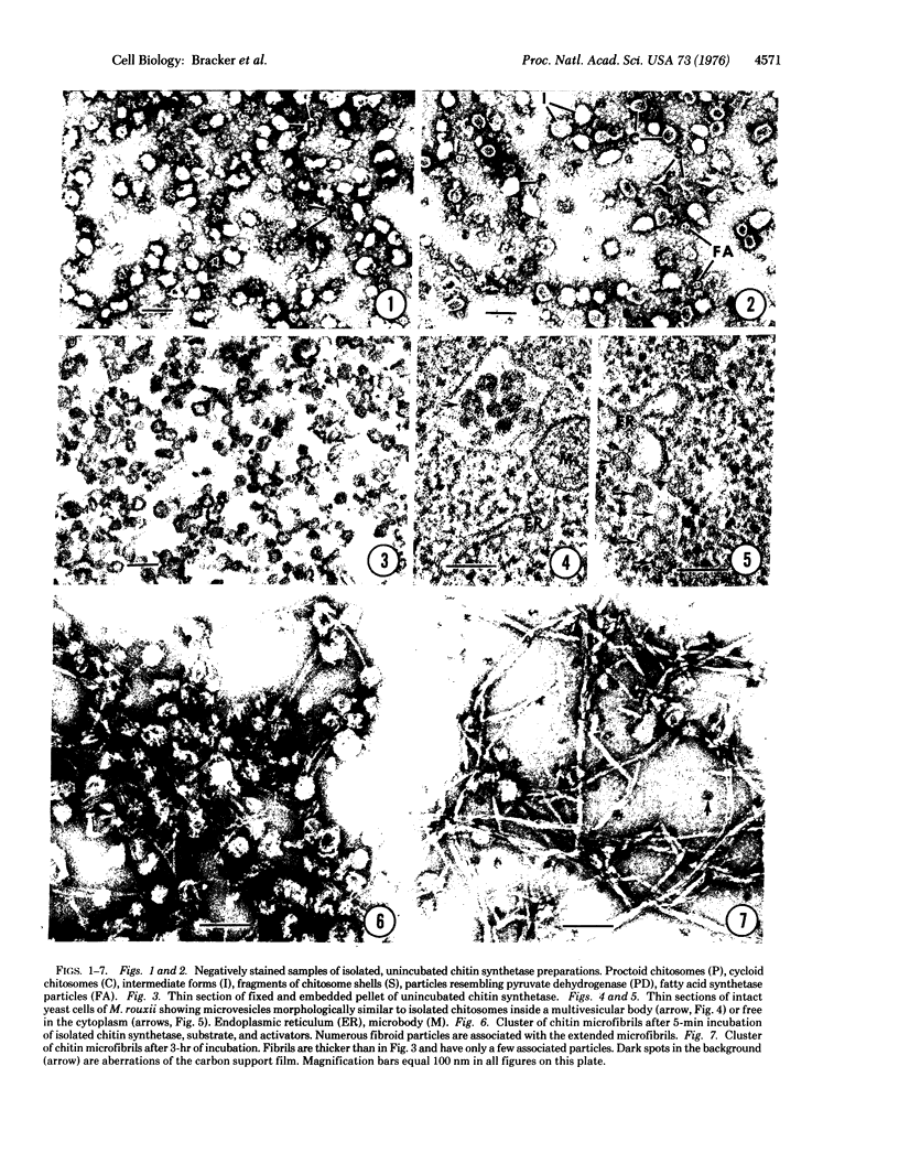
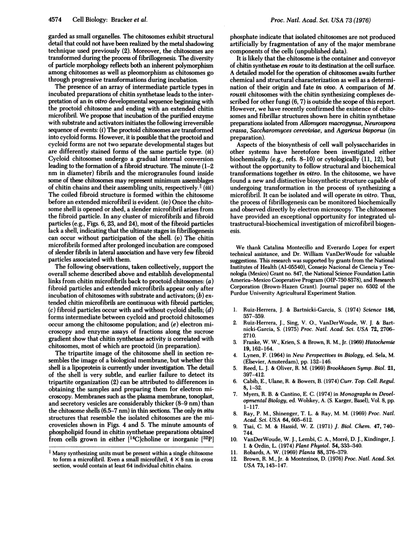
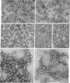
The fine structure of isolated chitin synthetase (UDP-2-acetamido-2-deoxy-D-glucose:chitin 4-beta-acetamido-deoxyglucosyltransferase; EC 2-4-1-16) particles (chitosomes) from Mucor rouxii and the elaboration of chitin microfibrils were studied by electron microscopy. Chitosomes are spheroidal, but often polymorphic, structures, mostly 40-70 nm in diameter. Their appearance after negative staining varies. Some reveal internal granular structure enclosed by a shell measuring 6-12 nm thick; others do not show internal structure but have a pronounced depression of the external surface. In thin sections, isolated chitosomes appear as microvesicular structures with a tripartite shell 6.5-7.0 nm thick. Morphologically similar structures can be seen in intact cells of M. rouxii. Isolated chitosomes undergo a seemingly irreversible series of transformations when substrate and activators are added. The internal structure changes, and a coiled microfibril (fibroid) appears inside the chitosome. The shell of the chitosome is opened or shed, and an extended microfibril arises from the fibroid particle. During prolonged incubation, the fibroid coils become less common and extended microfibrils appear thicker. We regard the chitosome as the cytoplasmic container and conveyor of chitin synthetase en route to its destination at the cell surface. Isolated chitosomes are well suited for integrated ultrastructural-biochemical studies of microfibril biogenesis in vitro.
Full text
PDF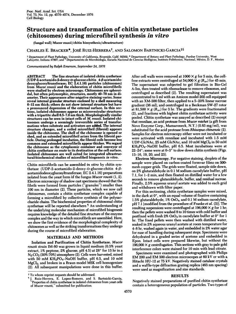
Images in this article
Selected References
These references are in PubMed. This may not be the complete list of references from this article.
- Brown R. M., Jr, Montezinos D. Cellulose microfibrils: visualization of biosynthetic and orienting complexes in association with the plasma membrane. Proc Natl Acad Sci U S A. 1976 Jan;73(1):143–147. doi: 10.1073/pnas.73.1.143. [DOI] [PMC free article] [PubMed] [Google Scholar]
- Cabib E., Ulane R., Bowers B. A molecular model for morphogenesis: the primary septum of yeast. Curr Top Cell Regul. 1974;8(0):1–32. doi: 10.1016/b978-0-12-152808-9.50008-0. [DOI] [PubMed] [Google Scholar]
- Franke W. W., Krien S., Brown R. M., Jr Simultaneous glutaraldehyde-osmium tetroxide fixation with postosmication. An improved fixation procedure for electron microscopy of plant and animal cells. Histochemie. 1969;19(2):162–164. doi: 10.1007/BF00281096. [DOI] [PubMed] [Google Scholar]
- Ray P. M., Shininger T. L., Ray M. M. ISOLATION OF beta-GLUCAN SYNTHETASE PARTICLES FROM PLANT CELLS AND IDENTIFICATION WITH GOLGI MEMBRANES. Proc Natl Acad Sci U S A. 1969 Oct;64(2):605–612. doi: 10.1073/pnas.64.2.605. [DOI] [PMC free article] [PubMed] [Google Scholar]
- Reed L. J., Oliver R. M. The multienzyme alpha-keto acid dehydrogenase complexes. Brookhaven Symp Biol. 1968 Jun;21(2):397–412. [PubMed] [Google Scholar]
- Ruiz-Herrera J., Bartnicki-Garcia S. Synthesis of cell wall microfibrils in vitro by a "soluble" chitin synthetase from Mucor rouxii. Science. 1974 Oct 25;186(4161):357–359. doi: 10.1126/science.186.4161.357. [DOI] [PubMed] [Google Scholar]
- Ruiz-Herrera J., Sing V. O., Van der Woude W. J., Bartnicki-Garcia S. Microfibril assembly by granules of chitin synthetase. Proc Natl Acad Sci U S A. 1975 Jul;72(7):2706–2710. doi: 10.1073/pnas.72.7.2706. [DOI] [PMC free article] [PubMed] [Google Scholar]
- Tsai C. M., Hassid W. Z. Solubilization and Separation of Uridine Diphospho-d-glucose: beta-(1 --> 4) Glucan and Uridine Diphospho-d-glucose:beta-(1 --> 3) Glucan Glucosyltransferases from Coleoptiles of Avena sativa. Plant Physiol. 1971 Jun;47(6):740–744. doi: 10.1104/pp.47.6.740. [DOI] [PMC free article] [PubMed] [Google Scholar]
- Van Der Woude W. J., Lembi C. A., Morré D. J. beta-Glucan Synthetases of Plasma Membrane and Golgi Apparatus from Onion Stem. Plant Physiol. 1974 Sep;54(3):333–340. doi: 10.1104/pp.54.3.333. [DOI] [PMC free article] [PubMed] [Google Scholar]