Abstract
AIM: To investigate the relationship between matrix metalloproteinase-2 (MMP-2) mRNA expression and clinicopathologic and urokinase-type plasminogen activator (uPA) system parameter and prognosis in human gastric cancer.
METHODS: Expression of MMP-2 mRNA, uPA, and uPA-R mRNA in tumor tissues and ≥5 cm adjacent normal tissues from 67 cases of gastric cancer was studied using RT-PCR and Northern blot respectively. Survival analyses were done using the Kaplan-Meier method.
RESULTS: The expression rates of MMP-2 mRNA, uPA and uPA-R mRNA in tumor tissues (31%, 41%, and 51%, respectively) were significantly higher than those in ≥5 cm adjacent tissues (19%, 11%, and 9%; χ2 = 4.59, 43.58, and 53.24 respectively, P<0.05, 0.0001, and 0.0001, respectively). Expression of MMP-2 mRNA was significantly correlated with lymph node metastasis (metastasis: 61.9%, no metastasis: 39.1%, χ2 = 7.61, P<0.05), Lauren’s classification of diffuse/mixed types: 54.2%, intestinal type: 26.3%, χ2 = 4.25, P<0.05, expression of uPA and uPA-R mRNA (uPA+: 55.1%, uPA-: 22.2% and uPA-R+: 54.9%, uPA-R-: 18.8%, χ2 = 5.72 and 6.40 respectively, P<0.05). Kaplan-Meier survival analysis of MMP-2 mRNA expression did not show significant difference in all 67 cases, but revealed an association of the expression of MMP-2 mRNA, uPA, and uPA-R mRNA with worse prognosis (P = 0.0083, 0.0160, and 0.0094, respectively).
CONCLUSION: MMP-2 may play an important role in the development of invasion and metastasis of gastric cancer.
Keywords: Gastric cancer, Matrix metalloproteinase-2, Urokinase-type plasminogen activator
INTRODUCTION
Gastric cancer is one of the leading malignancies in many countries. In spite of radical surgeries, such as sub-total or total gastrectomy combined with other modalities, its cure rate is quite low because of local invasion and metastasis, which are known to be associated with increased activities of several proteolytic enzymes, including matrix metalloproteinase (MMP) and plasminogen activators. MMP-2 (72 ku type IV collagenase) is a member of the MMP gene family which degrades the macromolecules of connective tissues and ECM, such as collagen, proteoglycans, laminin and fibronectin. Thus MMP-2 is believed to play an important role in tumor invasion and metastasis[1]. Our studies have shown that MMP-2 mRNA and protein are overexpressed in gastric cancer and related to the clinicopathologic parameters of cancer. In this context evaluation of MMP-2 expression in esophageal cancer, ovarian cancer, meningiomas and other tumors appears as a useful prognostic indicator[2-7]. MMP-2 has been detected preferentially on cells from advanced gastric carcinoma by immunohistochemistry and an association of MMP-2 with poor prognosis of cancer patients has been suggested[7].
There are two types of plasminogen activators, urokinase-type plasminogen activator (uPA) and tissue-type plasminogen activator. In particular, the uPA and its specific cell surface receptor uPA-R, play important roles in tumor invasion and metastasis. Tissue levels of uPA, uPA-R are higher in several malignant tumors than those of the corresponding adjacent normal tissues, such as gastric cancer and esophageal squamous cell carcinoma[8-10]. Many studies have shown that inhibition of uPA activity or uPA binding to uPA-R results in the suppression of tumor growth and reduces or abolishes formation of metastasis[11].
The current study was designed to examine the possible association between the expression of MMP-2 mRNA and clinicopathologic and uPA system parameters and prognosis in human gastric cancer.
MATERIALS AND METHODS
Samples
The samples of tumor tissue and ≥5 cm adjacent normal tissues from 67 gastric cancer patients were obtained from the Surgery Department of our hospital and People’s Hospital of Yunhe County, Zhejiang. Gastric tissues from 10 benign ulcer patients after partial gastrectomy were used as controls. The histological diagnosis was made by the pathologists of these two hospitals. Patients’ clinical features are shown in Table 1. The mean age was 62.2 years (SD 9.3, range 23-78 years), the male/female ratio was 1.79 (43/24). All samples were quenched in liquid nitrogen immediately after operation and then stored at -70 °C for study.
Table 1.
Relationship between MMP-2 mRNA expression and clinicopathologic and uPA system parameters.
| Variable | Case number | Expression of MMP-2 mRNA, n (%) | χ2 | P |
| Depth of invasion | ||||
| Early cancer | 11 | 3 (27.3) | 1.91 | >0.05 |
| Advanced cancer | 56 | 28 (50.0) | ||
| Differentiation | ||||
| Well-moderate | 16 | 6 (43.8) | 0.65 | >0.05 |
| Poor | 51 | 25 (47.1) | ||
| Lymph node metastasis | ||||
| Positive | 21 | 13 (61.9) | 7.61 | <0.01 |
| Negative | 46 | 18 (39.1) | ||
| Liver metastasis | ||||
| Positive | 8 | 6 (75.0) | 3.01 | >0.05 |
| Negative | 59 | 25 (42.4) | ||
| TNM stage | ||||
| I+II | 13 | 3 (23.1) | 3.49 | >0.05 |
| III+IV | 54 | 28 (51.9) | ||
| Lauren's classification | ||||
| Diffuse/mixed types | 48 | 26 (54.2) | 4.25 | <0.05 |
| Intestinal type | 19 | 5 (26.3) | ||
| uPA mRNA | ||||
| Positive | 49 | 27 (55.1) | 5.72 | <0.05 |
| Negative | 18 | 4 (22.2) | ||
| uPA-R mRNA | ||||
| Positive | 51 | 28 (54.9) | 6.40 | <0.05 |
| Negative | 16 | 3 (18.8) |
RT-PCR analysis of MMP-2 mRNA
dNTP, RNAsin and MMLV reverse transcriptase and Taq DNA were provided by Stratagene, La Jolla, CA, USA. MMP-2 primer pair was synthesized by Shanghai Cell Research Institute, Chinese Academy of Sciences and its sense and anti-sense were 5’-ACAAAGAGTGGCAGTG-CAA-3’ and 5’-CACGAGCAAAGGCATCATCC-3’ respectively. The expected size of MMP-2 product was 302 bp. Total RNA was extracted from frozen tissues by cesium chloride purifying method. A total amount of 20 μL reaction solution contained 5 μg RNA sample tissue, 1 mmol/L dNTP, 10 U RNAsin, 100 mmol/L Tris-HCl pH 8.4, 50 mmol/L KCl, 2.5 mmol/L MgCl2, 200 mg/mL BSA, 100 pole random six-polyoligonucleotide and 100 U MMLV reverse transcriptase. The reverse transcriptional condition was at 37 °C for 1 h, and at 95 °C for 5 min. Twenty microliters of cDNA reverse transcriptase product was put in PCR reaction solution containing 100 mmol/L MgCl2, 200 mg/mL BSA, 30 pmoL sense and anti-sense primers, and then 2 U Taq DNA polymerase was added in the solution. The PCR amplification conditions were: denaturation at 95 °C for 1 min, annealing at 65 °C for 1 min and extension at 72 °C for 1 min for 35 cycles. Ten microliters of DNA amplified product was subjected to electrophoresis in 4% agarose gel, stained with ethidium bromide and observed under ultraviolet light. The photographs of PCR results were used to measure the level of optical density (A) of MMP-2 cDNA bands by densitometry (Backman CD 2000).
Northern blot analysis of uPA and uPA-R mRNA
The probes of uPA and uPA-R cDNA encoding the PGEM recombinant plasmid of human uPA cDNA 387 bp Acc I-BgLII and uPAR cDNA 585 bp BamHI respectively were provided by Shanghai Sangon Biotechnology and β-actin DNA probe (1.0 kb) was provided by Huamei Biotechnology. All probes used in Northern blot analysis were labeled with α-32P-dCTP in Shanghai Sangon Biotechnology. Total RNA was isolated from frozen tumor tissue and ≥5 cm adjacent normal tissues using a guanidine isothiocyanate method[12]. Twenty-five micrograms of total RNA was fractionated in 0.8% agarose gel containing 1% formaldehyde at 30 V overnight, transferred to a nylon membrane and fixed by UV cross-linking. The filter was hybridized at 80 °C for 2 h with a radioactive labeled probe in ExpressHyb solution (Clontech), washed with 2×SSC/0.05% SDS for 40 min at room temperature and then with 0.1×SSC/0.1% SDS for 40 min at 50 °C. The probe was removed from the blot by incubating with 0.5% SDS in H2O at 90 °C for 10 min. The filter was then re-equilibrated in the ExpressHyb solution and reprobed with a new sequence. The hybridization signals were quantitated by scanning the autoradiograms with a laser densitometer (Computing Densitometer Model QTM970), and normalized to the quantity of mRNA and the specific activity of different probes.
Statistical analysis
The significance of differences in expression rates and A values among groups was determined by χ2 test and Student’s t-test respectively. Kaplan-Meier analysis was used to evaluate group oriented life-table curves which were confirmed by log-rank statistics. P<0.05 was considered statistically significant. All statistical analyses were performed using SPSS software.
RESULTS
Expression of MMP-2, uPA, and uPA-R mRNA in tumor and adjacent tissues
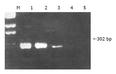
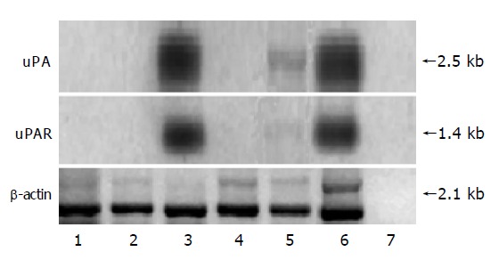
In 67 cases of gastric cancer, the positive rate of MMP-2 mRNA expression was significantly higher in tumor tissues (expressed in 31 cases) than in ≥5 cm adjacent normal ones (expressed in 19) (χ2 = 4.59, P<0.05, Figure 1). The A values of MMP-2 cDNA in 10 normal controls were very low and considered to be negative. The average A value of MMP-2 cDNA was 4.05 times higher in tumor tissues than in ≥5 cm adjacent normal tissues (t = 3.437, P<0.01). The positive rates of uPA and uPA-R mRNA expression were significantly higher in tumor tissues (expressed in 49 and 52 cases respectively) than in ≥5 cm adjacent normal (expressed in 11 and 9 respectively), (χ2 = 43.58 and 53.24 respectively, P<0.001). The average IOD value of uPA and uPA-R was 5.1 and 5.5 times higher in tumor tissues than in ≥5 cm adjacent normal ones (t = 3.841 and 4.026, P<0.001, Figure 2).
Figure 1.

Expression of MMP-2 mRNA in gastric cancerous tissues and tumor adjacent tissues. Lane M: DNA marker; lanes 1 and 2: gastric cancerous tissues where MMP-2 mRNA is overexpressed; lanes 3 and 4: tumor adjacent tissues; lane 5: normal control.
Figure 2.

Expression of uPA and uPA-R mRNA in gastric cancerous tissues and tumor adjacent tissues. Lanes 1 and 4: normal controls; lanes 2 and 5: tumor adjacent tissues; lanes 3 and 6: gastric cancerous tissues where uPA and uPA-R mRNA are overexpressed; lanes 7: empty control.
Relationship between MMP-2 mRNA expression and clinicopathologic and uPA system parameters (Table 1)
Although the expression rates of MMP-2 mRNA were lower in cases with early cancer, MMP-2 mRNA expression in tumor tissues correlated neither with depth of invasion, differentiation and liver metastasis nor with TNM stage. A significant correlation was seen, however, between lymph node metastasis, Lauren’s classification and positive expression of uPA and uPA-R mRNA (P<0.05).
Association between MMP-2mRNA expression and prognosis of patients
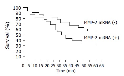
Kaplan-Meier survival analysis of MMP-2 mRNA detection did not show significant differences in 31 positive cases with a mean survival time (MST) of 30.41 mo and in 36 negative cases (MST: 46.51 months, P = 0.1321, Figure 3). Among the 48 cases of Lauren’s diffuse/mixed types, MST of 26 positive cases (25.05 mo) was significantly lower than that of 22 negative cases (51.77 mo, P = 0.0083). Among the 49 positive cases of uPA mRNA, MST of 27 positive cases (29.79 mo) was significantly lower than that of 22 negative cases (49.07 mo, P = 0.0160). Among the 51 positive cases of uPA-R mRNA, MST of 28 positive cases (25.82 mo) was significantly lower than that of 23 negative cases (52.11 mo, P = 0.0094).
Figure 3.

No significant differences between positive cases and negative cases in Kaplan–Meier survival analysis of MMP-2 mRNA.
DISCUSSION
Studies have shown that MMP-2 is mainly expressed in membrane of tumor cells while it cannot be detected in normal gastric mucosal cells and that MMP-2 positive cells are more numerous in poorly differentiated and advanced gastric cancers than in well differentiated and early gastric cancers[13-15]. Our previous study also revealed that MMP-2 mRNA and protein overexpressions are more significant in poorly differentiated and advanced gastric cancer cells than in well differentiated and early gastric cancer cells. In the current study, MMP-2 mRNA could not be detected in 10 cases of normal controls, demonstrating that in normal gastric tissues, MMP-2 is rarely expressed or the mRNA level is too low to be detected. Schwartz et al[16], reported that MMP-2 mRNA is expressed in invasive SK-GT1, SK-GT5 and SK-GT6 cell lines but not in noninvasive SK-GT2 and SK-GT4 cell lines. In ultrastructural study, MMP-2 mRNA is expressed markedly in cancer cells with rich false feet and rapid movement in culture, but insignificantly expressed in cancer cells with few false feet from unmetastatic and uninvasive gastric cancerous tissues, indicating that MMP-2 secretion is correlated with the invasion and metastasis of gastric cancer[17,18]. Studies have shown that downregulation of MMPs or reduction of MMP-2 expression results in inhibition of tumor growth and reduces or abolishes formation of metastasis[19,20]. In our study, the rates of positive MMP-2 mRNA expression were low in early gastric cancer with no liver metastasis and stages I and II gastric cancer, but there was no significant difference between MMP-2 mRNA expression and the degree of invasion, differentiation, liver metastasis and TNM stage of gastric cancer. However, the rates significantly increased in lymph node metastasis and diffuse/mixed types. This phenomenon implies that gastric cancer cells with more malignant and metastatic potential may secrete much more MMP-2 protein. Sier et al[21], reported that MMP-2 expression is significantly enhanced in gastric cancerous tissues compared with that in adjacent normal mucosal tissues. In our previous studies, adjacent normal mucosal tissues had lower MMP-2 mRNA positive expression and lower MMP-2 cDNA A than gastric cancerous tissues. This study also showed that MMP-2 mRNA expression and MMP-2 A were higher in gastric cancerous tissues than in ≥5 cm adjacent tissues. Although the levels of MMP-2 cDNA signals in tumor-adjacent tissues were lower than in tumor tissues, MMP-2 mRNA was overexpressed in tumor-adjacent tissues, suggesting that both gastric cancer cells and adjacent mesenchymal cells, including fibrocytes, endothelium cells, macrophages, and lymphocytes have the ability to secrete MMP-2. There may be information exchange between cancer cells and these mesenchymal cells through the dissolvable intercellular substances and membrane cement factors, and such information exchange may regulate the production of MMP-2. This may be very important in elucidating the mechanism of invasion and metastasis of cancer cells[22].
The role of MMP-2 protein and mRNA evaluation in the prognostic judgment of gastric cancer is still controversial. Mori et al[14], concluded that the expression of MT1-MMP may influence prognosis via tumor invasion of gastric wall and lymph node metastasis, and activation of MMP-2 may be clinically relevant to the progression of gastric carcinoma tumors. Caenazzo et al[15], suggested that the ratio of MT1-MMP and MMP-2 mRNA of gastric carcinoma tissue is a new preoperative molecular level prognostic factor for gastric carcinoma. Allgayer et al[5], showed that Kaplan-Meier survival analysis of immunohistochemical MMP-2 detection does not show significant differences in disease-free and overall survival of curatively resected patients. In diffuse types, however, a significant association of MMP-2 with disease-free and overall survival is revealed in curatively resected patients. These results demonstrate that there is an association of MMP-2 with prognosis of cancer patients. For diffuse gastric cancers, MMP-2 is a significant prognostic parameter; however, it is of no independent impact. In our study, the average survival period of 31 MMP-2 mRNA positive patients was shorter than that of negative ones, but the difference did not show statistical significance. In diffuse/mixed type, the average survival period was significantly shorter than negative ones, which was similar to the immunohistochemical results of Allgayer’s[5].
The uPA/uPA-R system not only takes part in tumor formation, but also plays an important role in tumor invasion and metastasis[23-26]. In this study, there was a significant difference in uPA and uPA-R mRNA expression between gastric cancer and adjacent tissues, and the IOD was significantly higher in cancer tissues. Some studies reported that the uPA and uPA-R protein activity in tumor center, tumor border and adjacent normal tissue determined by ELISA and immunohistochemistry decreases gradually, which is similar to the results of our study[23]. These studies show that either the protein or the mRNA level of uPA and/or uPA-R tends to decline following the decline of malignant degree of tumors. uPA-R as the membrane-bound center of the uPA system is able to concentrate on uPA activity by focusing on receptor-bound uPA and enzymes in the proteolytic cascade like MMP-2 may be activated, resulting in stroma protein hydrolysis and tumor cell invasion and metastasis[26-28]. Thus, MMP-2 and uPA/uPA-R system co-operate during tumor invasion and metastasis. Allgayer et al[5], reported a significant correlation between MMP-2 immunohistochemical detection and prognosis in cancers with uPA-R but not uPA overexpressed. While in our study, MMP-2 mRNA level in uPA and uPA-R positive patients increased significantly. In 49 uPA and 51 uPA-R mRNA positive patients, the survival time of MMP-2 positive patients was significantly shorter than that of negative ones, suggesting that MMP-2 can be designated as a prognostic factor when MMP-2 activated enzymes such as uPA/uPA-R are overexpressed[5,29]. In conclusion, MMP-2 mRNA expression may play important roles in gastric cancer formation, invasion, and metastasis.
References
- 1.Allgayer H, Babic R, Grützner KU, Beyer BC, Tarabichi A, Schildberg FW, Heiss MM. Tumor-associated proteases and inhibitors in gastric cancer: analysis of prognostic impact and individual risk protease patterns. Clin Exp Metastasis. 1998;16:62–73. doi: 10.1023/a:1006564002679. [DOI] [PubMed] [Google Scholar]
- 2.Yamashita K, Tanaka Y, Mimori K, Inoue H, Mori M. Differential expression of MMP and uPA systems and prognostic relevance of their expression in esophageal squamous cell carcinoma. Int J Cancer. 2004;110:201–207. doi: 10.1002/ijc.20067. [DOI] [PubMed] [Google Scholar]
- 3.Schmalfeldt B, Prechtel D, Härting K, Späthe K, Rutke S, Konik E, Fridman R, Berger U, Schmitt M, Kuhn W, et al. Increased expression of matrix metalloproteinases (MMP)-2, MMP-9, and the urokinase-type plasminogen activator is associated with progression from benign to advanced ovarian cancer. Clin Cancer Res. 2001;7:2396–2404. [PubMed] [Google Scholar]
- 4.Siddique K, Yanamandra N, Gujrati M, Dinh D, Rao JS, Olivero W. Expression of matrix metalloproteinases, their inhibitors, and urokinase plasminogen activator in human meningiomas. Int J Oncol. 2003;22:289–294. [PubMed] [Google Scholar]
- 5.Allgayer H, Babic R, Beyer BC, Grützner KU, Tarabichi A, Schildberg FW, Heiss MM. Prognostic relevance of MMP-2 (72-kD collagenase IV) in gastric cancer. Oncology. 1998;55:152–160. doi: 10.1159/000011850. [DOI] [PubMed] [Google Scholar]
- 6.Knappe UJ, Hagel C, Lisboa BW, Wilczak W, Lüdecke DK, Saeger W. Expression of serine proteases and metalloproteinases in human pituitary adenomas and anterior pituitary lobe tissue. Acta Neuropathol. 2003;106:471–478. doi: 10.1007/s00401-003-0747-5. [DOI] [PubMed] [Google Scholar]
- 7.Mönig SP, Baldus SE, Hennecken JK, Spiecker DB, Grass G, Schneider PM, Thiele J, Dienes HP, Hölscher AH. Expression of MMP-2 is associated with progression and lymph node metastasis of gastric carcinoma. Histopathology. 2001;39:597–602. doi: 10.1046/j.1365-2559.2001.01306.x. [DOI] [PubMed] [Google Scholar]
- 8.Kaneko T, Konno H, Baba M, Tanaka T, Nakamura S. Urokinase-type plasminogen activator expression correlates with tumor angiogenesis and poor outcome in gastric cancer. Cancer Sci. 2003;94:43–49. doi: 10.1111/j.1349-7006.2003.tb01350.x. [DOI] [PMC free article] [PubMed] [Google Scholar]
- 9.Park IK, Kim BJ, Goh YJ, Lyu MA, Park CG, Hwang ES, Kook YH. Co-expression of urokinase-type plasminogen activator and its receptor in human gastric-cancer cell lines correlates with their invasiveness and tumorigenicity. Int J Cancer. 1997;71:867–873. doi: 10.1002/(sici)1097-0215(19970529)71:5<867::aid-ijc27>3.0.co;2-3. [DOI] [PubMed] [Google Scholar]
- 10.Choi YK, Yoon BI, Kook YH, Won YS, Kim JH, Lee CH, Hyun BH, Oh GT, Sipley J, Kim DY. Overexpression of urokinase-type plasminogen activator in human gastric cancer cell line (AGS) induces tumorigenicity in severe combined immunodeficient mice. Jpn J Cancer Res. 2002;93:151–156. doi: 10.1111/j.1349-7006.2002.tb01253.x. [DOI] [PMC free article] [PubMed] [Google Scholar]
- 11.Holst-Hansen C, Johannessen B, Høyer-Hansen G, Rømer J, Ellis V, Brünner N. Urokinase-type plasminogen activation in three human breast cancer cell lines correlates with their in vitro invasiveness. Clin Exp Metastasis. 1996;14:297–307. doi: 10.1007/BF00053903. [DOI] [PubMed] [Google Scholar]
- 12.Sambrook J, Fritsch EF, Maniatis T. Molecular Cloning: A Laboratory Manual. 2nd Ed. New York: Cold Spring Harbor Laboratory Press; 1989. pp. 366–489. [Google Scholar]
- 13.Cai H, Kong ZR, Chen HM. Matrix metalloproteinase-2 and angiogenesis in gastric cancer. AiZheng. 2002;21:25–28. [PubMed] [Google Scholar]
- 14.Mori M, Mimori K, Shiraishi T, Fujie T, Baba K, Kusumoto H, Haraguchi M, Ueo H, Akiyoshi T. Analysis of MT1-MMP and MMP2 expression in human gastric cancers. Int J Cancer. 1997;74:316–321. doi: 10.1002/(sici)1097-0215(19970620)74:3<316::aid-ijc14>3.0.co;2-9. [DOI] [PubMed] [Google Scholar]
- 15.Caenazzo C, Onisto M, Sartor L, Scalerta R, Giraldo A, Nitti D, Garbisa S. Augmented membrane type 1 matrix metalloproteinase (MT1-MMP): MMP-2 messenger RNA ratio in gastric carcinomas with poor prognosis. Clin Cancer Res. 1998;4:2179–2186. [PubMed] [Google Scholar]
- 16.Schwartz GK, Wang H, Lampen N, Altorki N, Kelsen D, Albino AP. Defining the invasive phenotype of proximal gastric cancer cells. Cancer. 1994;73:22–27. doi: 10.1002/1097-0142(19940101)73:1<22::aid-cncr2820730106>3.0.co;2-o. [DOI] [PubMed] [Google Scholar]
- 17.Grigioni WF, D'Errico A, Fortunato C, Fiorentino M, Mancini AM, Stetler-Stevenson WG, Sobel ME, Liotta LA, Onisto M, Garbisa S. Prognosis of gastric carcinoma revealed by interactions between tumor cells and basement membrane. Mod Pathol. 1994;7:220–225. [PubMed] [Google Scholar]
- 18.Allgayer H, Heiss MM, Schildberg FW. Prognostic factors in gastric cancer. Br J Surg. 1997;84:1651–1664. [PubMed] [Google Scholar]
- 19.Zhang H, Morisaki T, Matsunaga H, Sato N, Uchiyama A, Hashizume K, Nagumo F, Tadano J, Katano M. Protein-bound polysaccharide PSK inhibits tumor invasiveness by down-regulation of TGF-beta1 and MMPs. Clin Exp Metastasis. 2000;18:343–352. doi: 10.1023/a:1010897432244. [DOI] [PubMed] [Google Scholar]
- 20.Denkert C, Siegert A, Leclere A, Turzynski A, Hauptmann S. An inhibitor of stress-activated MAP-kinases reduces invasion and MMP-2 expression of malignant melanoma cells. Clin Exp Metastasis. 2002;19:79–85. doi: 10.1023/a:1013857325012. [DOI] [PubMed] [Google Scholar]
- 21.Sier CF, Kubben FJ, Ganesh S, Heerding MM, Griffioen G, Hanemaaijer R, van Krieken JH, Lamers CB, Verspaget HW. Tissue levels of matrix metalloproteinases MMP-2 and MMP-9 are related to the overall survival of patients with gastric carcinoma. Br J Cancer. 1996;74:413–417. doi: 10.1038/bjc.1996.374. [DOI] [PMC free article] [PubMed] [Google Scholar]
- 22.Mizutani K, Kofuji K, Shirouzu K. The significance of MMP-1 and MMP-2 in peritoneal disseminated metastasis of gastric cancer. Surg Today. 2000;30:614–621. doi: 10.1007/s005950070101. [DOI] [PubMed] [Google Scholar]
- 23.Cho JY, Chung HC, Noh SH, Roh JK, Min JS, Kim BS. High level of urokinase-type plasminogen activator is a new prognostic marker in patients with gastric carcinoma. Cancer. 1997;79:878–883. [PubMed] [Google Scholar]
- 24.Plebani M, Herszènyi L, Carraro P, De Paoli M, Roveroni G, Cardin R, Tulassay Z, Naccarato R, Farinati F. Urokinase-type plasminogen activator receptor in gastric cancer: tissue expression and prognostic role. Clin Exp Metastasis. 1997;15:418–425. doi: 10.1023/a:1018454305889. [DOI] [PubMed] [Google Scholar]
- 25.Herouy Y, Aizpurua J, Stetter C, Dichmann S, Idzko M, Hofmann C, Gitsch G, Vanscheidt W, Schöpf E, Norgauer J. The role of the urokinase-type plasminogen activator (uPA) and its receptor (CD87) in lipodermatosclerosis. J Cutan Pathol. 2001;28:291–297. doi: 10.1034/j.1600-0560.2001.028006291.x. [DOI] [PubMed] [Google Scholar]
- 26.Krüger A, Soeltl R, Lutz V, Wilhelm OG, Magdolen V, Rojo EE, Hantzopoulos PA, Graeff H, Gänsbacher B, Schmitt M. Reduction of breast carcinoma tumor growth and lung colonization by overexpression of the soluble urokinase-type plasminogen activator receptor (CD87) Cancer Gene Ther. 2000;7:292–299. doi: 10.1038/sj.cgt.7700144. [DOI] [PubMed] [Google Scholar]
- 27.Heiss MM, Allgayer H, Gruetzner KU, Babic R, Jauch KW, Schildberg FW. Clinical value of extended biologic staging by bone marrow micrometastases and tumor-associated proteases in gastric cancer. Ann Surg. 1997;226:736–744; discussion 744-745. doi: 10.1097/00000658-199712000-00010. [DOI] [PMC free article] [PubMed] [Google Scholar]
- 28.Le DM, Besson A, Fogg DK, Choi KS, Waisman DM, Goodyer CG, Rewcastle B, Yong VW. Exploitation of astrocytes by glioma cells to facilitate invasiveness: a mechanism involving matrix metalloproteinase-2 and the urokinase-type plasminogen activator-plasmin cascade. J Neurosci. 2003;23:4034–4043. doi: 10.1523/JNEUROSCI.23-10-04034.2003. [DOI] [PMC free article] [PubMed] [Google Scholar]
- 29.Heiss MM, Babic R, Allgayer H, Gruetzner KU, Jauch KW, Loehrs U, Schildberg FW. Tumor-associated proteolysis and prognosis: new functional risk factors in gastric cancer defined by the urokinase-type plasminogen activator system. J Clin Oncol. 1995;13:2084–2093. doi: 10.1200/JCO.1995.13.8.2084. [DOI] [PubMed] [Google Scholar]


