Abstract
The purpose of this study was to investigate the effects of weight bearing at varying intervals in suppressing the progression of disuse muscle atrophy, while setting the total daily weight bearing time constant. Disuse muscle atrophy was induced by 2-week hindlimb suspension. Thirty male Wistar rats ( weight : 215 ± 8 g ) were divided into 6 groups ( 5 rats/group ) : control (CON), suspension alone (SUS), two sessions of 30-minute weight bearing at intervals of 4, 8, and 12 hours during suspension, and one session of 60-minute weight bearing at intervals of 24 hours during suspension. Weight bearing was done each day during the daytime. Histochemical staining, followed by morphometrical analysis using NIH Image, demonstrated that the cross-sectional area of type I fiber in SUS was 44% of that in CON, while in the weight bearing groups ranged from 62 to 70%. The proportion of type I fibers was lower in SUS and tended to increase as the interval of weight bearing become longer, indicating the effects of weight bearing at intervals of 12 and 24 hours. For both types I and II, the distribution of muscle fiber size indicated that weight bearing at longer intervals was more effective in keeping the cross-sectional areas of muscle fibers closed to that in CON. In conclusion, when suppressing the progression of disuse atrophy of rat soleus muscle by weight bearing of one hour per day, the results suggest that the weight bearing intervals is important factor.
Keywords: disuse muscle atrophy, weight bearing interval, soleus muscle
Bed rest plays an important role in the treatment of diseases. However, little movement and a decrease in the gravitation force applied in the direction of the axis of the trunk during bed rest can cause various functional disturbances which are called disuse syndrome16). In the field of space medicine, studies have been conducted concerning disuse syndrome induced under conditions of microgravity1)4)19). Although bed rest and stays in space differ in their background, their symptoms have many similarities. Therefore, knowledge of space medicine will also be useful in physical therapy.
In recent years, hindlimb suspension has been developed as an experimental model of disuse atrophy of the muscles17) and has been used in studying changes in skeletal muscles during stays in space and in seeking countermeasures against such atrophy8)10)24). Treadmill9)13)18), centrifugal5)6) and resistant exercises11)14)27) have been reported by many investigators to be countermeasures against disuse atrophy. Clinically, however, it is often difficult for long-term bed-resting patients to take exercise. Unlike in space where gravity can not be used as a therapeutic means, weight bearing is possible for patients on earth and can be utilized efficiently by physical therapists. It seems therefore clinically significant to study how to make efficient use of weight bearing to counteract disuse atrophy of the muscles.
Brown et al.3) reported that loss of muscle mass and strength during the unweighted period can be reduced by weight bearing for one hour each day. Widrick et al.26) reported that atrophy of type I fibers during a 2-week unweighted period could be reduced by loading with weights four times (10 minutes each time) per day. During the unweighted period, longer weight bearing will result in greater suppression of atrophy22). However, considering the reality of clinical situations, we think it unreasonable to advise bed-resting patients to bear weights for many hours. For this reason, we adopted one-hour daily weight bearing as a means of suppressing disuse atrophy. As we have described in our previous report32), one-hour weight bearing per day during a 2-week unweighted period suppressed the progression of atrophy although not completely. Following that study, we examined differences in the atrophy suppressing effect of different frequencies of weight bearing, to explore the optimum method of weight bearing. That study revealed that a single session of weight bearing per day was more effective than two sessions of weight bearing at intervals of 4 hours per day29). And our previous study of weight bearing frequency per week indicated that every day was more effective than every other day28)30). It was also suggested however that the intervals of weight bearing would more greatly determine the atrophy preventive effects than does the frequency of weight bearing. And various sessions of weight bearing can be used for this kind of attempt (e.g., four sessions of 15-minute weight bearing per day)2)26). In the present study, session number which were convenient for the physical therapists were selected.
The present study was therefore undertaken to assess the effects of weight bearing at varying intervals in suppressing the progression of disuse atrophy of muscles, while setting the total daily weight bearing time constant.
Methods
Thirty 8-week-old male Wistar rats (weight: 215 ± 8 g) were used for this experiment.
Disuse atrophy of muscles was induced by hindlimb suspension, that is, by applying a simplified jacket31) to the animal to keep the trunk and pelvis of the animal suspended for 2 weeks while its hindlimbs being unable to touch the floor. During suspension, each rat was able to move on his forelimbs and was allowed free access to food and water.
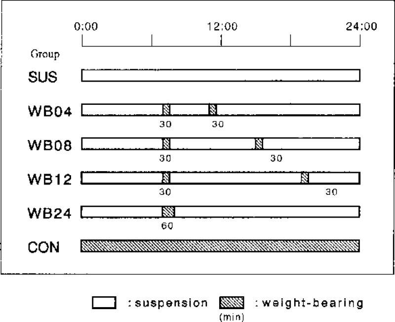
The rats were divided into 6 groups (5 rats/group) and individually housed in cages (280 × 440 × 180 mm). Animals which received 2-week suspension alone were assigned to the suspension group (SUS group). Animals which received no suspension were assigned to the control group (CON group). The other animals received 2-week suspension and weight bearing as follows : WB04 group (two sessions of 30-minute weight bearing at intervals of 4 hours during the suspension period), WB08 group (two sessions of 30-minute weight bearing at intervals of 8 hours), WB12 group (two sessions of 30-minute weight bearing at intervals of 12 hours), and WB24 group (one session of 60-minute weight bearing at internals of 24 hours). The rats were put on a reversed 12:12-h dark-light cycle. Weight bearing was induced by temporarily removing the suspension to allow the animal to bear its weight on all 4 of its limbs. This was done each day during the light cycle when rats are relatively inactive. Fig. 1 shows the daily weight bearing protocol for each group. The time from the start of one session of weight bearing to the start of the next session was deemed to be the weight bearing interval in this study. The interval of weight bearing was therefore 4+20 hours for WB04 group, 8+16 hours for WB08 group, and 12+12 hours for WB12 group. The total weight bearing time in the weight-bearing groups (WB04, WB08, WB12, and WB24 groups) was set at one hour per day. This study was undertaken pursuant to the guidelines for the care and use of laboratory animals in Takara-machi Campus of Kanazawa University.
Fig. 1.
Protocol of daily weight-bearing.
After 2 weeks of the experiment, pentobarbital sodium (50 mg/kg body weight) was injected intraperitoneally into each rat to effect anesthesia. The body weight was measured, and the soleus muscle was removed from each hindlimb. The wet weight of the muscles was measured. The muscles were then rapidly frozen in isopentane cooled by liquid nitrogen and stored at—80 °C until use.
Transverse frozen sections, 10 µm thick, were prepared for histochemical examination. The sections were subjected to ATPase staining (pH 10.6) to assess their muscle fiber types (I and II). The microscope pictures of the stained sections were later input into a computer (LC630, Macintosh) using a scanner (GT-9000ART, Epson), for analysis using image analyzing software (the public domain NIH Image program). The cross-sectional area of more than 200 muscle fibers in each muscle was measured, and the proportion of each type in number of muscle fiber was calculated.
Results were analyzed using a one-way analysis of variance (ANOVA). If significance was achieved (p<0.05), pairwise comparisons were performed using Scheffe's method.
Results
The wet weight of the soleus muscle was significantly smaller in SUS group and the weight bearing groups than in CON group, indicating that the unweighted period caused marked disuse atrophy of the muscles. The wet weight of the soleus muscle in WB04 and WB08 groups did not differ significantly from that in SUS group, while that in WB12 and WB24 groups was significantly greater than that in SUS group. Thus, weight bearing at intervals of 12 and 24 hours resulted in marked suppression of disuse atrophy of the muscle (Table 1). Since the wet weight of muscles is affected by body weight, we then analyzed changes in the relative weight of the soleus muscle. The weight of the soleus muscle relative to body weight was significantly greater in the weight bearing groups than in SUS group, indicating the effects of weight bearing. This parameter did not differ between WB24 and CON groups (Table 1).
Table 1. Muscle wet weight and muscle-to-body weight ratio.
| Group | Soleus weight (mg) | Soleus weight/body weight (mg/100 g) |
|---|---|---|
| SUS | 59.1 ± 9.5 | 29.8 ± 4.2 |
| WB04 | 69.3 ± 6.4 | 36.6 ± 3.8* |
| WB08 | 69.5 ± 7.3 | 37.3 ± 3.2* |
| WB12 | 72.4 ± 4.7* | 39.3 ± 1.8* |
| WB24 | 76.8 ± 8.0* | 41.2 ± 4.7* |
| CON | 124.5 ± 5.6* | 42.7 ± 2.2* |
Values are means ± SD.
p<0.05 (compared with SUS).
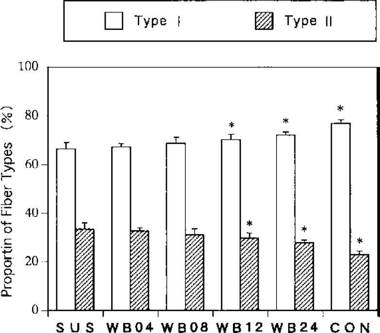
When the proportion of each type of muscle fiber was analyzed, the proportion of type I fibers was lower in SUS group and tended to increase as the interval of weight bearing became longer. This parameter in WB12 and WB24 groups was significantly higher than that in SUS group, indicating the effects of weight bearing at intervals of 12 and 24 hours (Fig. 2).
Fig. 2.
Percentage of muscle fiber types. Values are means ± SD. *p<0.05 (compared with SUS).
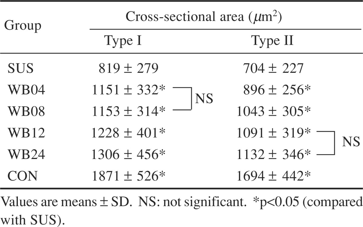
For both type I and II muscle fibers, the average cross- sectional area increased as the weight bearing interval became longer. This parameter for type I fiber in SUS group was 44% of that in CON group, while this parameter in the weight bearing groups ranged from 62 to 70% of that in CON group. This parameter did not differ significantly between WB04 and WB08 groups, while it differed significantly between any two of the other groups. The average cross-sectional area of type II fibers in SUS group was 42% of that in CON group, while this parameter in the weight bearing groups was 53–67% of that in CON group. In terms of this parameter, there was a significant difference between any two of all groups excluding WB12 and WB24 groups (Table 2).
Table 2. Cross-sectional area of muscle fibers.
 |
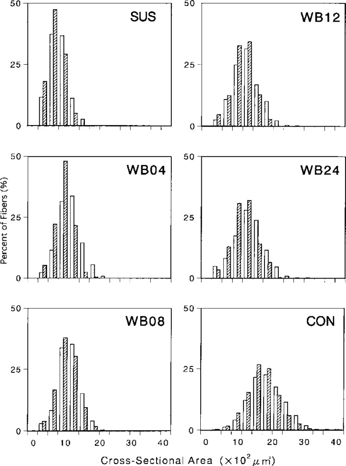
Fig. 3 graphically represents the distribution of cross- sectional areas of muscle fibers for all measured fibers. For both types I and II, the distribution was shifted to the left in SUS group, compared to CON group. The range of distribution was narrower in SUS group, indicating marked muscle atrophy. The distribution for weight bearing groups was intermediate between that for SUS group and that for CON group, indicating that progression of muscle atrophy was suppressed in weight bearing groups although not completely. The graph also shows that weight bearing at intervals of 12 and 24 hours is effective in keeping the cross-sectional areas of muscle fibers close to that in CON group.
Fig. 3.
Fiber size distribution in each group. The abbreviations are the same as in Fig. 2.
Discussion
In the present study, the effects of weight bearing on disuse atrophy of the muscles during unweighted periods were analyzed in relation to the intervals of weight bearing. The total weight bearing time was set at one hour per day. The effect of weight bearing in suppressing disuse atrophy is expected to increase as the weight bearing time becomes longer22). However, it is often difficult to advise bed-resting patients to bear weight for many hours. For this reason, we set the total weight bearing time at one hour per day, as in our previous studies28–30)32). Weight bearing was adopted because it is an easy method2) to use with long-term bed-resting patients and elderly patients. It seems unlikely that progression of atrophy can be suppressed completely by daily one-hour weight bearing. It is, however, likely that if atrophy can be suppressed to some extent, the time required for recovery will be shortened3). The soleus muscle was selected because this antigravity muscle is prone to the influence of unweighting, because the proportions of each type of muscle fiber constituting this muscle are relatively uniform, and because it serves as a key muscle in walking.
A number of reports have been published concerning the atrophy of rat soleus muscle caused by hindlimb suspension. The change of soleus muscle fibers from type I to II following suspension and the degree of decrease in the cross-sectional area of the fibers, observed in the present study, were approximately consistent with previous reports22)26). Because the relative weight of the soleus muscle, compared to body weight, did not differ between WB24 and CON groups, it seems likely that weight bearing allows quantitative prophylaxis of muscle atrophy. However, since some investigators have reported an increase in connective tissue and intramuscular fat following suspension3), a more detailed study is needed before making any conclusion. Regarding the intervals of weight bearing, our analysis of the proportions of each type of muscle fiber suggested the effects of weight bearing when the interval was 12 and 24 hours. The cross-sectional area of type I fibers did not differ between WB04 and WB08 groups. In a previous study28)30), weight bearing at intervals of 24 hours (i.e., once every day) was more effective than weight bearing at intervals of 48 hours (i.e., every other day). These results indicate that weight bearing at intervals of 12 and 24 hours is effective. And this suggests the importance of weight bearing intervals in the suppression of the progression of disuse muscle atrophy. The cross-sectional area of type II fibers, however, did not differ between WB12 and WB24 groups. This suggests that the responses to weight bearing vary between different types of muscle fibers.
During unweighted conditions, produced by hindlimb suspension, the ankle joint shows plantar flexion, and the soleus muscle becomes shortened position20). At the same time, the weight-supporting function is not required of the muscles. When weight is loaded during the unweighted period, the soleus is stretched, and its work to support body weight increases. These factors may be major ones involved in the effects of weight bearing on the soleus muscle15). During weight bearing, exercise by movement can also take place. Riley et al.19), however, have reported that rats after re-weighting moved little, when their responses were studied on video tape. Also in the present study, the animals moved little during the weight bearing periods, except for the short period immediately after the beginning of weight bearing. Therefore, little exercise effects are expected of weight bearing.
Factors which prevent or reduce muscle weakness include the method, intensity, duration and frequency of exercise. Regarding the effects of centrifugation in counteracting atrophy of the soleus muscle, it has been reported that the duration, frequency and muscular activity level are more important than the intensity of exercise5)6). In the present study, the total weight bearing time (one-hour per day) and the intensity (body weight) were kept constant, and the duration and frequency of weight bearing were changed, to identify the optimum weight bearing intervals. The study revealed that weight bearing at intervals of 12 and 24 hours is effective in suppressing the progression of atrophy. One possible reason for this result is the influence of inflammation process in muscular tissue12). Krippendorf et al.15) analyzed macrophage accumulation in the interstitial tissue and found that the degree of muscular degeneration caused by re-weighting was dependent on the duration of re-weighting. St.Pierre et al.23) examined the effects of re-weighting on the basis of the analysis of macrophage kinetics. They found that ED1 (cytoplasmic antigen) increased 2 days after the start of unweighting when the incidence of fiber necrosis was high (suggesting its relationship to necrosis), and that ED2 (surface membrane antigen) accumulated 4 and 7 days after the start of unweighting (suggesting its involvement in regeneration). Our previous immunohistochemical study29) also revealed marked macrophage accumulation in animals which bore weights at intervals of 4 hours. Schultz et al. 21) suggested that satellite cells serves as a sensitive and useful indicator of atrophic changes. They also say that changes in satellite cells appeared 12–24 hours after the beginning of unweighting. The experimental conditions in the present study were more complex, because cycles of unweighting and re-weighting periods were repeated every day. Other possible factors affecting the results of the present study include an increase in stress25) associated with the frequency of weight bearing and the effect of the autonomic nervous system7). Weight bearing at intervals of 12 hours is clinically difficult. Therefore, when atrophy of the soleus muscle is to be prevented by daily one hour weight bearing, it seems better to practice it in one session rather than dividing it in two.
The present study was designed to assess the effects of weight bearing on the skeletal muscles of lower extremities in relation to the weight bearing intervals. Needless to say, systemic conditions and bones can also affect the outcome of physical therapy. It also needs to be borne in mind that atrophy can not be completely prevented by short-term weight bearing alone. It seems therefore necessary to combine weight bearing with some other methods when counteracting atrophy of muscles efficiently. To this end, it is essential to evaluate the characteristics of muscle contraction, to analyze myosin heavy chains and to make a qualitative study of inflammation process. It is desirable to assess the effects of weight bearing on muscles other than the soleus muscle (e.g., gastrocnemius and extensor digitorum longus muscles), aged muscles and atrophic muscles.
In conclusion, when suppressing the progression of disuse atrophy of rat soleus muscle by weight bearing for the same amount of time per day, the weight bearing interval is important and weight bearing at intervals of 12 and 24 hours will be effective.
Acknowledgement.
This study was supported in part by Grant-in-Aid for Scientific Research (No. 08771113, 1996.), The Ministry of Education, Science, Sports and Culture, Japan.
References
- 1). Allen DL, Yasui W, et al. : Myonucler number and myosin heavy chain expression in rat soleus single muscle fibers after spaceflight. J Appl Physiol 81: 145–151, 1996. [DOI] [PubMed] [Google Scholar]
- 2). Alley KA, Thompson LV: Influence of simulated bed rest and intermittent weight bearing on single skeletal muscle fiber function in aged rats. Arch Phys Med Rehabil 78: 19–25, 1997. [DOI] [PubMed] [Google Scholar]
- 3). Brown M, Hasser EM: Weight-bearing effects on skeletal muscle during and after simulated bed rest. Arch Phys Med Rehabil 76: 541–546, 1995. [DOI] [PubMed] [Google Scholar]
- 4). Caiozzo VJ, Haddad F, et al. : Microgravity-induced transformations of myosin isoforms and contractile properties of skeletal muscle. J Appl Physiol 81: 123–132, 1996. [DOI] [PubMed] [Google Scholar]
- 5). D'Aunno DS, Thomason DB, et al. : Centrifugal intensity and duration as countermeasures to soleus muscle atrophy. J Appl Physiol 69: 1387–1389, 1990. [DOI] [PubMed] [Google Scholar]
- 6). D'Aunno DS, Robinson RR, et al. : Intermittent acceleration as a countermeasure to soleus muscle atrophy. J Appl Physiol 72: 428–433, 1992. [DOI] [PubMed] [Google Scholar]
- 7). Fagette S, Somody L, et al. : Central and peripheral sympathetic activities in rats during recovery from simulated weightlessness. J Appl Physiol 79: 1991–1997, 1995. [DOI] [PubMed] [Google Scholar]
- 8). Fitts RH, Metzger JM, et al. : Model of disuse: a comparison of hindlimb suspension and immobilization. J Appl Physiol 60: 1946–1953, 1986. [DOI] [PubMed] [Google Scholar]
- 9). Hauschka EO, Roy RR, et al. : Periodic weight support effects on rat soleus fibers after hindlimb suspension. J Appl Physiol 65: 1231–1237, 1988. [DOI] [PubMed] [Google Scholar]
- 10). Hauschka EO, Roy RR, et al. : Size and metabolic properties of single muscle fibers in rat soleus after hindlimb suspension. J Appl Physiol 62: 2338–2347, 1987. [DOI] [PubMed] [Google Scholar]
- 11). Herbert ME, Roy RR, et al. : Influence of one-week hindlimb suspension and intermittent high load exercise on rat muscles. Exp Neurol 102: 190–198, 1988. [DOI] [PubMed] [Google Scholar]
- 12). Kasper CE: Sarcolemmal disruption in reloaded atrophic skeletal muscle. J Appl Physiol 79: 607–614, 1995. [DOI] [PubMed] [Google Scholar]
- 13). Kasper CE, White TP, et al. : Running during recovery from hindlimb suspension induces transient muscle injury. J Appl Physiol 68: 533–539, 1990. [DOI] [PubMed] [Google Scholar]
- 14). Kirby CR, Ryan MJ, et al. : Eccentric exercise training as a countermeasure to non-weight-bearing soleus muscle atrophy. J Appl Physiol 73: 1894–1899, 1992. [DOI] [PubMed] [Google Scholar]
- 15). Krippendorf BB, Riley DA: Distinguishing unloading- versus reloading-induced changes in rat soleus muscle. Muscle Nerve 16: 99–108, 1993. [DOI] [PubMed] [Google Scholar]
- 16). Mita K: Disuse syndrome in space. Sogo Rihabiriteshon 25: 49–50, 1997. [Google Scholar]
- 17). Morey ER: Spaceflight and bone turnover: correlation with a new rat model of weightlessness. Bioscience 29: 168–172, 1979. [Google Scholar]
- 18). Pierotti DJ, Roy RR, et al. : Influence of 7 days of hindlimb suspension and intermittent weight support on rat muscle mechanical properties. Aviat Space Environ Med 61: 205–210, 1990. [PubMed] [Google Scholar]
- 19). Riley DA, Ellis S, et al. : In-flight and postflight changes in skeletal muscles of SLS-1 and SLS-2 spaceflown rats. J Appl Physiol 81: 133–144, 1996. [DOI] [PubMed] [Google Scholar]
- 20). Riley DA, Slocum GR, et al. : Rat hindlimb unloading: soleus histochemistry, ultrastructure, and electromyography. J Appl Physiol 69: 58–66, 1990. [DOI] [PubMed] [Google Scholar]
- 21). Schultz E, Darr KC, et al. : Acute effects of hindlimb unweighting on satellite cells of growing skeletal muscle. J Appl Physiol 76: 266–270, 1994. [DOI] [PubMed] [Google Scholar]
- 22). Someya F, Tachino K: Effect of various daily weight-bearing periods on rat soleus muscle during hindlimb suspension: histochemical and mechanical properties. Jpn J Rehabil Med 34: 410–417, 1997. [Google Scholar]
- 23). St Pierre BA, Tidball JG: Differential response of macrophage subpopulations to soleus muscle reloading after rat hindlimb suspension. J Appl Physiol 77: 290–297, 1994. [DOI] [PubMed] [Google Scholar]
- 24). Talmadge RJ, Roy RR, et al. : Distribution of myosin heavy chain isoforms in non-weight-bearing rat soleus muscle fibers. J Appl Physiol 81: 2540–2546, 1996. [DOI] [PubMed] [Google Scholar]
- 25). Thomason DB, Booth FW: Atrophy of the soleus muscle by hindlimb unweighting. J Appl Physiol 68: 1–12, 1990. [DOI] [PubMed] [Google Scholar]
- 26). Widrick JJ, Bangart JJ, et al. : Soleus fiber force and maximal shortening velocity after non-weight bearing with intermittent activity. J Appl Physiol 80: 981–987, 1996. [DOI] [PubMed] [Google Scholar]
- 27). Widrick JJ, Fitts RH: Peak force and maximal shortening velocity of soleus fibers after non-weight-bearing and resistance exercise. J Appl Physiol 82: 189–195, 1997. [DOI] [PubMed] [Google Scholar]
- 28). Yamazaki T: Effect of weight-bearing on disuse muscle atrophy in rats. Rigaku ryohogaku 23: 417–420, 1996. [Google Scholar]
- 29). Yamazaki T, Haida N, et al. : Effect of weight-bearing frequency per day in retarding disuse atrophy in rat soleus muscle. Rigaku ryoho janaru 30: 53–57, 1996. [Google Scholar]
- 30). Yamazaki T, Haida N, et al. : Effect of weight-bearing in prevention of disuse atrophy in rat hindlimb muscles: study of weight-bearing frequency in a week. Rigaku ryohogaku 22: 108–113, 1995. [Google Scholar]
- 31).Yamazaki T, Tachino K, et al. : Effect of short duration stretching under anesthesia in preventing disuse muscle atrophy in rats. Rigaku ryoho janaru 29: 135–138, 1995. [Google Scholar]
- 32).Yamazaki T, Tachino K, et al. : Effect of weight-bearing on disuse muscle atrophy in rats: study of weight-bearing time in a day. Memoirs Al Med Prof Kanazawa Univ 17: 63–67, 1993. [Google Scholar]


