Abstract
Arteritis due to Strongylus vulgaris is a well-known cause of colic in horses and donkeys. The current report describes a fatal incidence of arterial obstruction in cranial mesenteric artery caused by S. vulgaris infection in an adult donkey in which anthelmintic treatment was not regularly administered. Necropsy findings of the abdominal cavity revealed a complete cranial mesenteric arterial obstruction due to larvae of S. vulgaris, causing severe colic. To the authors’ knowledge, a complete cranial mesenteric arterial obstruction due to verminous arteritis has rarely been described in horses and donkeys. Based on recent reports of fatal arterial obstruction due to S. vulgaris infection in donkeys, it may be evident to consider acute colic caused by this pathogenic parasite a re-emerging disease in donkeys and horses.
Keywords: Colic, Strongylus vulgaris, Arterial obstruction
Introduction
Strongylus vulgaris has long been considered as one of the most prevalent and pathogenic parasites of the horse. Adult worms live in the cecum and right ventral colon. Fourth (L4) and fifth (L5) stage larvae are responsible for arteritis, necrosis, and fibrosis of the cranial mesenteric artery and its branches (1). Anthelmintic treatment strategies designed to control S vulgaris have been extremely successful in reducing prevalence, morbidity, and mortality from this parasite (2). There are 547134 Donkeys in Iran which most of them were used for agricultural work (www.ivo.org.ir). They were not given supplementary feed, but grazed around the village or in communal camps. However, little is known about the socioeconomics, health, nutrition, breeding or management of these animals. A surprisingly high prevalence of S vulgaris (33.3%) was found in a post-mortem survey in a recent study in Iranian donkeys (3).
This report describes a fatal incidence of arterial obstruction in cranial mesenteric artery caused by S. vulgaris infection in an adult donkey in which anthelmintic treatment was not regularly administered.
Case presentation
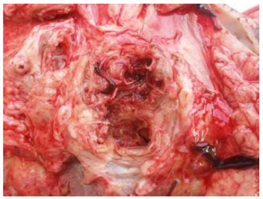
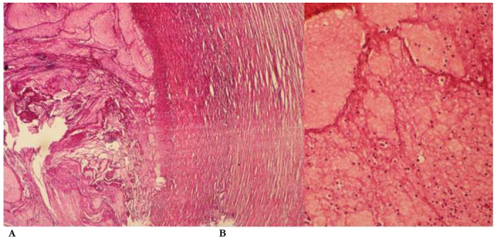
An 8-year-old donkey was referred to the Ferdowsi University of Mashhad veterinary teaching hospital, with a 24-hour history of colic and grazing on pastures. The donkey had shown signs of abdominal pain (rolling, pawing, flank watching, lying down); and already was treated with intravenous flunixin meglumine, mineral oil and water given by a nasogastric tube. At admission, the donkey was dull and did not exhibit overt signs of pain. Clinical examination revealed an elevated heart rate (60 beats/min) and pyrexia (rectal temperature 39.4 °C), dark pink mucous membranes with a capillary refill time of 3 seconds, increased skin turgor, and reduced intestinal sounds. Hematocrit was 40.6% and total protein was 64 g/L. Distended but flaccid loops of small intestine were detected on rectal palpation. Monitoring during the next few hours revealed a decreasing heart rate (50 beats/min) and rectal temperature (fluctuating between 38.2 °C and 38.4 °C), and the donkey seemed brighter. However, 3 days after admission, the donkey was died due to severe colic. Necropsy findings of the abdominal cavity revealed a complete cranial mesenteric arterial obstruction (Fig 1). In transverse section, the lumen of the cranial mesenteric artery was seen to contain reddish brown, loose, granular to fibrillar masses of thrombotic material. This was admixed with numerous grayish white, partly transparent nematode larvae, approximately 1-2 cm in length and 1 mm in thickness. Based on their size and intravascular location, they were interpreted to be fifth stage larvae of S. vulgaris. A segment of the arterial mesentery containing larva and its branches was excised and submitted for pathology. In histolopathological examination (Fig 2), many small to medium sized arteries were seen to be filled with thrombi and infiltration of inflammation cells. Moderate to large numbers of inflammatory cells dominated by neutrophils were present in the thrombus. Furthermore, the main lesions in other tissues such as liver, kidney and intestine were severe hemorrhage and inflammation.
Fig 1.

Cross section of cranial mesenteric artery showing strongylus vulgaris larva
Fig 2.

Histopathological findings in mesentric arteries. (A) Cranial mesentric artery with thrombus (H&E×40) (B) Close-up of neutrophil infiltrates in the vessel wall and thrombus (H&E×200)
Discussion
To the authors’ knowledge, a complete cranial mesenteric arterial obstruction due to verminous arteritis has rarely been described in horses and donkeys. In the current case, a complete section of the artery was obstructed due to arterial thrombi and strongylus vulgaris larva.
S. vulgaris has a long prepatent period (about 6–7 months) and anthelmintic drugs are able to remove both adult worms from the intestinal lumen and larvae from arteries. If reinfection occurs, larvae reach the cranial mesenteric arterial system branches 8 days post-infection, and remain there for about 3 months (4). Currently, Iranian donkeys are “randomly” treated, independently of epidemiological risk and faecal examination. If animals are grazing in an infected pasture, they can ingest larvae between treatments and migrating larvae can damage arterial walls. The findings of larvae in the current case indicate that the donkey was infected 2-4 months before appearance of clinical signs.
The frequent use of modern anthelmintic drugs has resulted in the selection of drug-resistant small strongyles and roundworms, leading to recognition of anthelmintic resistance among horse intestinal nematodes worldwide (2). Still, S vulgaris is considered fully susceptible to the anthelmintic drug classes available (5). To reduce the emergence of anthelmintic resistance, a selective approach of parasite management is encouraged. Strategic intestinal parasite management relies on treating only the horses, whereas donkeys are left untreated (6). The donkey owners were generally of the opinion that the animals do not get sick and consequently did not often treat them. Only few owners were aware that donkeys get worms, and no owners mentioned any occurrence of infectious diseases. This approach may render donkeys more prone to S. vulgaris infection, and leads to an increased strongyle eggs in pasture and need for monitoring of this highly pathogenic parasite (5).
Acute colic due to S. vulgaris usually affects young animals, as most horses and donkeys acquire relative immunity against the large strongyles as a consequence of natural infection (7). However, individual adult horses and donkeys may also be susceptible to this parasite, as demonstrated by the current case. The current case demonstrates the continued importance of this highly pathogenic parasite in donkeys and horses. In addition, information on basic donkey management and health were identified as needs. Provision of basic veterinary services and availability of training for owners about the correct treatment of their donkeys is also a priority identified by owners.
Acknowledgments
This work was carried out at the Hospital Reference Centre and Parasitology and Histopathology Section, Faculty of Veterinary Medicine, Ferdowsi University of Mashhad. We thank Mohammadnejad M. for photomicrographs and Eshrati H. and Azari GH. for assistance in parasite identification. The authors declare that there is no conflict of interests.
References
- 1.Duncan JL, Pirie HM. The life cycle of Strongylus vulgaris in the horse. Res Vet Sci. 1972;13:82–93. [PubMed] [Google Scholar]
- 2.Kaplan RM. Anthelmintic resistance in nematodes of horses. Vet Res. 2002;33:491–507. doi: 10.1051/vetres:2002035. [DOI] [PubMed] [Google Scholar]
- 3.Hosseini SH, Meshgi B, Eslami A, Bokai S, Sobhani M, Ebrahimi Samani R. Prevalence and biodiversity of helminth parasites in donkeys (Equus asinus) in Iran. Int J Vet Res. 2009;2:95–99. [Google Scholar]
- 4.McCraw BM, Slocombe JOD. Strongylus vulgaris in the horse: a review. Can Vet J. 1976;17:150–157. [PMC free article] [PubMed] [Google Scholar]
- 5.Reinemeyer CR, Nielsen MK. Parasitism and Colic. Vet Clin Equine. 2009;25:233–45. doi: 10.1016/j.cveq.2009.04.003. [DOI] [PubMed] [Google Scholar]
- 6.Coles GC. Anthelmintic resistance looking at the future: a UK perspective. Res Vet Sci. 2005;79:99–108. doi: 10.1016/j.rvsc.2004.09.001. [DOI] [PubMed] [Google Scholar]
- 7.Duncan JL, Pirie HM. The pathogenesis of single experimental infections with Strongylus vulgaris in foals. Res Vet Sci. 1975;18:82–93. [PubMed] [Google Scholar]


