Key Clinical Message
Giant cystic pheochromocytoma is a rare neuroendocrine tumor. The possibility of cystic pheochromocytoma should be considered for any peri-adrenal mass even in absence of characteristic symptoms and negative biochemical analysis. The key in the management of a case of cystic pheochromocytoma is the preoperative suspicion and the intraoperative crisis management.
Keywords: Adrenal tumor, cystic pheochromocytoma, liver abscess, management, neuroendocrine tumor
Introduction
Giant cystic pheochromocytoma is a rare neuroendocrine tumor; the manifestations of such cystic pheochromocytoma are varied and they seldom imitate liver abscess 1. The possibility of cystic pheochromocytoma should be considered for any peri-adrenal mass because of the high risk of surgery on unsuspected patients. Here, we present an unusual case report of giant cystic pheochromocytoma mimicking a liver abscess and its management in a limited resource patient.
Case Report
A 59-year-old woman, a known hypertensive on tablet amlodipine (2.5 mg/day) for last 3 years, reported to our emergency department with complaints of upper right abdominal discomfort for 4 days, and a history of high-grade fever for 2 days associated with vomiting.
An examination revealed that the patient had tachypnoea and blood pressure of 80/40 mmHg, in addition to feeble peripheral pulses and cold peripheries. An abdominal examination unveiled diffuse tenderness over right hypochondrium. She had leukocytosis (15,600 cells/cu.mm) with neutrophilia (9360 cells/cu.mm), hemoglobin (10 g/dL), and raised serum urea levels (40 mg/dL).
Sonography of the abdomen revealed possibilities of liver abscess or complex cyst with hemorrhage. In view of complex nature of the cyst, Serum Alpha Feto Protein (AFP) was sent and was within normal limits (5 ng/mL). As the patient opted for conservative management instead of definitive procedure, under parenteral antibiotic coverage, aspiration of the suspected cystic lesion was performed under sonographic guidance. A dark coffee-colored fluid of 300 mL was aspirated and sent for analysis.
The fluid culture showed no growth. Cytology indicated degenerated red blood cells with a background of lymphocytic infiltrate, and a few scattered hepatocytes consistent with liver abscess.
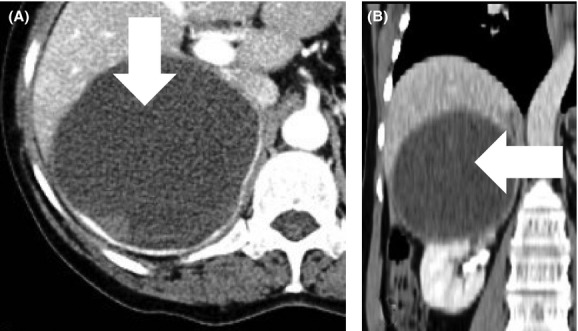
The case was further evaluated with contrast-enhanced computed tomography, which showed a large cystic lesion with thin enhancing wall and small nodular solid nonenhancing components existing within, in the right suprarenal region measuring 11.2 × 9.6 × 9.8 cm (Fig.1). It was reported as a simple adrenal cyst with probably a matured clot within the cyst or a complex cystic neoplasm of the right adrenal gland. In addition, the plasma-free metanephrines (20 pg/mL), urinary vanillylmandelic acid (6 mg/24 h), and metanephrine (30.5 μg/24 h) assays were within normal limits.
Figure 1.

Contrast-enhanced computed tomography showing the adrenal cyst in (A) axial view (B) coronal view.
In view of the size of the cyst, right adrenalectomy was performed through conventional open retroperitoneal approach. Intraoperatively, a cyst of about 11 × 9 × 9 cm was noted in the right supra renal region (Fig2A). The cyst was aspirated; the aspirate was similar to the dark coffee-colored aspirate carried out under sonographic guidance. The walls of the cyst were then removed along with the right adrenal gland and sent for histopathological examination (Fig2B). The intraoperative period was characterized by wide fluctuation in blood pressure (200/110 mmHg and 50/40 mmHg) requiring the use of supportive drugs (sodium nitroprusside and ephedrine, respectively). The patient's postoperative recovery was uneventful.
Figure 2.

(A) Intraoperative photograph, showing the adrenal cyst (black arrow) and the right kidney (white arrow); (B) Postoperative photograph of the adrenal cyst specimen; (C) Histopathology of cyst wall - H&E stain showing granular basophilic cells with variably sized nuclei and prominent nucleoli.
On gross analysis, the mass was completely cystic and contained dark brown-colored fluid with clots inside the cavity. The cavity of the cyst was multiloculated with smooth inner surface. The histopathology report revealed that the mass was composed of abundant basophilic to amphophilic, granular cytoplasm with large pleomorphic nuclei. The nuclei had prominent nucleolus and coarse chromatin (Fig2C). The specimen was finally reported as predominantly cystic pheochromocytoma with a PASS score of three. She is normotensive and is now doing well after 1 year of follow-up.
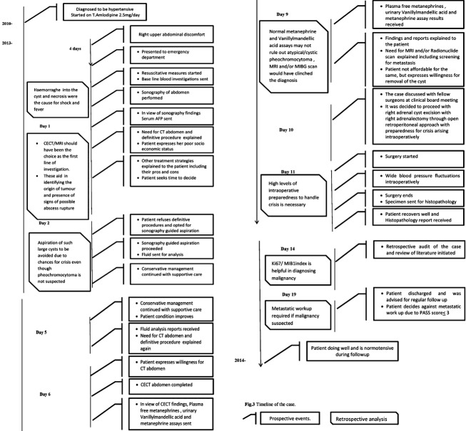
The summary of case management is presented in the form of a timeline (Fig.3).
Figure 3.
Timeline of the case.
Discussion
Cystic adrenal masses have been reported to have a minimal incidence of 0.064–0.18% in autopsy series; cystic masses account for only about 4–22% of all adrenal incidentalomas 2. Also, cystic pheochromocytoma is now being considered a rare variant which may not demonstrate the expected clinical, radiological, or biochemical features of pheochromocytoma. Moreover, they may have nonspecific abdominal symptoms as presenting complaints and may be confused with hepatic cysts and neoplasms 3. To date in the literature, only few cases of cystic pheochromocytoma have been reported. A review in 2008 found only 16 cases of cystic pheochromocytoma in the literature. Of the 16 cases, six patients were symptomatic during their presentation, and in the six cases, cystic pheochromocytoma was not thought until intraoperative hemodynamic instability occurred 4. Another review of 15 cases of cystic pheochromocytoma in 2008 revealed that a majority of them were female, half had no symptoms, and half had normal biochemical analysis 5. In several of the case reports reviewed, cystic pheochromocytoma has been wrongly diagnosed as liver or pancreatic tumor, with the correct diagnosis confirmed only during the surgery 4,6.
It has been postulated that when the neoplasm outgrows its blood supply, there is necrosis of the neoplasm followed by hemorrhage. Eventually, when the contents are liquefied and resorbed, cystic pheochromocytoma is developed. Patients may present in shock and/or with fever and leucocytosis as in our case; during episodes of hemorrhage into the neoplasm and during the necrosis stage.
Patients with cystic pheochromocytoma are often asymptomatic and yield negative plasma and urine biochemical analysis because the secreted catecholamines are metabolized within the neoplasm, and only a small amount, if any is released into the circulation and are not high enough to provide abnormal urinary values. However, the released catecholamines is sufficient to provoke the typical symptomatology like hypertension 7,8. Attaching too much importance to normal preoperative biochemical analysis of urine and plasma might misguide the surgeon from diagnosis.
On a contrast-enhanced computed tomography, they show rim enhancement and areas of low attenuation, in the range of 5–15 Hounsfield units 9. Although not performed, magnetic resonance imaging studies offer better clarity in identifying the nature of the cystic neoplasm; with hemorrhage appearing hypointense on both T1- and T2-weighted images and necrosis appearing hyperintense on T2-weighted images. The use of Metaiodobenzylguanidine (MIBG) radionuclide scan is especially helpful to identify atypical pheochromocytoma like the cystic pheochromocytoma and to differentiate them from benign adrenal cysts 10. The possibility of cystic pheochromocytoma should be considered for any peri-adrenal cystic lesions even though investigations are not suggestive of pheochromocytoma.
The diagnostic yield of fine-needle aspiration is also low in cystic pheochromocytoma as the needle passes through other normal tissue before reaching the cyst; thereby, interpreting the cytology specimen becomes tricky.
Surgical resection is the curative treatment for cystic pheochromocytoma, although laparoscopic adrenalectomy is effective, minimally invasive and safe; Traditional open surgery is the gold standard in the surgical management of giant cystic pheochromocytoma 11. As in most of the reported cases, diagnosis of a cystic pheochromocytoma is often an intraoperative surprise, the ability to tackle crisis during the intraoperative period is of immense importance. The preoperative suspicion could lessen the burden of surprise and in turn improve the outcome of the surgery from the surgeon, anesthetist, and patient's point of view. It is postulated that most of the metabolic products are proposed to be stored in the capsule and are released when isolating the mass during surgery; resulting in alarming fluctuations of blood pressure. Irrespective of the functional status of cystic pheochromocytoma, earlier dissection and ligation of central adrenal vein should be done during the resection for better control of blood pressure and blood loss 12.
Malignancy in a cystic pheochromocytoma is often indicated by radiological or intraoperative findings like an invasion of adjacent structures or metastasis 13. Histologic analysis cannot exclude malignancy on its own in cystic pheochromocytoma, a PASS score ≤3 is suggestive of benign cystic pheochromocytoma 14. Despite the fact that Ki67/MIB1 index is helpful in diagnosing malignancy in cystic pheochromocytoma, it is not diagnostic 15. If a locally advanced malignancy or in advanced malignancy where metastasis can be surgically approached is suspected in cystic pheochromocytoma, the treatment plan should include a multivisceral resection. This invariably reduces the mortality and the exposure of the cardiovascular system to catecholamines. In cases of advanced malignancy with unresectable local invasion or metastasis, palliation is the aim. Other treatment alternatives include chemotherapy, external beam radiation, radiofrequency ablation, and cryoablation. The possibility of treating metastasis with therapeutic doses of iodine-131 MIBG has also been evaluated 1.
After arriving at the diagnosis, a retrospective analysis of the management of this case revealed several pitfalls that could be avoided in the future. The presentation of the patient with high-grade fever, right hypochondrial tenderness, leucocytosis, shock, and a sonographic finding suggestive of liver abscess prompted us to a diagnosis of liver abscess with septic shock. Even though sonography is a good initial choice of investigation; computed tomography or magnetic resonance imaging would have been a better choice of investigation. Especially in cases like this, when large cyst or abscess is encountered computed tomography or magnetic resonance imaging provides better clarity in the origin and the presence of signs of possible abscess rupture, which would contraindicate any attempt at percutaneous aspiration. However, the socioeconomic status of the patient prevented her from undergoing a computed tomography at the time of admission forcing us to proceed with sonography and later an aspiration under sonographic guidance. Moreover, the cytology report in favor of liver abscess further misguided us. A discussion later with the pathologist reiterated the fact that aspiration cytology findings can at times be deceptive as in our case. In the setting of a cystic adrenal mass, a normal biochemical analysis literally made us exclude the possibility of pheochromocytoma. However, the intraoperative management of the patient, especially during the crisis was the keystone in the whole case management. Such intraoperative events could have been anticipated in advance if cystic pheochromocytoma was suspected preoperatively. Again, due to the cost factor, magnetic resonance imaging and/or MIBG scans, Ki67/MIB1 index and other metastatic work-up could not be done.
In conclusion, the key in the management of a case of cystic pheochromocytoma is the preoperative suspicion, intraoperative crisis management, and least importantly the preoperative investigations including imaging modalities. Although the low incidence of cystic pheochromocytoma could be attributed to the varied presentation, lack of suspicion and moreover the diagnostic difficulties encountered; all these factors contribute to faulty diagnosis and under reporting of the cystic pheochromocytoma. As such the prevalence of cystic pheochromocytoma could be even higher, which is yet to be ascertained 5.
Conflict of Interest
None declared.
References
- Costa SR, Cabral NM, Abhrão AT, Costa RB, Silva LM. Lupinacci RA. Giant cystic malignant pheochromocytoma invading right hepatic lobe: report on two cases. Sao Paulo Med. J. 2008;126:229–231. doi: 10.1590/S1516-31802008000400008. doi: 10.1590/S1516-31802008000400008. [DOI] [PMC free article] [PubMed] [Google Scholar]
- Bellantone R, Ferrante A, Raffaelli M, Boscherini M, Lombardi CP. Crucitti F. Adrenal cystic lesions: report of 12 surgically treated cases and review of the literature. J. Endocrinol. Invest. 1998;21:109–114. doi: 10.1007/BF03350324. PubMed ID: 9585385. [DOI] [PubMed] [Google Scholar]
- Grozinsky-Glasberg S, Szalat A, Benbassat CA, Gorshtein A, Weinstein R, Hirsch D. Clinically silent chromaffin-cell tumors: tumor characteristics and long-term prognosis in patients with incidentally discovered pheochromocytomas. J. Endocrinol. Invest. 2010;33:739–744. doi: 10.1007/BF03346680. doi: 10.1007/BF03346680. [DOI] [PubMed] [Google Scholar]
- Antedomenico E. Wascher RA. A case of mistaken identity: giant cystic pheochromocytoma. Curr. Surg. 2005;62:193–198. doi: 10.1016/j.cursur.2004.08.015. doi: 10.1016/j.cursur.2004.08.015. [DOI] [PubMed] [Google Scholar]
- Andreoni C, Krebs R, Bruna P, Goldman S, Kater C, Alves M, et al. Cystic phaeochromocytoma is a distinctive subgroup with special clinical, imaging and histological features that might mislead the diagnosis. BJU international. 2008;101(3):345–350. doi: 10.1111/j.1464-410X.2007.07370.x. doi: 10.1111/j.1464-410X.2007.07370.x. [DOI] [PubMed] [Google Scholar]
- Wu JS, Ahya SN, Reploeg MD, Singer GG, Brennan DC. Howard TK. Pheochromocytoma presenting as a giant cystic tumor of the liver. Surgery. 2000;128:482–484. doi: 10.1067/msy.2000.104113. doi: 10.1067/msy.2000.104113. [DOI] [PubMed] [Google Scholar]
- Crout JR. Sjoerdsma A. Turnover and metabolism of catecholamine in patients with pheochromocytoma. J. Clin. Invest. 1964;43:94–102. doi: 10.1172/JCI104898. doi: 10.1172/JCI104898. [DOI] [PMC free article] [PubMed] [Google Scholar]
- Sukumar S, Khanna V, Saheed CSM, Nair B, Kumar PG, Mathew G, et al. Cystic phaeochromocytomas: understanding a rare clinicopathologic variant. Indian J. Urol. 2008;24(Suppl. 2):S81. [Google Scholar]
- Munden R, Adams DB. Curry NS. Cystic pheochromocytoma: radiologic diagnosis. South. Med. J. 1993;86:1302–1305. doi: 10.1097/00007611-199311000-00029. PubMed ID: 8235793. [DOI] [PubMed] [Google Scholar]
- Wang Yuh-Feng, Cherng Shiou-Chi, Jen Tsu-Kang. Huang Wen-Sheng. Cystic pheochromocytoma-an unusual presentation of the adrenal mass. Ann. Nuli. Med. Sci. 2000;13:121–124. [Google Scholar]
- Staren ED. Prinz RA. Selection of patients with adrenal incidentalomas for operation. Surg. Clin. North Am. 1995;75:499–509. doi: 10.1016/s0039-6109(16)46636-3. PubMed ID: 7747255. [DOI] [PubMed] [Google Scholar]
- Changfu L, Chen Yongsheng. Teng Lichen. A case of clinically silent giant right pheochromocytoma and review of literature. Can. Urol. Assoc. J. 2012;6:E267–E269. doi: 10.5489/cuaj.11195. doi: org/ 10.5489/cuaj.11195. [DOI] [PMC free article] [PubMed] [Google Scholar]
- Norton AJ. Adrenal tumors, pheochromocytoma. In: DeVita VT Jr, Hellman S, Rosenberg SA, et al., editors. Cancer: principles and practice of oncology. 7th ed. Philadelphia: Lippincott Williams & Wilkins; 2005. pp. 1770–1778. [Google Scholar]
- Thompson LD. Pheochromocytoma of the adrenal gland scaled score (PASS) to separate benign from malignant neoplasms: a clinicopathologic and immunophenotypic study of 100 cases. Am. J. Surg. Pathol. 2002;26:551–566. doi: 10.1097/00000478-200205000-00002. PubMed ID: 11979086. [DOI] [PubMed] [Google Scholar]
- Strong VE, Kennedy T, Al-Ahmadie H, Tang L, Coleman J, Fong Y, et al. Prognostic indicators of malignancy in adrenal pheochromocytomas: clinical, histopathologic, and cell cycle/apoptosis gene expression analysis. Surgery. 2008;143:759–768. doi: 10.1016/j.surg.2008.02.007. doi: 10.1016/j.surg.2008.02.007. [DOI] [PubMed] [Google Scholar]


