

Abstract
Synthetic siRNA has been considered as a highly promising therapeutic agent for human diseases. However, clinical use of siRNA has been hampered by instability in the body and inability to deliver sufficient RNA interference compounds to the tissues or cells. To address this challenge, we present here a single siRNA nanocapsule delivery technology, which is achieved by encapsulating a single siRNA molecule within a degradable polymer nanocapsule with a diameter around 20 nm and positive surface charge. As proof-of-concept, since CCR5 is considered a major silencing target of HIV therapy, CCR5–siRNA nanocapsules were delivered into 293T cells and successfully downregulated the CCR5 RNA fused with mCherry reporter RNA. In the absence of human serum, nanocapsules and lipofectamine silenced expression of CCR5–mCherry expression to 8% and 15%, respectively. Such nanocapsules maintain the integrity of siRNA inside even after incubation with ribonuclease and serum for 1 h; under the same conditions, siRNA is degraded in the native form or when formulated with lipofectamine. In the presence of serum, CCR5–siRNA nanocapsules knocked down CCR5–mCherry expression to less than 15% while siRNAs delivered through lipofectamine slightly knocked down the expression to 55%. In summary, this work provides a novel platform for siRNA delivery that can be developed for therapeutic purposes.
RNA interference (RNAi) has been considered as a highly promising therapeutic agent for varieties of human diseases following upon the success of small molecule drugs and monoclonal antibodies.1,2 Viral and nonviral approaches have been adapted to overcome the delivery issue and stability of siRNA. Viral vector-mediated delivery of shRNA to cells allows endogenous synthesis of siRNA;3,4 however, immunogenicity, potential malignant transformation and high manufacturing cost may constrain their applications for human therapies.3,5,6 Various nonviral siRNA delivery systems were proposed, including cationic lipids, cell-penetrating peptides (CPPs) and cationic polymers.7,8 Cationic lipids, such as lipofectamine and lipid-like materials, are widely used for in vitro studies and showed great potential for in vivo gene silencing;9–16 however, further improvement in toxicity and efficiency are required to broaden their in vivo applications. For the CPPs-based approach, siRNA was assembled with CPPs or CPP bioconjugates into complexed particles with significantly improved delivery efficiency.17,18 Nevertheless, the formation of such an assembled structure was driven by weak noncovalent interactions; and these particles were generally unstable, particularly, against serum nucleases. For the cationic-polymer-based approach, siRNA was assembled with preformed cationic polymers mainly through the electrostatic interactions. The unique proton-sponge and membrane destabilization effect of cationic polymers provides the complexes with improved intracellular delivery efficiency.19–25 However, similar to the CPP-based approach, such systems based on self-assembly are also unstable in the bloodstream, which may readily dissociate and release their siRNA payload before they reach the cytoplasm of the target cells. Therefore, in spite of intensive efforts, the design and synthesis of an effective delivery vehicle for siRNA remains a challenge.
To address this challenge, we present herein a novel siRNA delivery technology. We previously described a polymer based nanocapsule technology for encapsulation of single protein molecules.26 We reasoned that since the size of proteins and siRNA molecules is similar, it may be possible to apply the nanocapsule technology to encapsulation of siRNAs. This is achieved by forming a degradable polymer shell around the surface of a single siRNA molecule with desirable size and charge. We expect that the polymer network provides siRNA resistance to nucleases, surface charge will allow effective delivery of siRNA to the target cells, and degradability will controllably release the siRNA. Our single siRNA nanocapsule platform, illustrated in Scheme 1, starts with a new monomer A (positively charged), crosslinker B, monomer C (neutral), and enriches these molecules around the surface of the siRNA (negatively charged) through electrostatic interaction and hydrogen bonding (Step I). Subsequent room temperature radical polymerization in an aqueous solution wraps each siRNA molecule with a thin shell of polymer (Step II), which effectively protects the siRNA from degradation by ribonucleases and serum. The hydrophilic monomer C is added to help the dispersion of nanocapsules. The crosslinker (B) is added to stabilize the polymer structure in the serum (pH~7.4). It is degradable at pH < 6 allowing release in the acidic endosomes. This unique responsive design provides the nanocapsules with outstanding stability in serum (pH ~ 7.4), enables effective endosomal escape due to proton-sponge effect (SI Figure 1) and cation-mediated membrane destabilization,21–25 and achieves effective release of the siRNA into the cytoplasm upon the degradation of the shell (Step IV).
Scheme 1.
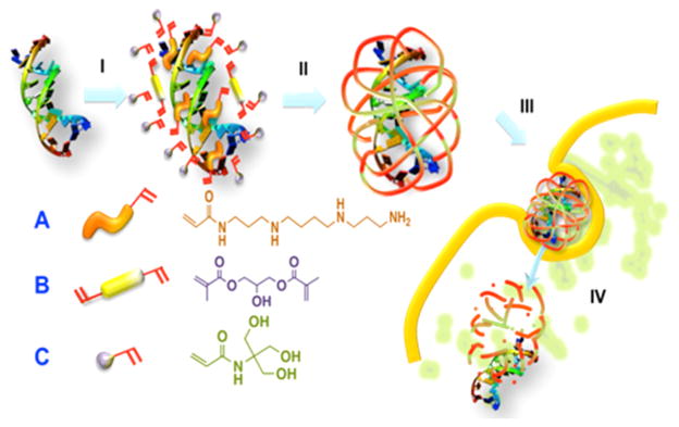
Schematic Illustration of the Synthesis and Delivery of Single-siRNA Nanocapsules: (I) Enriching of Monomers and Crosslinkers around the siRNA Molecule, (II) in sSitu Radical Polymerization Forming of siRNA Nanocapsules, (III) Intracellular Delivery, (IV) Releasing of siRNAs from the Nanocaspules
The siRNA nanocapsules were characterized using transmission electron microscopy (TEM). Figure 1A shows a representative TEM image of siRNA nanocapsules with the average diameter of 20 nm, which is consistent with the number of 24.6 nm obtained by light scattering (SI Figure 5). Interestingly, within each nanocapsule, a dark core with a diameter around 5 nm was clearly observed, which is likely a result of the preferred complex of siRNA with the tungsten-staining agent used for TEM observation. Since the average hydrodynamic diameter of a double-stranded siRNA (21 base pairs) is 4.2 nm, each of the nanocapsules likely only contains one siRNA molecule. To investigate the sensitivity of single siRNA nanocapsules against human serum and nucleases, siRNA complexed with lipofectamine and siRNA nanocapsules were incubated with nucleases and human serum for 1 h. After RNA extraction, agarose gel electrophoresis showed such nanocapsules could maintain the integrity of siRNA inside (Figure 1B), while siRNA is degraded at the same time in the native state or when formulated with lipofectamine.
Figure 1.
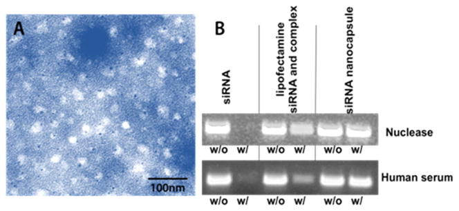
(A) TEM image of siRNA nanocapsules; (B) sensitivity of siRNA to nuclease and human serum. siRNA complexed with lipofectamine and siRNA nanocapsules were incubated for 1 h with nuclease (up) and human serum (down), respectively. Then, siRNAs were extracted from lipofectamine and nanocapsules with chloroform/0.1% SDS–0.5 M NaCl. Samples were run on 4% agarose gel and imaged with ImageQuant LAS4000.
siRNA nanocapsules can be effectively delivered to cells. The fluorescent image of HEK-293T cells after incubation with FITC-labeled siRNA for 4 h was shown in Figure 2A. The intense green fluorescence demonstrates delivery of the FITC-labeled siRNA nanocapsules. Flow cytometry of HEK-293 T cells transduced with FITC-labeled siRNA nanocapsules confirms the results of optical fluorescence and demonstrates successful delivery of fluorescence-labeled siRNA nanocapsule (Figure 2B).
Figure 2.

Internalization of single siRNA nanocapsules. (A) Fluorescence image (up) and optical image (down) of HEK-293T cells (scale bar = 10 μm); (B) Flow cytometry of HEK-293T cells. Cells were dosed with FITC-labeled siRNA nanocapsules at 100 nM for 4 h. Then nanocapsules were removed and cells were washed 3 times with PBS. After trypsinization, cells were pictured with Leica Zeiss Axio Observer and also analyzed by a flow cytometer.
CWR cells stably expressing luciferase were used to test the gene-silencing efficacy of single siRNA nanocapsules (Figure 3). Cells treated with luciferase siRNA nanocapsules showed a significant decrease in the luciferase activity especially at concentrations above 50 nM, while cells treated with control siRNA nanocapsules did not exhibit significant decrease in the luciferase activity. The siRNA nanocapsules did not show obvious cytotoxicity at the concentration of siRNA below 200 nM. At 300 nM, the viability of cells treated with siRNA nanocapsules was slightly reduced to about 75%.
Figure 3.
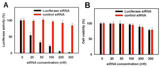
(A) Knockdown of luciferase gene expression in CWR cells using luciferase siRNA nanocapsules and control siRNA nanocapsules. (B) Cell viability after treatment of siRNA nanocapsules. CWR cells were treated with siRNA nanocapsules at 0, 20, 50, 100, 200 and 300 nM for 4 h at 37 °C in serum-free medium. Then media were changed to DMEM with 10% Bovine Fetal Serum. After 48 h, the luciferase activity was determined using a 96-well plate reader.
We used 293T cells expressing an mRNA fusion of CCR5–mCherry to demonstrate potential applications of siRNA nanocapsules in HIV therapy. CCR5 is the primary HIV-1 co-receptor, essential for HIV-1 infection.27–30 A recent case study suggests that knockdown of CCR5 can result in substantial attenuation of HIV-1 replication and a favorable clinical course. We previously established a potent shRNA against CCR5, termed sh100527 (5′-GAGCAAGCTCAGTTTACACC-3′). This shRNA was selected to maintain potency at low expression levels, minimizing cytotoxic effects.31 We constructed the sequence of the corresponding siRNA for nanocapsules.
As proof of concept for siRNA nanocapsules to down-regulate CCR5 expression, we co-transduced 293T cells with siRNA nanocapsules and the plasmid expressing a fusion of CCR5 and mCherry reporter gene sequence. Cells were treated with siRNA nanocapsules at 200 nM for 4 h at 37 °C in serum-free medium. Then, medium was changed to DMEM with 10% Bovine Fetal Serum. After 48 h, mCherry expression in 293T cells treated with CCR5–siRNA nanocapsules is much dimmer than that in 293T cells treated with control-siRNA nano-capsules targeted to EGFP sequence (Figure 4A). We further quantitatively compared siRNA nanocapsules and siRNA lipofectamine for downregulation of CCR5 through flow cytometry. In the absence of human serum, nanocapsules and lipofectamine (Invitrogen) silenced expression of CCR5–mCherry expression to 8% and 15%, respectively. However, in the presence of serum, CCR5–siRNA nanocapsules still knocked down CCR5–mCherry expression to less than 15% while siRNAs delivered by lipofectamine only slightly knocked down the expression of CCR5–mCherry to 55% (Figure 4B). Therefore, compared with lipofectamine, nanocapsules can provide extra protection and stabilization to siRNAs against human serum nucleases.
Figure 4.

Knockdown of CCR5 RNA fused with mCherry in 293T cells through siRNA nanocapsules. (A) Fluorescence images of HEK-293T cells treated with CCR5 siRNA nanocapsules (up) and control siRNA nanocapsules (down) in serum-free medium (scale bar = 10 μm); (B) flow cytometry of HEK-293T cells treated with siRNA nanocapsules or siRNA lipofectamine complex in serum-free medium or 50% human serum medium. Cells were treated with siRNA nanocapsules or siRNA lipofectamine complex at 200 nM for 4 h at 37 °C in different cell culture medium. Then mediums were changed to DMEM with 10% Bovine Fetal Serum. After 48 h, the images were taken or flow cytometry was run. The relative CCR5–mCherry expression (%) was calculated from the mean fluorescence.
In conclusion, we have developed a single siRNA nano-capsule delivery technology, which is achieved by encapsulating a single siRNA molecule within a very small polymer nanocapsule. The small size of the single siRNA nanocapsules, which is one of the smallest siRNA delivery vehicles,22,32–36 leads to a high delivery efficiency, and possibly a high diffusional rate through tissues or organs. Importantly, the polymer encapsulation greatly enhances stability against nuclease and serum compared with the commercial transfection agent lipofectamine. The technology provides precise control in structure and a high yield of encapsulation of siRNA. We believe these advances will provide new opportunities in the RNAi therapeutic space for treatment of human diseases.
Supplementary Material
Acknowledgments
This work was supported by a seed fund from UCLA CNSI, postdoc fellowship award and mentorship award from UCLA AIDS Institute and the UCLA Center for AIDS Research (AI28697).
Footnotes
Notes
The authors declare no competing financial interest.
ASSOCIATED CONTENT
Experimental details for monomer synthesis, preparation and characterization of siRNA nanocapsules, cell culture, cytotoxicity analysis, and cellular images. This material is available free of charge via the Internet at http://pubs.acs.org.
References
- 1.Cheng K, Mahato RI. Mol Pharmaceutics. 2009;6:649. doi: 10.1021/mp900094j. [DOI] [PubMed] [Google Scholar]
- 2.Van Stry M, Oguin TH, Cheloufi S, Vogel P, Watanabe M, Pillai MR, Dash P, Thomas PG, Hannon GJ, Bix M. J Virol. 2012;86:4151. doi: 10.1128/JVI.05303-11. [DOI] [PMC free article] [PubMed] [Google Scholar]
- 3.Li M, Bauer G, Michienzi A, Yee J, Lee N, Kim J, Li S, Castanotto D, Zaia J, Rossi J. Mol Ther. 2003;8:196. doi: 10.1016/s1525-0016(03)00165-5. [DOI] [PubMed] [Google Scholar]
- 4.Miyagishi M, Taira K. Nat Biotechnol. 2002;20:497. doi: 10.1038/nbt0502-497. [DOI] [PubMed] [Google Scholar]
- 5.Liang M, Pariente N, Morizono K, Chen I. J Gene Med. 2009;11:185. doi: 10.1002/jgm.1290. [DOI] [PMC free article] [PubMed] [Google Scholar]
- 6.Grimm D, Streetz K, Jopling C, Storm T, Pandey K, Davis C, Marion P, Salazar F, Kay M. Nature. 2006;441:537. doi: 10.1038/nature04791. [DOI] [PubMed] [Google Scholar]
- 7.Timko BP, Whitehead K, Gao WW, Kohane DS, Farokhzad O, Anderson D, Langer R. Annu Rev Mater Res. 2011;41:1. [Google Scholar]
- 8.Whitehead KA, Langer R, Anderson DG. Nat Rev Drug Discovery. 2009;8:129. doi: 10.1038/nrd2742. [DOI] [PMC free article] [PubMed] [Google Scholar]
- 9.Schroeder A, Levins CG, Cortez C, Langer R, Anderson DG. J Intern Med. 2010;267:9. doi: 10.1111/j.1365-2796.2009.02189.x. [DOI] [PMC free article] [PubMed] [Google Scholar]
- 10.Love KT, Mahon KP, Levins CG, Whitehead KA, Querbes W, Dorkin JR, Qin J, Cantley W, Qin LL, Racie T, Frank-Kamenetsky M, Yip KN, Alvarez R, Sah DWY, de Fougerolles A, Fitzgerald K, Koteliansky V, Akinc A, Langer R, Anderson DG. Proc Natl Acad Sci USA. 2010;107:1864. doi: 10.1073/pnas.0910603106. [DOI] [PMC free article] [PubMed] [Google Scholar]
- 11.Cho SW, Goldberg M, Son SM, Xu QB, Yang F, Mei Y, Bogatyrev S, Langer R, Anderson DG. Adv Funct Mater. 2009;19:3112. doi: 10.1002/adfm.200900519. [DOI] [PMC free article] [PubMed] [Google Scholar]
- 12.Akinc A, Goldberg M, Qin J, Dorkin JR, Gamba-Vitalo C, Maier M, Jayaprakash KN, Jayaraman M, Rajeev KG, Manoharan M, Koteliansky V, Rohl I, Leshchiner ES, Langer R, Anderson DG. Mol Ther. 2009;17:872. doi: 10.1038/mt.2009.36. [DOI] [PMC free article] [PubMed] [Google Scholar]
- 13.Akinc A, Zumbuehl A, Goldberg M, Leshchiner ES, Busini V, Hossain N, Bacallado SA, Nguyen DN, Fuller J, Alvarez R, Borodovsky A, Borland T, Constien R, de Fougerolles A, Dorkin JR, Jayaprakash KN, Jayaraman M, John M, Koteliansky V, Manoharan M, Nechev L, Qin J, Racie T, Raitcheva D, Rajeev KG, Sah DWY, Soutschek J, Toudjarska I, Vornlocher HP, Zimmermann TS, Langer R, Anderson DG. Nat Biotechnol. 2008;26:561. doi: 10.1038/nbt1402. [DOI] [PMC free article] [PubMed] [Google Scholar]
- 14.Mahon KP, Love KT, Whitehead KA, Qin J, Akinc A, Leshchiner E, Leshchiner I, Langer R, Anderson DG. Bioconjugate Chem. 2010;21:1448. doi: 10.1021/bc100041r. [DOI] [PMC free article] [PubMed] [Google Scholar]
- 15.Leuschner F, Dutta P, Gorbatov R, Novobrantseva TI, Donahoe JS, Courties G, Lee KM, Kim JI, Markmann JF, Marinelli B, Panizzi P, Lee WW, Iwamoto Y, Milstein S, Epstein-Barash H, Cantley W, Wong J, Cortez-Retamozo V, Newton A, Love K, Libby P, Pittet MJ, Swirski FK, Koteliansky V, Langer R, Weissleder R, Anderson DG, Nahrendorf M. Nat Biotechnol. 2011;29:1005. doi: 10.1038/nbt.1989. [DOI] [PMC free article] [PubMed] [Google Scholar]
- 16.Nguyen DN, Mahon KP, Chikh G, Kim P, Chung H, Vicari AP, Love KT, Goldberg M, Chen S, Krieg AM, Chen JZ, Langer R, Anderson DG. Proc Natl Acad Sci USA. 2012;109:E797. doi: 10.1073/pnas.1121423109. [DOI] [PMC free article] [PubMed] [Google Scholar]
- 17.Broderick JA, Zamore PD. Gene Ther. 2011;18:1104. doi: 10.1038/gt.2011.50. [DOI] [PMC free article] [PubMed] [Google Scholar]
- 18.Manavella PA, Koenig D, Weigel D. Proc Natl Acad Sci USA. 2012;109:2461. doi: 10.1073/pnas.1200169109. [DOI] [PMC free article] [PubMed] [Google Scholar]
- 19.Lee JS, Green JJ, Love KT, Sunshine J, Langer R, Anderson DG. Nano Lett. 2009;9:2402. doi: 10.1021/nl9009793. [DOI] [PMC free article] [PubMed] [Google Scholar]
- 20.Siegwart DJ, Whitehead KA, Nuhn L, Sahay G, Cheng H, Jiang S, Ma ML, Lytton-Jean A, Vegas A, Fenton P, Levins CG, Love KT, Lee H, Cortez C, Collins SP, Li YF, Jang J, Querbes W, Zurenko C, Novobrantseva T, Langer R, Anderson DG. Proc Natl Acad Sci USA. 2011;108:12996. doi: 10.1073/pnas.1106379108. [DOI] [PMC free article] [PubMed] [Google Scholar]
- 21.Akinc A, Thomas M, Klibanov AM, Langer R. J Gene Med. 2005;7:657. doi: 10.1002/jgm.696. [DOI] [PubMed] [Google Scholar]
- 22.Boussif O, Lezoualch F, Zanta MA, Mergny MD, Scherman D, Demeneix B, Behr JP. Proc Natl Acad Sci USA. 1995;92:7297. doi: 10.1073/pnas.92.16.7297. [DOI] [PMC free article] [PubMed] [Google Scholar]
- 23.Miyata K, Nishiyama N, Kataoka K. Chem Soc Rev. 2012;41:2562. doi: 10.1039/c1cs15258k. [DOI] [PubMed] [Google Scholar]
- 24.Sonawane ND, Szoka FC, Verkman AS. J Biol Chem. 2003;278:44826. doi: 10.1074/jbc.M308643200. [DOI] [PubMed] [Google Scholar]
- 25.Wagner E. Acc Chem Res. 2012;45:1005. doi: 10.1021/ar2002232. [DOI] [PubMed] [Google Scholar]
- 26.Yan M, Du J, Gu Z, Liang M, Hu Y, Zhang W, Priceman S, Wu L, Zhou Z, Liu Z, Segura T, Tang Y, Lu Y. Nat Nanotechnol. 2010;5:48. doi: 10.1038/nnano.2009.341. [DOI] [PubMed] [Google Scholar]
- 27.An D, Donahue R, Kamata M, Poon B, Metzger M, Mao S, Bonifacino A, Krouse A, Darlix J, Baltimore D, Qin F, Chen I. Proc Natl Acad Sci US A. 2007;104:13110. doi: 10.1073/pnas.0705474104. [DOI] [PMC free article] [PubMed] [Google Scholar]
- 28.An D, Qin F, Auyeung V, Mao S, Kung S, Baltimore D, Chen I. Mol Ther. 2006;14:494. doi: 10.1016/j.ymthe.2006.05.015. [DOI] [PMC free article] [PubMed] [Google Scholar]
- 29.Hutter G, Nowak D, Mossner M, Ganepola S, Müssig A, Allers K, Schneider T, Hofmann J, Kücherer C, Blau O, Blau I, Hofmann W, Thiel E. N Engl J Med. 2009;360:692. doi: 10.1056/NEJMoa0802905. [DOI] [PubMed] [Google Scholar]
- 30.Kumar P, Ban H, Kim S, Wu H, Pearson T, Greiner D, Laouar A, Yao J, Haridas V, Habiro K, Yang Y, Jeong J, Lee K, Kim Y, Kim S, Peipp M, Fey G, Manjunath N, Shultz L, Lee S, Shankar P. Cell. 2008;134:577. doi: 10.1016/j.cell.2008.06.034. [DOI] [PMC free article] [PubMed] [Google Scholar]
- 31.Shimizu S, Hong P, Arumugam B, Pokomo L, Boyer J, Koizumi N, Kittipongdaja P, Chen A, Bristol G, Galic Z, Zack J, Yang O, Chen I, Lee B, An D. Blood. 2010;115:1534. doi: 10.1182/blood-2009-04-215855. [DOI] [PMC free article] [PubMed] [Google Scholar]
- 32.Dohmen C, Edinger D, Frohlich T, Schreiner L, Lachelt U, Troiber C, Radler J, Hadwiger P, Vornlocher HP, Wagner E. Acs Nano. 2012;6:5198. doi: 10.1021/nn300960m. [DOI] [PubMed] [Google Scholar]
- 33.Gary DJ, Lee H, Sharma R, Lee JS, Kim Y, Cui ZY, Jia D, Bowman VD, Chipman PR, Wan L, Zou Y, Mao GZ, Park K, Herbert BS, Konieczny SF, Won YY. Acs Nano. 2011;5:3493. doi: 10.1021/nn102540y. [DOI] [PMC free article] [PubMed] [Google Scholar]
- 34.DeRouchey J, Schmidt C, Walker GF, Koch C, Plank C, Wagner E, Radler JO. Biomacromolecules. 2008;9:724. doi: 10.1021/bm7011482. [DOI] [PubMed] [Google Scholar]
- 35.Meyer M, Philipp A, Oskuee R, Schmidt C, Wagner E. J Am Chem Soc. 2008;130:3272. doi: 10.1021/ja710344v. [DOI] [PubMed] [Google Scholar]
- 36.Shimizu H, Hori Y, Kaname S, Yamada K, Nishiyama N, Matsumoto S, Miyata K, Oba M, Yamada A, Kataoka K, Fujita T. J Am Soc Nephrol. 2010;21:622. doi: 10.1681/ASN.2009030295. [DOI] [PMC free article] [PubMed] [Google Scholar]
Associated Data
This section collects any data citations, data availability statements, or supplementary materials included in this article.