Abstract
This case report describes the chiropractic management of a 30-year-old female patient with severe postpartum pelvic pain secondary to pubic symphysis diastasis. No literature was found on the chiropractic management of postpartum symphysis pubis diastasis. The existing literature concerning chiropractic care for symphysis pubis dysfunction during pregnancy is limited and indicates a potential benefit. Separation of the pubic symphysis may include ligamentous injury to the sacroiliac joints and may lead to chronic pain. Pubic symphysis separation of 17 millimeters was present on digital radiograph. Management consisted of chiropractic adjustments, trigger point release, electrical stimulation, moist heat, sacroiliac belt, and specific stabilizing exercises. The patient’s pain improved immediately following treatment on the initial visit. Pain was reduced from 8/10 VAS at the first visit to 2/10 at the fourth visit. She was able to resume normal activities and reached a final pain level of 1/10. The diastasis was reduced by 7 millimeters at 14-weeks post radiograph for a final separation of just under 10 millimeters. Collaboration between obstetricians, midwives and chiropractors may be warranted.
Keywords: chiropractic, diastasis, spinal manipulation, postpartum pelvic pain, symphysis pubis
Abstract
Cette étude de cas décrit le traitement chiropratique d’une patiente de 30 ans souffrant de douleurs pelviennes post-partum secondaires à une symphyse pubienne avec diastasis. Aucun ouvrage n’a été trouvé sur le traitement chiropratique d’une symphyse pubienne post-partum avec diastasis. Les ouvrages au sujet des soins chiropratiques d’un dysfonctionnement symphyse pubienne durant la grossesse sont rares et indiquent un bienfait potentiel. La séparation de la symphyse pubienne peut entraîner une lésion ligamenteuse à l’articulation sacro-iliaque et causer des douleurs chroniques. Une radiographie numérique montre une séparation de la symphyse pubienne de 17 mm. Le traitement consistait à des ajustements chiropratiques, à un relâchement de points gâchettes, à de la stimulation électrique, à une chaleur humide, à une ceinture sacroiliaque et à des exercices adaptés de stabilisation. La douleur de la patiente a diminué immédiatement après le traitement de la première rencontre. La douleur est passée de 8/10 à l’EVA à la première rencontre à 2/10 à la quatrième rencontre. La patiente a réussi à reprendre ses activités habituelles et son niveau de douleur a diminué à 1/10. Le diastasis a diminué de 7 mm 14 semaines après la radiographie pour une séparation définitive inférieure à 10 mm. On recommande une collaboration entre les obstétriciens, les sage-femmes et les chiropraticiens.
Keywords: chiropratique, diastasis, manipulation vertébrale, douleur pelvienne post-partum, symphyse pubienne
Introduction
Symphysis pubis diastasis is a rare cause of pelvic pain in pregnancy but may be underdiagnosed.1,2 It is a complication of pregnancy and vaginal delivery in which the pubic symphysis separates, resulting in acute pelvic pain, and may lead to severe long-term consequences.1 This separation may occur during delivery from a rapid birth, forceps delivery, in late pregnancy or post-natal. The incidence has been variously estimated from 1 out of 300 to 1 of 30,000.1–4 The pubic articulation should not exceed 8 mm in non-pregnant adults or 10 mm in children.5 Bahlman and colleagues found pubic symphysis width in pregnant women increased from a mean of 4 mm to a mean of 7 mm at term, with an average increase in pubic symphysis width of 3 mm during pregnancy.6 Garagiola found a mean pubic symphysis width of 6.5 mm in women within 24 hours of uncomplicated vaginal delivery, with widths ranging from 3 to 11 mm.7
Diagnosis of diastasis is made based on symptoms and radiographic examination. The patient presents with pain and swelling. Crepitus may be present on walking. Visible deformity may be present on physical examination. Pubic symphysis separation greater than 10 to 13 millimeters on radiograph is diagnostic and represents a subdislocation. A diastasis greater than 14 millimeters indicates attendant damage to the sacroiliac joint.6 Conservative treatment is recommended, with surgery sometimes necessary for separations greater than 25 millimeters.4 Surgical treatment may consist of debridement or fusion. Surgical management with plate and screws necessitates cesarean section in the event of future pregnancy. In the absence of appropriate care, diastasis may lead to chronic pain. Surgical management interferes with breast-feeding due to analgesics, antibiotics, and thromboembolic prophylaxis.8 Scriven et al found a direct relation between permanence of the pubic separation and chronic pain. There does not seem to be a consensus on the natural history of pubic symphysis diastasis nor does the literature suggest that this condition is self-limiting. Pubic symphysis diastasis can lead to incontinence, dyspareunia, chronic pain and / or disability and there is little evidence to guide the clinician.9 It is uncertain what percentage of patients will end up with poor long-term outcomes. Out of a series of nine patients with diastasis treated by Scriven and colleagues, four were pain free at last follow up, one required surgical fusion, two ended up with severe disability (one in a wheelchair), and the rest had some combination of low back pain or pubic pain and / or dyspareunia.1 Due to the rarity of the condition and limited literature available, what constitutes appropriate treatment for pubic symphysis diastasis remains controversial.10
Since there is a lack of literature on the treatment of postpartum pubic symphysis diastasis, the author reviewed the literature on the conservative treatment of postpartum pelvic pain in general to aid in his clinical decision making. The evidence of treatment effectiveness for postpartum pelvic pain is weak.11 Various conservative treatments, including physiotherapy, exercises and sacroiliac belt, have been recommended but there does not seem to be a consensus. Nillson-Wikmar and colleagues compared home exercises, in-office exercises and sacroiliac belt in women with postpartum pelvic pain, finding improvement in all three groups with no significant difference between the groups. The authors concluded that home or in-office exercise provided no additional benefit beyond that achieved by a non-elastic sacroiliac belt and giving information.12 In contrast, Depledge and associates concluded that sacroiliac belts did not add to the results achieved with exercise and advice for symphysis pubis dysfunction.13 Haugland and colleagues found no significant difference between a control group and those treated with intervention for pregnancy related pelvic pain. The intervention group was given education on ergonomics, exercises, pain management, and advice for daily life movement, pelvic belt/crutches, and information about delivery. The control group was free to seek other advice or interventions so it is possible that they may have received benefit from other treatment or information. However, the utility of care was rated higher by patients in the intervention group.14 Stuge and associates compared specific stabilizing exercises with physical therapy without specific stabilizing exercises in a long-term study on postpartum pelvic pain. The group that received specific stabilizing exercises demonstrated lower levels of pain and disability 2 years after delivery.11
A literature search was conducted using the Index to Chiropractic Literature and PubMed. A search on PubMed using the keywords, “chiropractic symphysis pubis diastasis,” “chiropractic symphysis pubis dysfunction” and “chiropractic postpartum pelvic pain” was performed. No previous literature was found concerning the chiropractic care of a patient with diastasis. Two articles describing three cases of pelvic pain during pregnancy were found.
Panarello reported on the case of a 32-year-old female with severe pubic and groin pain that began when she was 28 weeks pregnant. The pain caused sleep disturbance and difficulty sitting. The patient reported some pain relief following the first chiropractic adjustment and was able to sleep that night. She was pain free by the fifth adjustment. The patient had an uncomplicated vaginal delivery, with no recurrence of pubic pain.15
Howell reported on two cases of symphysis pubis dysfunction with successful chiropractic management. Treatment included soft tissue therapy, pregnancy support belt, side-lying mobilizations, pelvic blocks and instrument-assisted pubic symphysis adjustments. Postpartum rehabilitation exercises were done to restore muscular endurance, control and pelvic stability. The two patients were given home care advice to apply ice, stay active, move as a unit, stretch, use a pillow between the knees while sleeping, take regular breaks from sitting and do pelvic floor (Kegel) exercises. Both patients were 30 weeks pregnant. Both patients were mostly pain free on long-term follow up (eleven months for one patient and twelve months for the other).16
Intervention and Outcome
The author has obtained the patient’s written consent to publish this case report. Patient care, including radio-graphic procedures were rendered in compliance with applicable law.
A 30-year-old female presented with severe pelvic pain seven days after giving birth to her first child by normal vaginal delivery at home with midwife attending. Her midwife referred her for chiropractic care. She had prior treatment by her primary care physician, who also practiced obstetrics. She was diagnosed with pelvic separation and was treated with ibuprofen.
The patient described constant dull ache at the pelvic area with the pain feeling crushing at times. She indicated pain at the pubic and sacroiliac areas on both sides. She rated the pain 8/10 visual analog scale (VAS). She reported: a) that the pain interfered with walking and lifting either leg, b) crepitus at the sacroiliac area with walking, c) pain at the lower back and radiating to both thighs posteriorly, worse on the right, and d) paresthesia and swelling at both legs with prolonged standing or sitting. The pain did not follow a dermatomal pattern.
The patient’s past medical history included left knee surgery and tonsillectomy. She had a past history of T11 fracture. The patient was married and worked as a missionary. Hobbies included biking, tennis and walking. Family history included hypertension. Review of systems was negative other than the chief complaint and associated symptoms already described. Medications included ibuprofen as necessary.
The patient was 162.5 cm height and weighed 59 kg. Vital signs: pulse was 76, blood pressure was 137/89 and respiration rate was 20. She was alert and oriented to person, place, and time, with appropriate mood and affect. The patient appeared in pain. Ambulation was impaired to the point that she required assistance from her husband to walk more than a few steps and her movements were antalgic. Straight leg raise was negative. Lower extremity motor was grade 5/5. Deep tendon reflexes were 2+ at the lower extremities. Dermatome testing at the lower extremities was normal. All lumbosacral ranges of motion were limited. There was tenderness and hypertonicity at the right sacroiliac area. Palpation revealed asymmetry and restriction at L4, L5, sacrum and both sacroiliac joints. Leg length inequality was evaluated with the patient in the prone and supine positions with knees extended, which revealed a right short leg in the prone position and a left short leg in the supine position. Various estimates exist for the specificity, sensitivity, reliability, and clinical significance of leg length inequality. Shambaugh and colleagues demonstrated inter and intraexaminer reliability of the Derifield-Thompson test for leg length inequality to less than 3mm.17 In a review of the literature relevant to leg length inequality measures Mannello found that no definitive conclusions could be made because of the variation in estimates of reliability.18 Knutson investigated the relationship between supine leg length asymmetry and self-reported back pain in a group of 74 volunteers and found sensitivity of 74%, specificity 78%, and positive predictive value of 82%.19 Schneider et al studied the interexaminer reliability of prone leg length analysis using two chiropractors to examine 45 patients. The authors found 82% interexaminer reliability for determining the short leg in the prone position with knees extended, with 67% agreement for the amount of leg length inequality. The authors concluded that the two clinicians showed good reliability in determining which leg was short but poor reliability in measuring the exact amount of the leg length discrepancy.20 Triano and colleagues reviewed the literature concerning methods used by chiropractors to determine the site for manipulation, screening 2594 titles, with 201 articles meeting the inclusion criteria. The authors found high quality evidence supporting the use, with limitations, of leg length inequality to assess the pelvis, while the literature has not demonstrated validity for relationship to symptoms.21
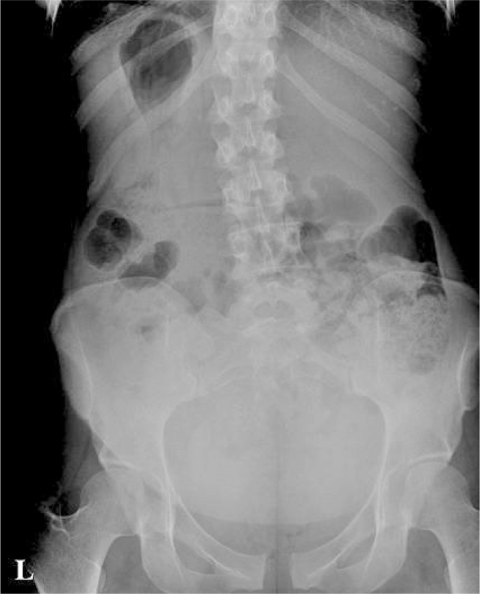
The author reviewed the radiology report from prior AP pelvic radiograph (single view) taken by the patient’s primary care physician at 5 days post-partum using a source-image distance (SID) of 40 inches. The radiologist’s report indicated a 24 mm diastasis at the public symphysis and indicated likely ligamentous injury to the sacroiliac joints. The author consulted the limited literature available and in the author’s opinion further imaging was necessary to assess the safety and appropriateness of chiropractic treatment. This was due to the severity of the patient’s pain, the failure of symptoms to improve with prior medical treatment, in consideration of the risk of developing incontinence, dyspareunia, chronic pain and disability, and due to the possible need for immediate surgical referral in the event that the diastasis had increased. AP and lateral digital radiographs were taken of the lumbopelvic region at 7 days post-partum with the patient standing at a 40 inch SID and collimated to 14 x 17 inches. Gonadal shielding was not used because it would have obscured part of the area of interest. For the purposes of this paper the patient marker and date (part of the overlay) were removed to maintain patient confidentiality. There were no bony abnormalities and no congenital anomalies were present. The lumbar lordosis was increased with anterior pelvic tilt. There was a mild left convex scoliosis at the lumbar spine. A 17 mm pubic symphysis separation was noted (Figure 1).
The patient was diagnosed with postpartum diastasis of the pubic symphysis, accompanied by injury to the sacroiliac joints and segmental dysfunction of the lumbar, sacral and pelvic regions. Transcutaneous electrical nerve stimulation (TENS) and moist heat therapy were applied to the lower lumbar and sacral regions to modulate pain.
The analgesic effect of TENS for acute pain has been consistently demonstrated by randomized controlled trials but remains controversial for specific conditions such as low back pain due to poor study designs and small sample sizes.22 A review by French et al found limited evidence for the common practice of applying superficial heat or cold for low back pain, while moderate evidence in a small number of trials supported the use of superficial heat for the short-term reduction of pain and disability in patients with acute or subacute low back pain. The addition of exercise further reduced pain and improved function.23 Low force chiropractic adjustments of L4, L5, sacrum, left and right innominate bones were done using an Activator (chiropractic percussive instrument). Contact points were the lumbar mamillary processes, sacral apex and ischial tuberosity. The patient was fitted for a sacroiliac (SI) belt. She was instructed on specific stabilizing therapeutic exercises, which were done in-office and instructions were given for home exercises. Exercises included Kegel’s, pelvic tilt and bridge, progressing to core strengthening using a stability ball. Myofascial trigger points were found at the hip flexors by palpatory examination with the patient in a supine position with the hips slightly flexed. The location of the trigger points on either side was ascertained as the iliopsoas due to location inferior to the inguinal ligament and was confirmed by asking the patient to actively flex the hips. Trigger points were identified by palpation of a localized tender point within a taut band of muscle and the observance of a local twitch response.24 Manual compression was applied until a release was felt and reduced tenderness was perceived by the patient. No treatment was applied directly to the pubic symphysis. The patient’s pain improved immediately following treatment on the initial visit and was reduced to 2/10 VAS at the fourth visit. She was able to drive herself for the first time in approximately one month after three weeks of care.
After five weeks of chiropractic care, the patient had a consultation with an orthopedic surgeon. She indicated that her condition was improving but continued to limit her ability to stand, walk and bend and prevented sexual intercourse. She reported that chiropractic care, moist heat, and ibuprofen provided pain relief. The orthopedist ordered an AP pelvic radiographs with and without SI belt, at a 40 inch SID. The diastasis was reduced to 12 mm without the SI belt and 8 mm with the SI belt. The orthopedist recommended continuing use of SI belt, noting that the SI belt was effective in reducing the diastasis.
After six weeks of chiropractic care, the patient reported progressive functional improvement, with reduced difficulty getting up from sitting, less difficulty walking and climbing stairs, and less difficulty lying on her side.
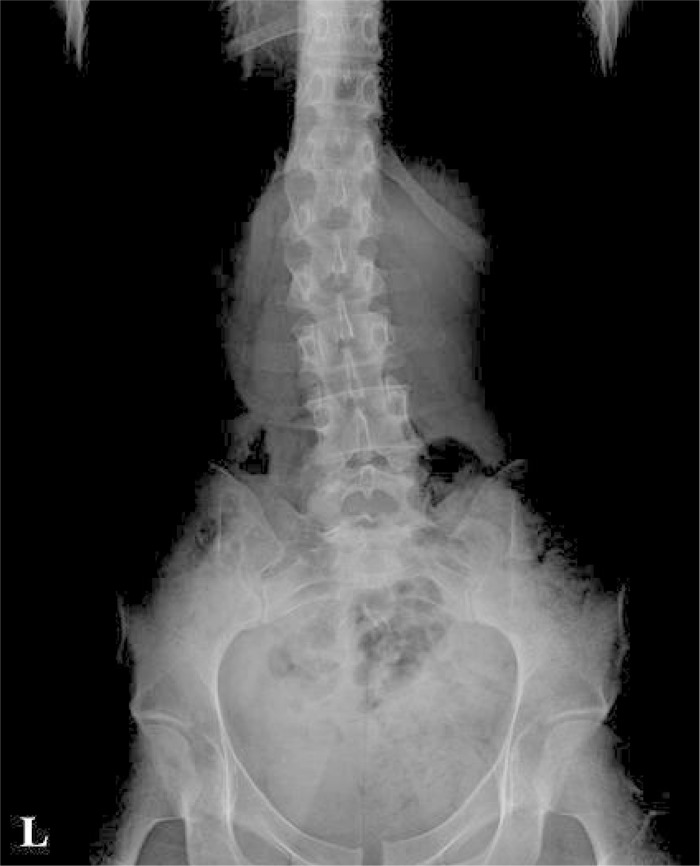
The patient returned for a follow up appointment at approximately nine weeks and was treated using Activator chiropractic adjustment of L5, sacrum, and the sacroiliac joints and home exercise recommendations. She continued with home exercises and the SI belt. At a fourteen-week follow up, she rated her pain 1/10 VAS and reported generally feeling better with return to normal activity. She reported soreness and tightness at the groin on squatting. A post AP lumbopelvic radiograph was taken per patient request and because the patient expressed apprehension to returning to physical activity without additional imaging. This radiograph indicated pubic symphysis separation of just under 10 mm (Figure 2) (without the SI belt). The patient was encouraged to gradually return to normal activity and was released to return as needed.
Discussion
Various physiotherapy methods have been recommended for postpartum pelvic pain but the evidence of benefit is weak. This patient’s care included specific stabilizing exercises, home care advice and sacroiliac belt, in accordance with approaches described in the existing literature. Additionally, she was treated using specific chiropractic adjustments to reduce joint dysfunction at the lumbar, sacral and pelvic areas, using an ischial tuberosity contact to correct anterior superior pelvic misalignment. In this case chiropractic management appears to have helped reduce pain, reduce pubic symphysis separation, and facilitate a return to normal activities.
The author suggests future research to investigate chiropractic care for patients with pubic symphysis diastasis.
Conclusion
In this case, the patient reported that chiropractic care was effective in reducing pain associated with pubic symphysis diastasis. While the author acknowledges that there is no way to know what intervention, if any, helped in this patient’s improvement or whether reduction in pain and decreased diastasis resulted from natural history, the potential for long-term pain and disability and the scarcity of existing literature indicate the need for further investigation. Collaboration between chiropractors, midwives, and obstetricians should be encouraged.
Figures 1 and 2, both without SI belt, were taken at the author's office.
Figure 1:
Pre radiograph showing 17 mm separation at pubic symphysis.
Figure 2:
Post radiograph at 14 weeks, showing reduced separation at pubic symphysis to just under 10 mm.
Acknowledgments
Thank you to John Hart, DC, MHSc, Assistant Director of Research at Sherman College of Chiropractic, for his assistance with revising this manuscript.
Footnotes
There are no disclaimers.
The author has obtained the patient’s written consent to publish the case. There was no external funding or support.
References
- 1.Scriven MW, Jones DA, McKnight L. The importance of pubic pain following childbirth: a clinical ultrasonographic study of diastasis of the pubic symphysis. JR Soc Med. 1995;88:28–30. [PMC free article] [PubMed] [Google Scholar]
- 2.Musumeci R, Villa E. Symphysis pubis separation during vaginal delivery with epidural anaesthesia. Reg Anaesth. 1994;19:289–91. [PubMed] [Google Scholar]
- 3.Senechal PK. Symphysial pubis separation during childbirth. J Am Board Fam Pract. 1994;7(2):141–4. [PubMed] [Google Scholar]
- 4.Parker J, Bhattacharjee M. Peripartum diastasis of the symphysis pubis. N Engl J Med. 2009 2009 Nov 5;361:1886. doi: 10.1056/NEJMicm0807117. [DOI] [PubMed] [Google Scholar]
- 5.Muecke EC, Currarino G. Congenital widening of the pubic symphysis: associated clinical disorders and roentgen anatomy of affected bony pelves. Am J Roentgenol Radium Ther Nucl Med. 1968 May;103(1):179–85. [PubMed] [Google Scholar]
- 6.Bahlmann F, Merz E, Macchiella D, Weber G. Ultrasound imaging of the symphysis fissure for evaluating damage to the symphysis in pregnancy and postpartum. Z Geburtshilfe Perinatol. 1993 Jan-Feb;197(1):27–30. [PubMed] [Google Scholar]
- 7.Garagiola DM, Tarver RD, Gibson L, Rogers RE, Wass JL. Anatomic changes in the pelvis after uncomplicated vaginal delivery: a CT study on 14 women. AJR Am J Roentgenol. 1989 Dec;153(6):1239–41. doi: 10.2214/ajr.153.6.1239. [DOI] [PubMed] [Google Scholar]
- 8.Pedrazzini A, Bisaschi R, Borzoni R, Simonini D, Guardoli A. Post partum diastasis of the pubic symphysis: a case report. Acta Biomed. 2005 Apr;76(1):49–52. [PubMed] [Google Scholar]
- 9.Shippey S, Roth J, Gaines R. Pubic symphysis diastasis with urinary incontinence: collaborative surgical management. Int Urogynecol J. 2013 Oct;24(10):1757–9. doi: 10.1007/s00192-013-2120-0. Epub 2013 May 15. [DOI] [PubMed] [Google Scholar]
- 10.Hou Z, Riehl JT, Smith WR, Strohecker KA, Maloney PJ. Severe postpartum disruption of the pelvic ring: report of two cases and review of the literature. Patient Saf Surg. 2011;5:2. doi: 10.1186/1754-9493-5-2. Published online 2011 January 13. [DOI] [PMC free article] [PubMed] [Google Scholar]
- 11.Stuge B, Laerum E, Kirkesola G, Vøllestad N. The efficacy of a treatment program focusing on specific stabilizing exercises for pelvic girdle pain after pregnancy: a randomized controlled trial. Spine (Phila Pa 1976) 2004 Feb 15;29(4):351–9. doi: 10.1097/01.brs.0000090827.16926.1d. [DOI] [PubMed] [Google Scholar]
- 12.Nilsson-Wikmar L, Holm K, Oijerstedt R, Harms-Ringdahl K. Effect of three different physical therapy treatments on pain and activity in pregnant women with pelvic girdle pain: a randomized clinical trial with 3, 6, and 12 months follow-up postpartum. Spine (Phila Pa 1976) 2005 Apr 15;30(8):850–6. doi: 10.1097/01.brs.0000158870.68159.d9. [DOI] [PubMed] [Google Scholar]
- 13.Depledge J, McNair PJ, Keal-Smith C, Williams M. Management of symphysis pubis dysfunction during pregnancy using exercise and pelvic support belts. Phys Ther. 2005 Dec;85(12):1290–300. [PubMed] [Google Scholar]
- 14.Haugland KS, Rasmussen S, Daltveit AK. Group intervention for women with pelvic girdle pain in pregnancy. A randomized controlled trial. Acta Obstet Gynecol Scand. 2006;85(11):1320–6. doi: 10.1080/00016340600780458. [DOI] [PubMed] [Google Scholar]
- 15.Panarello SR. Symphysis pubis subluxation: pre and post partum chiropractic care. J Clinical Chiropr Ped. 2005;6(3):432–435. [Google Scholar]
- 16.Howell ER. Pregnancy-related symphysis pubis dysfunction management and postpartum rehabilitation: Two case reports. J Can Chiropr Assoc. 2012 Jun;56(2) Online access only p1-2-111. [PMC free article] [PubMed] [Google Scholar]
- 17.Shambaugh P1, Sclafani L, Fanselow D. Reliability of the Derifield-Thompson test for leg length inequality, and use of the test to demonstrate cervical adjusting efficacy. J Manipulative Physiol Ther. 1988 Oct;11(5):396–9. [PubMed] [Google Scholar]
- 18.Mannello DM. Leg length inequality. J Manipulative Physiol Ther. 1992 Nov-Dec;15(9):576–90. [PubMed] [Google Scholar]
- 19.Knutson GA. Incidence of foot rotation, pelvic crest unleveling, and supine leg length alignment asymmetry and their relationship to self-reported back pain. J Manipulative Physiol Ther. 2002 Feb;25(2):110E. doi: 10.1067/mmt.2002.121414. [DOI] [PubMed] [Google Scholar]
- 20.Schneider M1, Homonai R, Moreland B, Delitto A. Interexaminer reliability of the prone leg length analysis procedure. J Manipulative Physiol Ther. 2007 Sep;30(7):514–21. doi: 10.1016/j.jmpt.2007.07.001. [DOI] [PubMed] [Google Scholar]
- 21.Triano JJ, Budgell B, Bagnulo A, Roffey B, Bergmann T, Cooperstein R, Gleberzon B, Good C, Perron J, Tepe R. Review of methods used by chiropractors to determine the site for applying manipulation. Chiropr Man Therap. 2013 Oct 21;21(1):36. doi: 10.1186/2045-709X-21-36. [DOI] [PMC free article] [PubMed] [Google Scholar]
- 22.DeSantana JM1, Walsh DM, Vance C, Rakel BA, Sluka KA. Effectiveness of transcutaneous electrical nerve stimulation for treatment of hyperalgesia and pain. Curr Rheumatol Rep. 2008 Dec;10(6):492–9. doi: 10.1007/s11926-008-0080-z. [DOI] [PMC free article] [PubMed] [Google Scholar]
- 23.French SD, Cameron M, Walker BF, Reggars JW, Esterman AJ. A Cochrane review of superficial heat or cold for low back pain. Spine (Phila Pa 1976) 2006 Apr 20;31(9):998–1006. doi: 10.1097/01.brs.0000214881.10814.64. [DOI] [PubMed] [Google Scholar]
- 24.Gerwin RD. Diagnosis of myofascial pain syndrome. Phys Med Rehabil Clin N Am. 2014 May;25(2):341–55. doi: 10.1016/j.pmr.201401.011. Epub 2014 Mar 18. [DOI] [PubMed] [Google Scholar]


