Abstract
Alpha-glucosidase inhibitors currently form an important basis for developing novel drugs for diabetes treatment. In our preliminary tests, the ethyl acetate fraction of Phlomis tuberosa extracts showed significant α-glucosidase inhibitory activity (IC₅₀ = 100 μg/mL). In the present study, a combined method using Sepbox chromatography and thin-layer chromatography (TLC) bioautography was developed to probe α-glucosidase inhibitors further. The ethyl acetate fraction of P. tuberosa extracts was separated into 150 individual subfractions within 20 h using Sepbox chromatography. Then, under the guidance of TLC bioautography, 20 compounds were successfully isolated from these fractions, including four new diterpenoids [14-hydroxyabieta-8,11,13-triene-11-carbaldehyde-18-oic-12-carboxy-13-(1-hydroxy-1-methylethyl)-lactone (1), 14-hydroxyabieta-8,11,13-triene-17-oic-12-carboxy-13-(1-hydroxy-1-methylethyl)-lactone (2), 14,16-dihydroxyabieta-8,11,13-triene-15,17-dioic acid (3), and phlomisol (15,16-eposy-8,13(16),14-labdatrien-19-ol) (4)], and 16 known compounds. Activity estimation indicated that 15 compounds showed more potent α-glucosidase inhibitory effects (with IC50 values in the range 0.067–1.203 mM) than the positive control, acarbose (IC50 = 3.72 ± 0.113 mM). This is the first report of separation of α-glucosidase inhibitors from P. tuberosa.
Introduction
Diabetes is one of the most important health issues facing the world in the 21st century [1]. Alpha-glucosidase inhibitors have been used to lower blood glucose for about 20 years now. These compounds show reasonably good efficacy comparable with other oral blood glucose-lowering drugs and, in some parts of the world, are the most commonly prescribed oral diabetes medication, especially in Asian countries [2]. In recent years, investigations of herbal medicines have become increasingly important in the search for new, effective, and safe α-glucosidase inhibitors for diabetes treatment [3].
Phlomis tuberosa L. is an Asian folk medicine used as a general roborant for intoxication, tuberculosis, pulmonary and cardiovascular diseases, and rheumatoid arthritis [4]. In preliminary tests in which we screened α-glucosidase inhibitors from medicinal herbs, the ethyl acetate fraction of P. tuberosa extracts showed significant inhibitory activity against α-glucosidase (IC50 = 100 μg/mL). Repeated column chromatography over silica gel is frequently used for purification of bioactive compounds from medicinal herbs. However, this separation method can be a tedious process requiring long time frames and large volumes of organic solvents; irreversible adsorption of samples onto the solid phase, which sometimes results in reductions or disappearance of active compounds, may also occur [5]. Therefore, better purification strategies to assure the efficacy and reliability of α-glucosidase inhibitor identification from P. tuberosa are necessary.
Automated HPLC/SPE/HPLC coupled separation using a Sepbox system is a standard technology for separating compounds from natural resources; the technique allows automatic processing of samples by up to 30 times faster than conventional processes [6]. An extract can be fractionated into 100–300 substances composed of 1–4 compounds using Sepbox chromatography within less than 30 h [7]. Thin-layer chromatography (TLC) bioautography combines chromatographic separation with in situ biological activity determination, allows the direct and rapid localization of active compounds in complex extracts [8]. A TLC bioautographic method has been established to detect α-glucosidase inhibitors in plant extracts [9]. Because of its unique benefit of simultaneous chromatographic fractionation and bioactivity screening, Sepbox chromatography coupled with TLC bioautography is speculated to be an attractive strategy for rapid identification of α-glucosidase inhibitors from P. tuberosa.
In the present study, Sepbox chromatography and TLC bioautography led to the isolation of four new diterpenoids (1, 2, 3, 4), two known diterpenoids (5, 6), three known phenylethanoid glycosides (7, 8, 9), three known flavonoids (10, 11, 12), four known iridoids (13, 14, 15, 16), and four other compounds (17, 18, 19, 20) from P. tuberosa. The α-glucosidase inhibitory activities of the isolated compounds were subsequently determined.
Materials and Methods
General
The automated HPLC/SPE/HPLC experiment was performed using a Sepbox 2D-2000 (Sepiatec, Germany). Silica gel 60 F254TLC plates (MN, Germany) were used for TLC analysis. HPLC analysis was performed using an Agilent 1100 HPLC system (Agilent Technologies, Santa Clara, USA) consisting of a G1312A QuatPump, a G1314A DAD detector, a G1313A autosampler, and a G1332A degasser equipped with an Agilent Zorbax SB-C18 column (250 mm × 4.6 mm, i.d. 5 m). Compound purification was performed using a LC-3000 Semi-preparation gradient HPLC system (Beijing Tong Heng Innovation Technology Co., Ltd., China) equipped with a UV-vis detector and a YMC-Pack PRO C18 column (250 mm × 10 mm, i.d. 5 μm; YMC, Japan). Identification of isolated compounds was performed using 1H NMR, 13C NMR, and MS. NMR spectra were obtained on a Bruker Avance 400 or 600 NMR spectrometer (Bruker Inc., Bremen, Germany). HRESIMS spectra were obtained using a Waters UPLC Premier QTOF spectrometer, and UPLC-MS analysis was performed on an Acquity Waters ultra-performance liquid chromatographic system equipped with a Waters UPLC column (Acquity UPLC BEH C18 1.7 μm, 2.1 mm × 50 mm) and a Micromass ZQ 2000 ESI mass spectrometer. α-d-Glucosidase, 2-naphthyl-α-d-glucopyranoside, Fast Blue B salt, 4-nitrophenyl α-glucopyranoside, and acarbose were purchased from Sigma-Aldrich (St. Louis, MO, USA). All solvents used for chromatography and extraction were of HPLC grade and obtained from Fisher Scientific (Fair Lawn, NJ, USA). All other chemicals used were of analytical grade and applied without further purification.
Plant material
Roots of P. tuberosa were collected from Shangdu Town, Inner Mongolia Autonomous Region, China (latitude/longitude, 42°15′11″N/115°59′52″E), in November 2011 and authenticated by Prof. A.O. Wuliji (Inner Mongolia University for the Nationalities). A voucher specimen (No. kgcs-120515) was deposited at Shanghai R&D Center for Standardization of Traditional Chinese Medicines, Shanghai, China.
Ethics
No specific permissions were required for the described field studies. The locations are neither privately owned nor protected by the Chinese government. No endangered or protected species were sampled.
Sample preparation
Air-dried, chopped roots (1.0 kg) of P. tuberosa were extracted thrice using 10 L of 95% ethanol under reflux for 2.5 h. The extracts were combined and evaporated to dryness at 60°C under reduced pressure. The resulting residue was resuspended in distilled water and partitioned thrice in a separatory funnel with an equal volume of ethyl acetate each time. The ethyl acetate layers were combined and then further dried in vacuo at ambient temperature for 24 h to produce the ethyl acetate fraction, which was stored at -20°C prior to Sepbox chromatography separation.
Sepbox chromatography separation
The Sepbox system combines the advantages of HPLC and SPE and was coupled to an HPLC/SPE/HPLC setup to allow two-dimensional separation. In the Sepbox 2D-2000 system used in this study, a crude ethyl acetate fraction of P. tuberosa (5 g) was absorbed onto C4 reverse-phase resin (4 g) and initially separated into 15 fractions using a C4 reverse phase HPLC column. These fractions were transferred to 15 SPE trap columns. Fractions trapped in each SPE column were passed through a C18 RP HPLC column for subsequent separation; here, elution was performed with solvents different from those used during the first fractionation. Through monitoring of HPLC peaks from UV (254 nm) and ELSD detectors, a total of 150 individual subfractions (named Pt1–150) were collected. The separation conditions are shown in S1 Table.
TLC analysis
In the present work, a high-performance thin-layer chromatographic system (CAMAG, Switzerland) equipped with an automatic TLC Sampler 4 and a Reprostar 3 with a 12-bit charge-coupled device camera for photo-documentation and controlled by WinCATS-4 software was used. An aliquot of the subfraction solution (20 μL), the ethyl acetate fraction of P. tuberosa extract (5 mg/mL, 20 μL), acarbose methanol solution (1 mg/mL, 4 μL), or individual pure isolate methanol solution (2.5 mM, 4 μL) was deposited directly (as spots or bands) onto the TLC plates. After migration of the samples with appropriate solvents or no migration, the plates were inspected under ultraviolet light (366 nm) and then subjected to bioautographic assay as described below.
TLC bioautographic assay: TLC bioautographic assay was carried out as described previously in terms of reaction principle and response system [9] but with slight modifications. First, the concentration of α-glucosidase was reduced from 10 U/mL to 2.5 U/mL. Second, the plate was dipped into the reaction solutions (including the enzyme solution, the substrate solution, and the coloration solution), instead of being sprayed with them. Third, the conditions for enzyme incubation were changed from room temperature for 60 min to 37°C for 30 min. Briefly, α-d-glucosidase (750 U) was dissolved in 75 mL of buffer solution (10.25 g of sodium acetate in 250 mL with addition of 0.1 M acetic acid to pH 7.5). The stock solution was kept at -20°C. A stock solution of 25 mL was diluted with the same buffer solution to 100 mL as the enzyme solution, and a substrate solution of 1 mg/mL 2-naphthyl-α-d-glucopyranoside in ethanol was prepared. To probe active spots (or bands) on the TLC plate, the plate was first dipped in the substrate solution, dried under a stream of cold air for complete removal of the solvent, and then dipped into the enzyme solution. For enzyme incubation, the plate was laid flat on plastic plugs in a plastic tank containing a little water, avoiding direct contact between the water and the plate. A cover was placed on the tank to keep the atmosphere humid and incubation was performed at 37°C for 30 min. To detect the active enzyme, the TLC plate was dipped in a solution of Fast Blue B salt (150 mg) in distilled water (100 mL) prepared immediately before use to produce a white spot under a purple background after 5 min.
HPLC analysis
Subfractions showing α-glucosidase inhibitory activity in bioautographic assay were analyzed by an Agilent 1100 HPLC system, and subfractions determined to contain two or above compounds in HPLC analysis were further purified by Semi-preparative HPLC. The isolated pure compounds (from both direct Sepbox chromatography fractionation and additional Semi-preparative HPLC purification) as well as the ethyl acetate fraction of P. tuberosa extracts were separately analyzed by an Agilent 1100 HPLC system. The mobile phase was a mixture of water (A) and acetonitrile (B), and the detailed gradient program is given in S2 Table. The flow rate was 1 mL/min, and the effluents were monitored by a DAD detector at 280 nm.
α-Glucosidase inhibitory activity assay
α-Glucosidase inhibitory activity assay was performed as reported previously [10] with minor modifications. α-Glucosidase was obtained from Saccharomyces cerevisiae and dissolved in potassium phosphate buffer (pH 6.8) to achieve a concentration of 0.26 U/mL. A total of 50 μL of the test sample (1–200 μg/mL extract, 0.01–2 mM pure compounds) was mixed with 50 μL of α-glucosidase. After incubation at 37°C for 10 min, 100 μL of the substrate, 4-nitrophenyl α-glucopyranoside (5 mM, in potassium phosphate buffer) was added to the mixture. The enzymatic reaction was allowed to proceed at 37°C for 20 min and stopped by addition of 100 μL of 0.2 M NaCO3. 4-Nitrophenol absorptions were subsequently measured at 405 nm using a spectrophotometer. The percentage inhibition of α-glucosidase was calculated as inhibition rate (%) = 100 × [1 - (A sample–A s-blank)/(A control–A blank)], where A sample represents the absorbance of the reaction system containing the test sample, enzyme, and substrate, A s-blank represents the absorbance of the reaction system containing the test sample and substrate but without enzyme, A control represents the absorbance of the reaction system containing the enzyme and substrate but without test sample, and A blank represents the absorbance of the reaction system containing the substrate but without test sample and enzyme. A carbose was used as a positive control. The IC50 value was defined as the concentration of α-glucosidase inhibitor required to inhibit 50% of α-glucosidase activity under the assay conditions. Experiments were performed in triplicate, and all results are expressed as mean ± SEM.
Results and Discussion
Screening of α-glucosidase inhibitors from P. tuberosa
A method combining Sepbox chromatography and TLC bioautography was developed to identify α-glucosidase inhibitors from P. tuberosa. Sepbox chromatography separated a total of 150 subfractions (designated Pt1–Pt150) from the ethyl acetate fraction of P. tuberosa extracts within 20 h, after which modified TLC autographic assay was used to examine the α-glucosidase inhibitory activity of each subfraction. As shown in S1 Fig., compared with the previously reported TLC autographic assay [9], the modified assay showed a darker background color, thereby allowing easier detection of active spots. In the bioautographic chromatograms, both the ethyl acetate fraction of P. tuberosa extract and acarbose solution produced white spots, which indicates their α-glucosidase inhibitory activities. At least nine spots (corresponding to subfractions Pt15, Pt35, Pt76, Pt102, Pt103, Pt104, Pt120, Pt121, and Pt133) in the chromatograms showed α-glucosidase inhibitory activities.
Analyses of the HPLC data revealed that subfractions Pt15, Pt35, Pt102, and Pt120 contained only one major compound and the five other subfractions (Pt76, Pt103, Pt104, Pt121 and Pt133) contained less than four compounds. Pt15, Pt35, Pt102, and Pt120 were individually collected and concentrated, yielding 4.52 mg of compound 5, 25.7 mg of compound 7, 9.10 mg of compound 15, and 4.83 mg of compound 4, respectively. The five other subfractions were individually collected and further purified by preparative HPLC to produce compound 1 (11.24 mg), compound 2 (3.40 mg), compound 3 (3.01 mg), compound 6 (1.78 mg), compound 8 (4.96 mg), compound 9 (3.78 mg), compound 10 (2.11 mg), compound 11 (5.08 mg), compound 12 (6.01 mg), compound 13 (5.12 mg), compound 14 (9.15 mg), compound 16 (2.16 mg), compound 17 (5.62 mg), compound 18 (1.69 mg), compound 19 (1.32 mg), and compound 20 (3.14 mg). The origins of these compounds are shown in Table 1.
Table 1. Origins and α-glucosidase inhibitory activities of the pure isolated compounds.
| Active fraction | Compound | IC50 (mM) |
|---|---|---|
| Pt15 | 5 | 0.482 ± 0.019 |
| Pt35 | 7 | 0.518 ± 0.017 |
| Pt76 | 8 | 1.203 ± 0.032 |
| 9 | - | |
| 12 | 0.562 ± 0.021 | |
| Pt102 | 15 | - |
| Pt103 | 1 | 0.379 ± 0.016 |
| 2 | 0.624 ± 0.026 | |
| 3 | 0.721 ± 0.037 | |
| Pt104 | 6 | 0.210 ± 0.010 |
| 14 | - | |
| 16 | - | |
| Pt120 | 4 | 0.067 ± 0.003 |
| Pt121 | 17 | 0.229 ± 0.017 |
| 18 | 0.283 ± 0.013 | |
| 19 | 0.255 ± 0.013 | |
| 20 | 0.371 ± 0.015 | |
| Pt133 | 10 | - |
| 11 | 0.428 ± 0.018 | |
| 13 | - | |
| Positive control | Acarbose | 3.72 ± 0.113 |
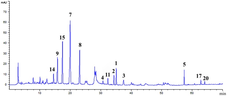
HPLC analysis of the ethyl acetate fraction indicated that most of the major compounds shown in the HPLC profile could be separated by Sepbox chromatography (Fig. 1). Following HPLC, the purities of compounds 1, 2, 3, 4, 5, 6, 7, 8, 9, 10, 11, 12, 13, 14, 15, 16, 17, 18, 19, and 20 were 99.1%, 98.3%, 97.7%, 92.8%, 96.6%, 92.4%, 98.1%, 92.5%, 91.8%, 95.3%, 94.1%, 92.5%, 92.7%, 98.2%, 97.3%, 92.5%, 98.5%, 91.1%, 92.0%, and 99.0%, respectively. This work is the first to report the simultaneous separation of 20 compounds from P. tuberosa.
Fig 1. HPLC chromatogram of the ethyl acetate fraction of P. tuberosa extracts.
Peaks with numbers represent isolated compounds.
Structural determination of isolated compounds
The 20 isolated compounds were identified by 1H and 13C NMR analyses and compared with published data.
Compound 1 was obtained as a white amorphous powder; [α]20 D + 0.255 (c 1.8, MeOH); UV (MeOH) λmax (log ε) 218 (1.78), 242 (1.11), 302 (0.46), and 362 (0.28) nm (S2 Fig.). Its molecular formula C20H22O6 indicated 11 degrees of unsaturation, as deduced from its HRESIMS spectrum at m/z 357.1329 ([M–H]-, calcd. 357.1338; S3 Fig.). The IR spectrum of 1 (S4 Fig.) showed hydroxyl (3399 cm-1), carbonyl (1745 and 1697 cm-1), and phenyl (1587 and 1243 cm-1) groups. The 1H NMR spectrum of 1 (S5 Fig.) showed two methyl singlets at δH 1.32 (3H, s, H-19) and δH 1.35 (3H, s, H-20) and one aldehyde proton at δH 10.9 (1H, s, H-15). The 13C NMR and DEPT spectra of 1 (S6 Fig.) indicated a total of 20 carbon signals comprising two methyls, six methylenes, one methine, one aldehyde, and ten quaternary carbons. The relevant 1H and 13C NMR data are given in Table 2. The spectroscopic characteristics described above resemble those of an abietane diterpene with a tetracyclic system. HMBC correlations between H-20 (δH 1.35) and C-1 (δC 40.7), C-5 (δC 54.8), C-9 (δC 151.5), and C-10(δC 42.1); between H-3 (δH 2.21) and C-1 (δC 40.7); and between H-19 (δH 1.32) and C-3 (δC 38.2), C-4 (δC 44.8), and C-5(δC 54.8) support the abietane diterpene skeleton. Correlations between the aldehyde group (δH 10.9) and C-9 (δC 151.5), C-11 (δC 131.8) were also observed in the HMBC spectrum (S8 Fig.). Thus, the structure of 1 was established as 14-hydroxyabieta-8,11,13-triene-11-carbaldehyde-18-oic-12-carboxy-13-(1-hydroxy-1-methylethyl)-lactone.
Table 2. 1H and 13C NMR data of compounds 1–4 in CD3OD (400 MHz and 100 MHz for 1H and 13C NMR, respectively; δ in ppm, J values in Hz).
| Position | 1 | 2 | 3 | 4 | ||||
|---|---|---|---|---|---|---|---|---|
| δH | δC | δH | δC | δH | δC | δH | δC | |
| 1 | 2.21 m, 1.28 td (13.2, 3.6) | 40.7 | 2.38 d (12.6), 1.41 td (13.2, 3.0) | 41.1 | 2.73 d (12.0), 1.43 m | 37.5 | 1.91 m, 1.88 m | 36.8 |
| 2 | 2.02 m, 1.55 m | 21.1 | 2.06 m, 2.11 m | 21.2 | 2.08 m, 1.63 d (14.0) | 20.9 | 1.26 m | 38.2 |
| 3 | 2.21 d (13.2), 1.08 td (13.8, 4.2) | 38.2 | 2.26 d (13.2), 1.13 td (13.2, 3.6) | 38.6 | 2.22 d (12.8), 1.12 td (13.6, 3.6) | 38.4 | 1.73 m, 1.66 m | 19.7 |
| 4 | - | 44.8 | - | 44.8 | - | 45.0 | - | 38.4 |
| 5 | 1.44 d (11.4) | 54.8 | 1.55 d (12.6) | 53.2 | 1.52 d (12.4) | 53.6 | 1.31 dd (12.8, 1.6) | 54.1 |
| 6 | 2.32 dd (14.4, 7.2), 2.03 m | 20.6 | 2.33 dd (13.8, 6.6), 1.65 d (13.8) | 21.3 | 2.32 dd (14.0, 7.2), 2.13 m | 20.8 | 1.65 m, 1.47 m | 20.3 |
| 7 | 3.04 dd (18.6, 4.8), 2.63 ddd (19.8, 13.2, 7.2) | 28.6 | 3.08 dd (18.0, 4.8), 2.57 ddd (18.6, 12.6, 6.6) | 27.5 | 3.06 dd (18.8, 5.6), 2.58 ddd (19.3, 12.6, 7.3) | 28.2 | 2.04 dd (11.2, 6.8), 1.98 d (6.4) | 34.9 |
| 8 | - | 132.8 | - | 131.6 | - | 134.0 | - | 127.5 |
| 9 | - | 151.5 | - | 153.3 | - | 147.2 | - | 141.2 |
| 10 | - | 42.1 | - | 40.3 | - | 41.6 | - | 40.0 |
| 11 | 131.8 | 7.41 s | 114.7 | 6.73 s | 99.9 | 2.26 m, 2.11 m | 30.2 | |
| 12 | - | 130.1 | - | 129.9 | - | 121.2 | 2.44 m | 26.8 |
| 13 | - | 124.8 | - | 125.0 | - | 123.2 | 126.8 | |
| 14 | - | 152.0 | - | 150.2 | - | 157.9 | 6.31 d (0.8) | 111.7 |
| 15 | 10.9 s | 198.8 | - | - | - | - | 7.37 t (1.6) | 143.9 |
| 16 | - | 172.4 | - | 174.3 | - | 171.5 | 7.27 s | 139.6 |
| 17 | 5.30 d (15.0), 5.27 d (15.0) | 69.6 | 5.26 s | 69.6 | 5.35 d (14.4), 5.29 d (14.4) | 59.0 | 1.60 s | 19.8 |
| 18 | - | 181.1 | - | 181.3 | - | 181.5 | 4.24 d (9.6), 3.80 d (9.2) | 71.8 |
| 19 | 1.32 s | 29.5 | 1.34 s | 29.2 | 1.32 s | 29.6 | 1.02 s | 27.7 |
| 20 | 1.35 s | 21.4 | 1.20 s | 23.9 | 1.35 s | 21.3 | 0.99 s | 21.2 |
Compound 2 was obtained as a white amorphous powder; [α]20 D + 0.087 (c 1.0, MeOH); UV (MeOH) λmax (log ε) 210 (1.73), 254 (0.62), and 296 (0.29) nm (S9 Fig.). Its molecular formula C19H22O5 was determined based on its HRESIMS spectrum at m/z 329.1370 ([M–H]-, calcd. 329.1362; S10 Fig.). The IR spectrum of 2 (S11 Fig.) showed hydroxyl (3429 cm1), carbonyl (1735 and 1686 cm-1), and phenyl (1591 and 1262 cm-1) groups. 1H and 13C NMR data (Table 2; S12 and S13 Figs.) revealed the presence of two methyls [δH 1.20 (3H, s, H-20) and δH 1.34 (3H, s, H-19)], one lactone group [δC 174.3 (C-16)], one carboxylic group [δC 181.3 (C-18)], and one aromatic proton [δH 7.41 (1H, s) and δC 114.7]. These data, together with other spectroscopic characteristics, suggested that 2 is an abietane diterpenoid. HMBC correlations between H-20 (δH 1.20) and C-1 (δC 41.1), C-9 (δC 153.3), and C-10 (δC 40.3) supported this inference; the only difference between 1 and 2 is that the proton at C-11 in 2 is replaced by an aldehyde group in 1, as deduced from HMBC correlations of H-11 (δH 7.41) with C-8 (δC 131.6), C-10 (δC 40.3), C-12 (δC 129.9), C-14 (δC 150.2), and C-16 (δC 174.3) (S15 Fig.). Thus, 2 was determined to be an abietane norditerpene, namely, 14-hydroxyabieta-8,11,13-triene-17-oic-12-carboxy-13-(1-hydroxy-1-methylethyl)-lactone.
Compound 3 was obtained as a yellow amorphous powder; [α]20 D + 0.3 (c 2.3, MeOH); UV (MeOH) λmax (log ε) 217 (1.12), 257 (0.29), and 306 (0.20) nm (S16 Fig.). Its molecular formula was determined from its HRESIMS spectrum at m/z 349.2349 to be C19H24O6 ([M + H]+, calcd. 349.2355; S17 Fig.), which is 18 mass units larger than that of 2; the difference in molecular formulas observed corresponds to one additional oxygen atom and two hydrogen atoms. The IR spectrum of 3 (S18 Fig.) showed hydroxyl (3434 cm-1), carbonyl (1735 and 1654 cm-1), and phenyl (1466 and 1287 cm-1) groups. 1H and 13C NMR data (Table 2; S19 and S20 Figs.) revealed the presence of two methyls [δH 1.35 (3H, s, H-20), and δH 1.32 (3H, s, H-19)], one lactone group [δC 171.5 (C-16)], one carboxylic group [δC 181.5 (C-18)], one aromatic hydrogen δH 6.73 (1H, s), and carbon signals at δC 99.9. HMBC correlations between H-20 (δH 1.35) and C-1 (δC 37.5), C-9 (δC 147.2), and C-10 (δC 41.6) and between H-11 (δH 6.73) and C-8 (δC 134.0), C-10 (δC 41.6), C-12 (δC 121.2), C-14 (δC 157.9), and C-16 (δC 171.5) confirmed the tetracyclic abietane diterpene skeleton of 3. HMBC correlations from H-17 (δH 5.35, 5.29) to C-12 (δC 121.2), C-13 (δC 123.2), C-14 (δC 157.9), and C-16 (δC 171.5) were also observed (S23 Fig.). According to the 1H and 13C NMR data shown in Table 2, 3 is very similar to 2. Therefore, 3 was identified as 14,16-dihydroxyabieta-8,11,13-triene-15,17-dioic acid.
Compound 4 was obtained as a white amorphous powder; [α]20 D + 0.005 (c 0.68, MeOH); UV (MeOH) λmax (log ε) 204 (0.22) nm (S24 Fig.). Its molecular formula was determined to be C20H30O2 by its HRESIMS spectrum at m/z 303.3067 ([M + H]+, calcd. 303.3059; S25 Fig.). The IR spectrum of 4 (S26 Fig.) showed hydroxyl (3432 cm-1) and carbonyl (1655 and 1630 cm-1) groups. The 1H NMR spectrum of 4 (S27 Fig.) showed three methyl singlets at δH 0.99 (3H, s, H-20), 1.02 (3H, s, H-19), and 1.60 (3H, s, H-17). 13C NMR and DEPT spectra (S28 Fig.) indicated a total 20 carbon signals composing three methyls, seven methylenes, three methines, one oxygen-bearing methylene, and five quaternary carbons. The relevant 1H and 13C NMR data are given in Table 2. The spectroscopic characteristics described above resemble those of a labdane diterpene with a tricyclic system [11]. HMBC correlations between H-20 (δH 0.99) and C-5 (δC 54.1), C-9 (δC 141.2), and C-10(δC 40.0); between H-19 (δH 1.02) and C-2 (δC 38.2), C-4 (δC 38.4), and C-5(δC 54.1); between H-17 (δH 1.60) and C-7 (δC 34.9), C-8(δC 127.5), and C-9 (δC 141.2); between H-11 (δH 2.26, 1.29) and C-8 (δC 127.5), C-9 (δC 141.2), and C-10(δC 40.0); between H-3 (δH 1.73, 1.66) and C-19 (δC 27.7); and between H-18 (δH 4.24, 3.80) and C-4 (δC 38.5), C-19 (δC 27.7), and C-5(δC 54.1) supported the labdane diterpene skeleton. Correlations between H-14 (δH 6.31) and C-13 (δC 126.8); between H-15 (δH 7.37) and C-13 (δC 126.8); and between H-16 (δH 7.27) and C-13 (δC 126.8) were also observed in the HMBC spectrum (S30 Fig.). Thus, 4 was determined to be 15,16-eposy-8,13(16),14-labdatrien-19-ol and named phlomisol.
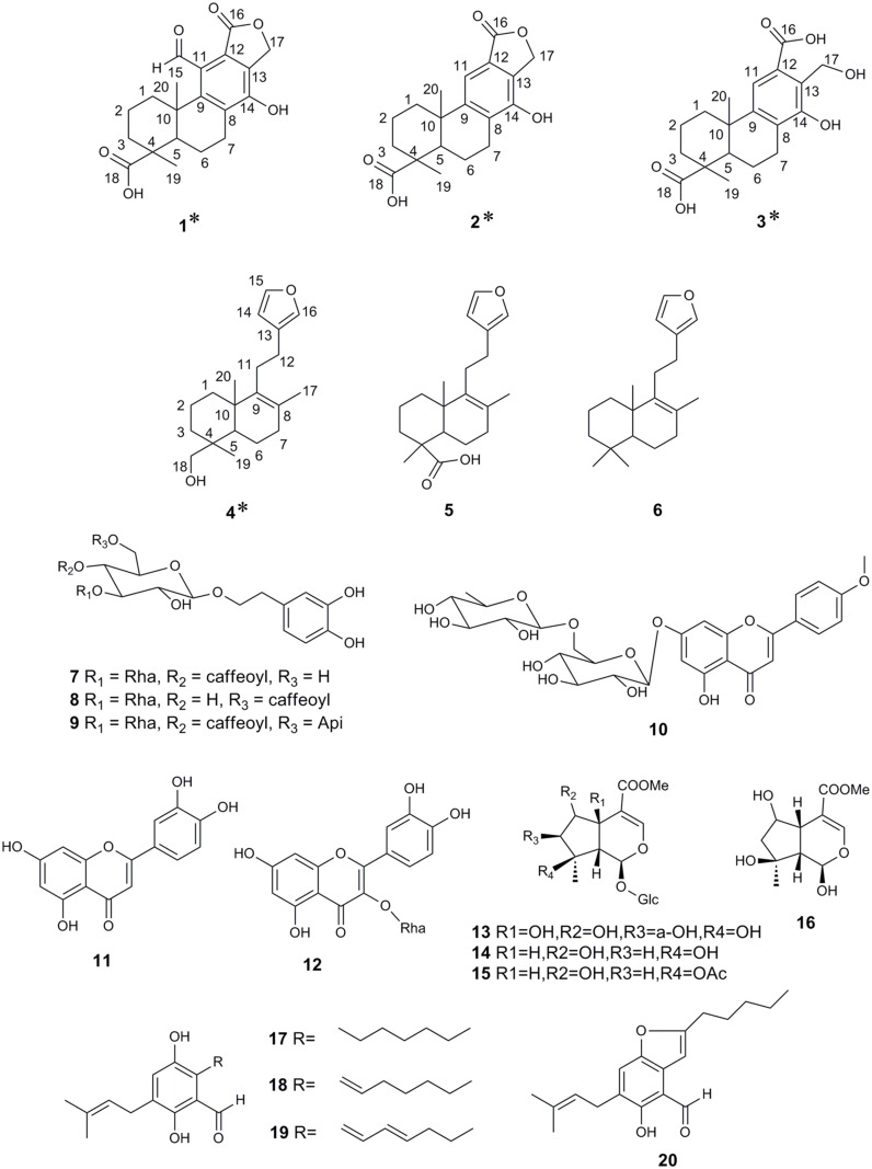
Comparison of physical and spectral findings with published data allowed identification of compounds 5 to 20 as phlomisoic acid (5) [11], 15,16-eposy-8,13(16),14-labdatrien (6) [12], acteoside (7), isoacteoside (8) [13], forsythoside B (9) [14], acacetin-7-rutinoside (10) [15], luteolin (11) [16], quercetin-3-rhamnoside (12) [17], phlomiol (13) [18], shanzhiside methyl ester (14), 8-O-acetylshanzhiside methyl ester (15) [19], 8-O-acetylshanzhigenin methyl ester (16) [20], flavoglaucin (17), tetrahydroauroglaucin (18), dihydroauroglaucin (19) [21], 2-(2′,3-epoxy-1′-heptenyl)-6-hydroxy-5-(3″-methyl-2″-butenyl) benzaldehyde (20) [22]. The structures of the 20 isolated compounds are shown in Fig. 2. Compounds 1 to 4 are original natural compounds, compounds 6, 10, and 17–20 are isolated from the genus Phlomis for the first time, and compounds 5, 8, 11–13, 15, and 16 are isolated from P. tuberosa for the first time.
Fig 2. Structures of compounds 1–20 isolated from P. tuberosa.
Asterisks indicate new compounds.
α-Glucosidase inhibitory activity of the isolated compounds
Another TLC bioautographic assay was conducted to confirm the α-glucosidase inhibitory activity of the isolated compounds. The ethyl acetate fraction of P. tuberosa extracts and 20 individual pure compounds were migrated with the appropriate solvent, inspected under UV 366 nm, and then subjected to bioautographic assay (S31 Fig.). In the bioautographic chromatograms, 15 of the compounds, including compounds 1–8, 10–12, and 17–20, were active components corresponding to the active spots of the ethyl acetate fraction of P. tuberosa extracts. This result indicates that the α-glucosidase inhibitory components of P. tuberosa can be obtained by an integrated Sepbox chromatography prefractionation and TLC bioautograph-guided separation strategy. However, we note that the data in S31 Fig. are not quantitative.
The inhibitory capacity of these compounds was quantitatively estimated by conventional α-glucosidase inhibitory activity assay because this method is considered to be the most direct and reliable method for determining α-glucosidase inhibitory capacity thus far [10]. Results showed that compounds 1–8, 10–12, and 17–20 exhibited much stronger α-glucosidase inhibitory activities (with IC50 values in the range 0.067–1.203 mM) than the positive control acarbose (IC50 = 3.72 ± 0.113 mM; Table 1). Previous toxicity studies have also demonstrated that compounds 7, 10, and 12 are nontoxic [23–25], which indicates their potential use as antidiabetes drugs. Toxicological tests of other compounds have yet to be performed.
Conclusions
A simple, rapid, and effective Sepbox chromatography method coupled to TLC bioautography was established to probe active ingredients with α-glucosidase inhibition capability from P. tuberosa. Twenty compounds, including four new compounds, were isolated; six of these compounds were first isolated from the genus Phlomis. Among the compounds obtained from P. tuberosa, the four new compounds and 11 other compounds showed significant α-glucosidase inhibition activities. These compounds demonstrated much higher α-glucosidase inhibitory capacities than acarbose, which indicates their potential use as alternative medicines for diabetes mellitus.
Supporting Information
Pt: ethyl acetate fraction of P. tuberosa (5 mg/mL, 20 μL); Acar: acarbose (1 mg/mL, 4 μL); 1–150: 150 subfractions (20 μL) obtained by Sepbox chromatography separation. Subfractions with α-glucosidase inhibitory activity are boxed in yellow frames.
(TIF)
(TIF)
(TIF)
(TIF)
(TIF)
(TIF)
(TIF)
(TIF)
(TIF)
(TIF)
(TIF)
(TIF)
(TIF)
(TIF)
(TIF)
(TIF)
(TIF)
(TIF)
(TIF)
(TIF)
(TIF)
(TIF)
(TIF)
(TIF)
(TIF)
(TIF)
(TIF)
(TIF)
(TIF)
(TIF)
Twenty microliters of the ethyl acetate fraction (Pt, 5 mg/mL) of P. tuberosa and 4 μL of the pure isolates (2.5 mM) were applied as bands on TLC plates. The first plate was eluted with petroleum ether/ethyl acetate (10:1), the second plate was eluted with trichloromethane/methanol (12:1), and the third plate was eluted with ethyl acetate/methanol/water (15:2:1).
(TIF)
(DOC)
(DOC)
Data Availability
All relevant data are within the paper and its Supporting Information files.
Funding Statement
The authors gratefully acknowledge the Program for Changjiang Scholars and Innovative Research Team in University (IRT1071) for the financial support. The funders had no role in study design, data collection and analysis, decision to publish, or preparation of the manuscript.
References
- 1. Hanefeld M (2007) Cardiovascular benefits and safety profile of acarbose therapy in prediabetes and established type 2 diabetes. Cardiovasc Diabetol 6: 20 [DOI] [PMC free article] [PubMed] [Google Scholar]
- 2. Standl E, Schnell O (2012) Alpha-glucosidase inhibitors 2012–cardiovascular considerations and trial evaluation. Diabetes Vasc Dis Res 9: 163–169. 10.1177/1479164112441524 [DOI] [PubMed] [Google Scholar]
- 3. Misbah H, Aziz AA, Aminudin N (2013) Antidiabetic and antioxidant properties of Ficus deltoidea fruit extracts and fractions. BMC Comp Alt Med 13: 118. [DOI] [PMC free article] [PubMed] [Google Scholar]
- 4. Calis I, Kirmizibekmez H, Ersoz T, Donmez AA, Gotfredsen CH, et al. (2005) Iridoid glucosides from Turkish Phlomis tuberosa . Z Naturforsch B 60: 1295. [Google Scholar]
- 5. Li JW- H, Vederas JC (2009) Drug discovery and natural products: end of an era or an endless frontier? Science 325: 161–165. 10.1126/science.1168243 [DOI] [PubMed] [Google Scholar]
- 6. Bhandari M, Anil B, Bhandari A (2011) Sepbox technique in natural products. J Young Pharm 3: 226–231. 10.4103/0975-1483.83771 [DOI] [PMC free article] [PubMed] [Google Scholar]
- 7. Park JS, Yang HO, Cha JW, Maharaj VJ, Chung SK (2011) Rapid Identification of Bioactive Compounds Reducing the Production of Amyloid β-Peptide (AB) from South African Plants Using an Automated HPLC/SPE/HPLC Coupling System. Biomol Ther 19: 90–96. [Google Scholar]
- 8. Cheng Z, Wu T (2013) TLC Bioautography: High Throughput Technique for Screening of Bioactive Natural Products. Comb Chem High T Scr 16: 531–549. [DOI] [PubMed] [Google Scholar]
- 9. Simões-Pires CA, Hmicha B, Marston A, Hostettmann K (2009) A TLC bioautographic method for the detection of α-and β-glucosidase inhibitors in plant extracts. Phytochem Anal 20: 511–515. 10.1002/pca.1154 [DOI] [PubMed] [Google Scholar]
- 10. Kim J- S, Hyun TK, Kim M- J (2011) The inhibitory effects of ethanol extracts from sorghum, foxtail millet and proso millet on α-glucosidase and α-amylase activities. Food Chem 124: 1647–1651. 21740770 [Google Scholar]
- 11. Katagiri M, Ohtani K, Kasai R, Yamasaki K, Yang C- R, et al. (1994) Diterpenoid glycosyl esters from Phlomis younghusbandii and P. medicinalis roots. Phytochem 35: 439–442. [DOI] [PubMed] [Google Scholar]
- 12. Nishizawa M, Yamada H, Hayashi Y (1987) Cyclization control of ambliofuran analog: effective total synthesis of (±)-baiyunol. J Org Chem 52: 4878–4884. [Google Scholar]
- 13. Kanchanapoom T, Kasai R, Yamasaki K (2002) Phenolic glycosides from Markhamia stipulata . Phytochem 59: 557–563. [DOI] [PubMed] [Google Scholar]
- 14. Çaliş İ, Hosny M, Khalifa T, Rüedi P (1992) Phenylpropanoid glycosides from Marrubium alysson . Phytochem 31: 3624–3626. [DOI] [PubMed] [Google Scholar]
- 15. Zhang T, Zhou J, Wang Q (2007) Flavonoids from aerial part of Bupleurum chinense DC. Biochem Syst Ecol 35: 801–804. [Google Scholar]
- 16. Xu M, Shen L, Wang K (2009) A new biflavonoid from Daphniphyllum angustifolium Hutch. Fitoterapia 80: 461–464. 10.1016/j.fitote.2009.06.006 [DOI] [PubMed] [Google Scholar]
- 17. Li Y- L, Li J, Wang N- L, Yao X- S (2008) Flavonoids and a new polyacetylene from Bidens parviflora Willd. Molecules 13: 1931–1941. [DOI] [PMC free article] [PubMed] [Google Scholar]
- 18. Modaressi M, Delazar A, Nazemiyeh H, Fathi-Azad F, Smith E, et al. (2009) Antibacterial iridoid glucosides from Eremostachys laciniata . Phytother Res 23: 99–103. 10.1002/ptr.2568 [DOI] [PubMed] [Google Scholar]
- 19. Takeda Y, Matsumura H, Masuda T, Honda G, Otsuka H, et al. (2000) Phlorigidosides A–C, iridoid glucosides from Phlomis rigida . Phytochem 53: 931–935. [DOI] [PubMed] [Google Scholar]
- 20. Zhang C- Z, Li C, Feng S- I, Shi J- G (1991) Iridoid glucosides from Phlomis rotata . Phytochem 30: 4156–4158. [Google Scholar]
- 21. Gao J, León F, Radwan MM, Dale OR, Husni AS, et al. (2011) Benzyl derivatives with in vitro binding affinity for human opioid and cannabinoid receptors from the fungus Eurotium repens . J Nat Prod 74: 1636–1639. 10.1021/np200147c [DOI] [PMC free article] [PubMed] [Google Scholar]
- 22. Li D- L, Li X- M, Li T- G, Dang H- Y, Proksch P, et al. (2008) Benzaldehyde derivatives from Eurotium rubrum, an endophytic fungus derived from the mangrove plant Hibiscus tiliaceus . Chem Pharm Bull 56: 1282–1285. [DOI] [PubMed] [Google Scholar]
- 23. Y-l Liu, W-j He, L Mo, M-f Shi, Y-y Zhu, et al. (2013) Antimicrobial, anti-inflammatory activities and toxicology of phenylethanoid glycosides from Monochasma savatieri Franch. ex Maxim. J Ethnopharmacol 149: 431–437. 10.1016/j.jep.2013.06.042 [DOI] [PubMed] [Google Scholar]
- 24. Nassar M, Aboutabl E, Maklad Y, El-Khrisy E, Osman A (2009) Chemical constituents and bioactivities of Malabaila suaveolens . Pharmacogn Res 1: 342. [Google Scholar]
- 25. Hanamura T, Aoki H (2008) Toxicological evaluation of polyphenol extract from acerola (Malpighia emarginata DC.) fruit. J Food Sci 73: T55–T61. 10.1111/j.1750-3841.2008.00708.x [DOI] [PubMed] [Google Scholar]
Associated Data
This section collects any data citations, data availability statements, or supplementary materials included in this article.
Supplementary Materials
Pt: ethyl acetate fraction of P. tuberosa (5 mg/mL, 20 μL); Acar: acarbose (1 mg/mL, 4 μL); 1–150: 150 subfractions (20 μL) obtained by Sepbox chromatography separation. Subfractions with α-glucosidase inhibitory activity are boxed in yellow frames.
(TIF)
(TIF)
(TIF)
(TIF)
(TIF)
(TIF)
(TIF)
(TIF)
(TIF)
(TIF)
(TIF)
(TIF)
(TIF)
(TIF)
(TIF)
(TIF)
(TIF)
(TIF)
(TIF)
(TIF)
(TIF)
(TIF)
(TIF)
(TIF)
(TIF)
(TIF)
(TIF)
(TIF)
(TIF)
(TIF)
Twenty microliters of the ethyl acetate fraction (Pt, 5 mg/mL) of P. tuberosa and 4 μL of the pure isolates (2.5 mM) were applied as bands on TLC plates. The first plate was eluted with petroleum ether/ethyl acetate (10:1), the second plate was eluted with trichloromethane/methanol (12:1), and the third plate was eluted with ethyl acetate/methanol/water (15:2:1).
(TIF)
(DOC)
(DOC)
Data Availability Statement
All relevant data are within the paper and its Supporting Information files.


