CASE PRESENTATION
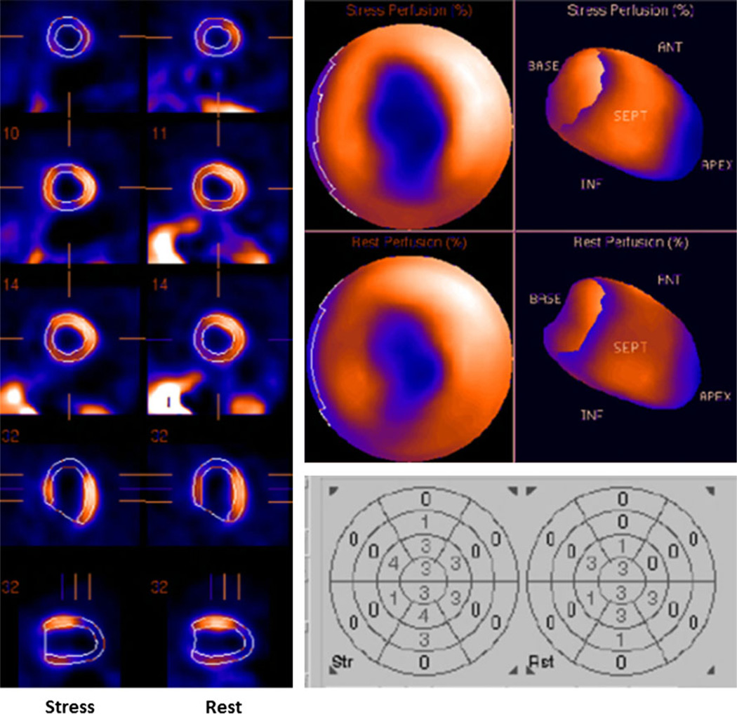
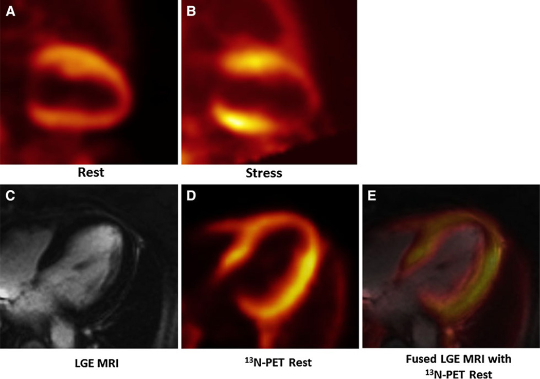
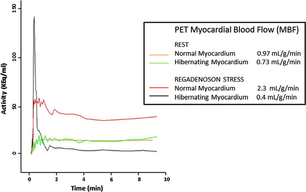
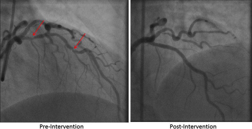
Intermittent ischemia and stunning appear to characterize hibernating myocardium.1,2 In this report, we demonstrate the variable flow abnormalities that may underlie these phenomena. A 72-year-old woman underwent a rest/vasodilator stress ECG-gated 99mTc-SPECT myocardial perfusion imaging (MPI). Image interpretation was consistent with a large anterior/anteroapical infarction with borderzone ischemia (Figure 1). Three days later, repeat rest/vasodilator MPI using 13N-ammonia and late gadolinium enhancement imaging (LGE) was performed simultaneously on a PET/MR (Biograph mMR, Siemens) as part of a research protocol to compare the accuracy of ischemia detection between SPECT and PET/MR. PET/MR MPI revealed extensive and severe anterior/anteroapical ischemia without infarction with resting and post-stress anteroapical hypokinesis (Figure 2A, B; Supplementary Movies 1–3). LGE PET/MR images showed no evidence of infarction in the area of ischemia detected by PET/MR (Figure 2C–E). The constellation of these findings allowed us to speculate that there was myocardial hibernation. Of note, absolute MBF calculated from the PET data demonstrated near similar resting flow values between the hibernating and normal myocardium with a paradoxical reduction in flow in the hibernating territory at stress, indicative coronary steal (Figure 3). Subsequent coronary angiography confirmed high grade LAD disease (Figure 4).
Figure 1.
Rest/vasodilator stress 99mTc-SPECT MPI. Left panel Representative tomographic images demonstrating an extensive persistent perfusion abnormality in the anterior wall extending to the anteroapex, suggesting infarction surrounded by borderzone ischemia. Top right Bull’s eye views (stress—top and rest—bottom) confirming similar findings. Bottom right Numeric scores for stress and rest perfusion defects. Summed stress severity score (SSS) = 27, summed rest severity score (SRS) = 17, summed difference severity score (SDS) = 8.
Figure 2.
13N-ammonia and late gadolinium enhancement (LGE) PET/MR. A, B Two-chamber long axis views of rest (A) and stress (B) 13N-ammonia PET/MR MPI demonstrating a predominantly reversible anterior/anteroapical perfusion defect consistent with ischemia. C LGE imaging shows no delayed contrast uptake in the anterior/anteroapical wall, indicating the absence of infarction. D 13N-ammonia PET demonstrating mild anterior and apical hypoperfusion at rest. E Fused PET perfusion and MR LGE images show no LGE in the area of resting hypoperfusion detected by PET.
Figure 3.
Quantification of myocardial blood flow (MBF) by PET. Rest and stress MBF was quantified in mL/g/minute using the 13N-ammonia PET data sets. At rest, MBF in normal and hibernating myocardium were comparable (0.97 vs 0.73 mL/g/minute). With stress MBF in hibernating myocardium was paradoxically reduced compared to resting blood flow (from 0.73 to 0.4 mL/g/minute), indicative of a coronary artery steal phenomenon.
Figure 4.
Coronary angiography demonstrates high grade left anterior descending (LAD) disease. Areas of LAD stenosis were intervened with two drug eluting stents, with restoration of TIMI grade 3 flow after intervention.
This case provides evidence of the various types of flow abnormalities that characterize myocardial hibernation. They include resting hypoperfusion, impaired vasodilator capacity, and coronary steal. These flow abnormalities, either individually or collectively, can result in myocardial ischemia. However, their intermittent nature will lead to myocardial stunning (normal resting perfusion with abnormal resting wall motion). The finding of variable resting flow values based on the differences in the relative SPECT and PET/MR MPI studies is particularly noteworthy as it may impact the accuracy of SPECT and PET viability studies that are based on relative flow assessment.
Supplementary Material
Footnotes
Electronic supplementary material The online version of this article (doi:10.1007/s12350-013-9757-4) contains supplementary material, which is available to authorized users.
Disclosure
Dr. Agus Priatna is employed by Siemens Medical Solutions USA.
References
- 1.Conversano A, Walsh JF, Geltman EM, Perez JE, Bergmann SR, Gropler RJ. Delineation of myocardial stunning and hibernation by positron emission tomography in patients with advanced coronary artery disease. Am Heart J. 1996;131:440–450. doi: 10.1016/s0002-8703(96)90521-9. [DOI] [PubMed] [Google Scholar]
- 2.Camici PG, Rimoldi OE. Myocardial blood flow in patients with hibernating myocardium. Cardiovasc Res. 2003;57:302–311. doi: 10.1016/s0008-6363(02)00716-2. (Review). [DOI] [PubMed] [Google Scholar]
Associated Data
This section collects any data citations, data availability statements, or supplementary materials included in this article.


