Abstract
We report a rare case of left paraduodenal hernia in patient with symptoms of abdominal subobstruction treated successful with laparoscopic management in urgent situation that have reduced the length of stay and postoperative pain. Internal hernia is only 1% of the causes of abdominal obstruction and the left paraduodenal hernia about 50% of them; it is a congenital defect that derive from malrotation and abnormal mesenteric adhesion. The modern imaging techniques help for the correct diagnosis despite difficult identification of the pathology for the various clinical presentation. The treatment of choice is the surgical intervention; the laparoscopic approach is rarely described in literature but it can reduce the morbidity, postoperative pain and the length of hospital stay.
Keywords: Paraduodenal, Hernia, Obstruction, Laparoscopic, Congenital
Introduction
Internal hernia result from protrusion of one or more abdominal viscera through an intraparietal opening with the herniated viscera remaining inside the peritoneal cavity. Intraperitoneal openings may be normal (such as the foramen of Winslow) or abnormal (paraduodenal fossa, ileocecal fossa) and the origins include congenital or acquired defects (inflammation, infectious process, trauma, postoperative defect). Internal hernias are infrequent with the 0,2–0,9% cases of intestinal obstruction and with 0,5–4,1% cases of intestinal obstruction caused by hernia. They are divided into distinct subgroups based on the localization: the paraduodenal is a most common type with an overall incidence of 53%, followed by pericecal (13%), Foramen of Winslow and transmesenteric (8%), intersigmoid, supravescical and pelvic (6%), transomental (1–4%) ones (1–4). During the last years the incidence of transmesenteric and transmesocolic, derived from new surgical procedures, has been increasing (5). The paraduodenal hernia, also called congenital mesentericoparietal hernia, is an herniation of small bowel into a sac derived from a fold of peritoneum found at the terminal or 4th portion of duodenum. The most frequently encountered fossae are: inferior of Treitz (60%), combinated superior and inferior (30%), superior (5%), fossa of Landzert (2%) and fossa of Waldeyer (1%) (4, 6).
There are two type of congenital paraduodenal hernia: left-sided, which is more common (75%), and right sided, which is very uncommon (25%). Despite is a congenital defect, due to malrotation of midgut that form a potential space near the ligament of Treitz, the mean age of diagnosis is the 4th–6th decade of life; usually men are 3 times more affected than women (7).
We want to discuss about one case of paraduodenal hernia treated successfully with laparoscopic approach.
Case report
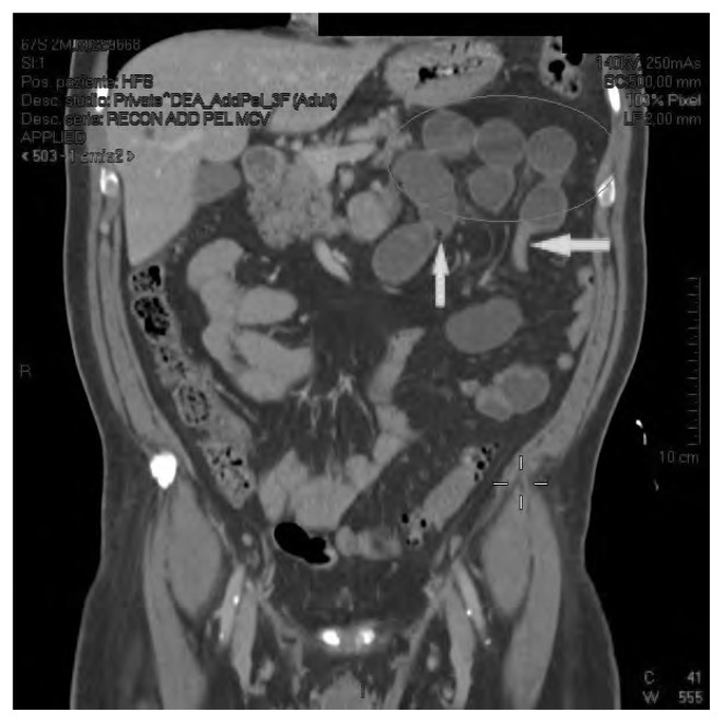
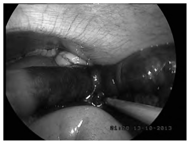
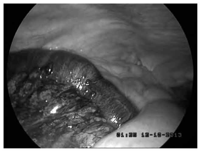
We present a case of a 67 year old man who was admitted to Emergency Department with one day of acute abdominal pain, distension, diarrhoea and vomit that were not reduced by painkillers administration; he had an history of hypertension and diverticulosis without any surgical intervention. The blood tests were negative and the abdominal radiography demonstrated upper bowel air fluid levels. He underwent observation for some hours and, because of the clinical conditions were worsening, barium was administered: the radiography showed a left air fluid levels and progression of contrast. An abdominal CT confirmed a small bowel loops distension with levels and two caliber reductions in left hypocondrium. A diagnosis of internal hernia was made and was indicated a surgical intervention (Fig. 1). We have chosen a laparoscopic approach with four trocar: the first sovraumbilicar was introduced with open technique, and the other were located two in left side and one in epigastric region. The exploration of the cavity demonstrated loops distension and left paraduodenal hernia that was easily reduced without incision of the neck of the sac (Fig. 2). The ileal loop had ischemic signs but, after hot peritoneal washings, peristalsis and bowel color improved and the resection was not needed (Fig. 3). A peritoneal drain was positioned. The postoperative period was uneventful without local and general complication. The patient started to eat in 2nd postoperative day and was discharged in 4th.
Fig. 1.
TC image.
Fig. 2.
Laparoscopic Image before treatment.
Fig. 3.
Laparoscopic image after treatment.
Discussion
Paraduodenal hernia was first defined by Treitz in 1857 and the first classification in right and left was done in 1889 by Jonnesco. Andrews first described the currently accepted mechanism of formation as a type of malrotation. During the period between the 5th and 11th weeks of gestation there is a midgut rotation and the fusion of mesentery with the posterior abdominal structures from the ligament of Treitz to the right iliac fossa (3, 6, 8). Right and left paraduodenal hernia are separate entities, differing in anatomical position and also in embryologic origin: the right hernia leads to the entrapment of the intestine by the mesentery of the cecum and colon because the prearterial limb of the duodenal-jejunal loop fails to rotate around the superior mesenteric artery forming the fossa of Waldeyer; the left one occurs if the small intestine invaginates the connective tissue beyond the descending mesocolon, after failing to fully rotate counterclockwise around the superior mesenteric artery forming a Lanzert’s fossa. This is bound medially by a portion of ascending duodenum and inferiorly by the inferior mesenteric vein proper or a branch to the left colic that form a neck of the sac; the anterior edge of the fossa is the mesocolon and the duodeno-jejunal junction is anterior to this sac (9).
A clinical presentation is various: it could begin with symptoms of acute obstruction, recurring abdominal pain (43%) or can be asymptomatic for all the life. Between 10% and 50% of internal hernia are discovered during unrelated abdominal surgeries or imaging exams and autopsy (10). The pain is often postprandial and may be relieved when the patient is supine for retroperitoneal mass effect. The non specific symptoms are often mistakenly attributed to biliary disease, gastritis or gastroesophageal reflux. The laboratory findings are frequently inconclusive. The physical examination is usually not revealing unless the hernia is large enough to produce on abdominal mass, located on left upper quadrants, and the eccentric abdominal distention situated on the corresponding side of the hernia (6, 8). External hernia symptomatology is easy reliable to the swelling but in internal one this correlation is not present; besides the small content is mostly liquid and obstruction less likely develops in these type of hernia. The multiplicity of clinical presentation can make difficult the diagnosis delaying the surgical intervention and increasing the complications such as occlusion, necrosis and perforation (6).
An hypothesis of internal hernia should be considered for patient with signs and symptoms of intestinal obstruction, particularly in absence of inflammatory intestinal disease, external hernia or previous laparotomy (1).
The first correct preoperative diagnosis of a paraduodenal hernia was made by Kummer in 1921, who described its presence by a barium swallow. Later, Taylor in 1930 diagnosed a case of right paraduodenal hernia by radiologic appearances (4).
Advent of the modern imaging technique like computerized tomography has increased the probability of correct preoperative diagnosis of paraduodenal hernia, which was once difficult because of its non specific presentation (11).
About 10–15% of cases are discovered preoperatively. The abdomen radiography can give information regarding the intestinal segment involved and the extension of the intestinal obstruction; a gastrointestinal series with barium may show dilated loops of small bowel in the upper quadrant, delay of contrast or the point of obstruction (5, 9). Ultrasonography may demonstrate an abdominal mass or internal tubular cysts that change shape over time and after ingestion of fluid. The celiac and superior mesenteric arteriography, often done for other reasons, can demonstrate a displaced spleen and jejunal arteries displaced upward to the left (10). Abdominal CT is a gold standard to provide the correct diagnosis: the hernia have a characteristic appearance of a formation include clustering of small bowel loops, a saclike mass with encapsulation at the ligament of Treitz, duodeno-jejunal junction depression, mass effect on the posterior stomach wall, engorgement and crowding of the mesentery vessels with frequent right displacement of the main mesenteric trunk, anterior and upward displacement of the inferior mesenteric vein that lie in the ventral circumference of the hernia orifice and depression of the transverse colon (6).
Once diagnosed, left paraduodenal hernias should be surgically repaired because they carry an approximately 50% lifetime risk of complication. The mortality rate associated with paraduodenal hernia is not clear, but it has been stated to be 20–50%, due to large proportion of patient with intestinal obstruction and ischemia requiring emergency surgery (8). The surgical management include reduction of herniated structures, resection of ischemic intestinal segment and closure of hernia orifice that is generally indicated for prevention of recurrence of hernia through abnormal orifice. Prosthesis placement in defect repair is not common: mesh implants are reserved in large defect or recurrent hernia (1, 6, 12). As in our case, often the reduction of the hernia is easy, but when the neck of the sac is small and obscured by adhesion it is difficult to identify accurately. In this case the hernia sac should be opened by an incision into an avascular area of the mesentery of the descending colon or in the inferior border of the hernia opening to the right side of the mesenteric vessels. When there is a tight or obscured hernia ring, the inferior mesenteric vein has to been sacrificed. The defect is removed with simple closure or with a wide opening of the sac making it continuous with the peritoneal cavity; removal of the sac remains controversial because it is part of the mesocolon and may lead to colonic vascular impairment. Due to the fact that the hernia can reduce spontaneously preoperatively and because all peritoneal space are not always routinely examined intraoperatively, they can go undiagnosed (10, 13).
There are few reports in literature, with the total number reported case being less than five hundred, but the case treated with laparoscopic approach are about twenty (13, 14) (Table 1).
Table 1.
REPORTED CASE OF PARADUODENAL HERNIA.
| Author and year | Number of cases | Type | Emergency or Elective surgery | Length of stay | Complications |
|---|---|---|---|---|---|
| Erdas et al. (2013) | 1 | right | emergency | 4 | Conversion for bowel distension |
| Hussein et al. (2012) | 1 | left | emergency | 3 | - |
| Parmar et al. (2010) | 1 | left | elective | 3 | - |
| Khalaileh et al. (2010) | 1 | left | emergency | 3 | - |
| Bittner et al. (2009) | 1 | right | emergency | 1 | - |
| Uchiyama et al. (2009) | 1 | left | elective | 7 | - |
| Jeong et al. (2008) | 2 | left | emergency | 5 | - |
| Palanivelu et al. (2008) | 4 | 1right | 1 emergency | 3 | 1 vomiting and ileus |
| 3 left | 3 elective | - | |||
| Shoji ae al. (2007) | 1 | left | elective | ? | - |
| Moon et al. (2006) | 1 | left | emergency | 1 | - |
| Fukunaga et al. (2004) | 1 | left | elective | 6 | - |
| Rollins et al. (2004) | 1 | left | elective | 3 | - |
| Antedomenico et al. (2003) | 1 | right | emergency | 3 | - |
| Uematsu et al. (1998) | 1 | left | elective | 8 | - |
Laparoscopic repair would be expected to reduce postoperative pain, morbidity and the hospital stay: in the literature the mean time of hospitalization is 3–5 days and the postoperative complication are about 5% considering the 18 cases in literature (17–26).
Beside the laparoscopic approach has provided the abilities of both verification of diagnosis and simultaneous surgical intervention in cases that could not be diagnosed by radiologic method and in suspicion of internal hernia (3, 11, 14–16). During the urgent intervention the laparoscopic approach is more difficult for the bowel loops distension that may reduce the operative space and hamper the surgical movements with more risk of lesions.
Conclusion
The paraduodenal hernia is a rare case of intestinal obstruction that should be considered in case of recurrent abdominal pain and obstruction without any history of surgical intervention, external hernia or inflammatory disease. As in our case the fast diagnosis can reduce the complications such as obstruction, necrosis and perforation: CT is a gold standard methodic for a correct diagnosis. The surgical intervention is the treatment of choice, also in asymptomatic cases, because it reduces the urgent surgery and complications related to hernia, that appear in almost half of cases. The laparoscopic approach, that is more difficult in urgency, is suggested because can reduce morbidity, postoperative pain and length of stay.
References
- 1.Husain A, Bhat S, Roy AK, Sharma V, Dubey SA, Faridi MS. Internal hernia through paraduodenal recess with acute intestinal obstruction: a case report. Indian J Surg. 2012;74:354–355. doi: 10.1007/s12262-011-0243-4. [DOI] [PMC free article] [PubMed] [Google Scholar]
- 2.Al Khyatt W, Aggarwal S, Birchall J, Rowlands TE. Acute intestinal obstruction secondary to left paraduodenal hernia: a case report and literature review. World J Emer Surg. 2013;8:5. doi: 10.1186/1749-7922-8-5. [DOI] [PMC free article] [PubMed] [Google Scholar]
- 3.Akbulut S. Case report. Unusual cause of intestinal obstruction: left paraduodenal hernia. Case Rep Med. 2012 doi: 10.1155/2012/529246. ID 529246. [DOI] [PMC free article] [PubMed] [Google Scholar]
- 4.Nuno Guzman C, Arroniz Jauregui J, Hernandez Gonzalez C, Reyes Macias F, Nava Garibaldi R, Guerrero Diaz F, Martinez Chavez J, Solis Ugalde J. Right paraduodenal hernia in an adult patient: diagnostic approach and surgical management. Case Rep Gastroenterol. 2011;5:479–486. doi: 10.1159/000331033. [DOI] [PMC free article] [PubMed] [Google Scholar]
- 5.Fernandez Rey CL, Martinez Alvarez C, Concejo Cutoli P. Acute abdomen secondary to left paraduodenal hernia: diagnostic by multislice computer tomography. Rev Esp Enferm Dig. 2011;1:38–39. doi: 10.4321/s1130-01082011000100008. [DOI] [PubMed] [Google Scholar]
- 6.Manfredelli S, Zitelli A, Pontone S, Leonetti G, Marcantonio M, Forte A, Alberto A, Mancini R. Rare small bowel obstruction: right paraduodenal hernia. Case report. Int J Surg Case Rep. 2013;4:412–415. doi: 10.1016/j.ijscr.2012.11.027. [DOI] [PMC free article] [PubMed] [Google Scholar]
- 7.Amodio PM, Alberti A, Bigonzoni E, Piciollo M, Fortunati T, Alberti D. Ernia paraduodenal sinistra. Case report e review della letteratura. Chir It. 2008;60:721–724. [PubMed] [Google Scholar]
- 8.Tun MY, Choi YM, Choi SK, Kim SJ, Ahn SI, Kim KR. Left paraduodenal hernia presenting with atypical symptoms. Yonsei Med J. 2010;51:787–789. doi: 10.3349/ymj.2010.51.5.787. [DOI] [PMC free article] [PubMed] [Google Scholar]
- 9.Moon CH, Chung MH, Lin KM. Diagnostic laparoscopy and laparoscopic repair of a left paraduodenal hernia can shorten hospital stay. JSLS. 2006;10:90–93. [PMC free article] [PubMed] [Google Scholar]
- 10.Kuzinkovas V, Haghighi K, Singhal R, Andrews NJ. Paraduodenal hernia: a rare cause of abdominal pain. Can J Surg. 2008;51:127–128. [PMC free article] [PubMed] [Google Scholar]
- 11.Parmar BPS, Parman RS. Laparoscopic management of left paraduodenal hernia. J Minm Access Surg. 2010;6:122–124. doi: 10.4103/0972-9941.72601. [DOI] [PMC free article] [PubMed] [Google Scholar]
- 12.Fukada T, Mukai H, Shimamura F, Furukawa T, Miyazaki M. A causal relationship between right paraduodenal hernia and superior mesenteric artery syndrome: a case report. Medical Case Reports. 2010;4:159. doi: 10.1186/1752-1947-4-159. [DOI] [PMC free article] [PubMed] [Google Scholar]
- 13.Downes R, Cawich SO. A case of a paraduodenal hernia. Int J Surg Case Rep. 2010;1:19–21. doi: 10.1016/j.ijscr.2010.06.003. [DOI] [PMC free article] [PubMed] [Google Scholar]
- 14.Hussein M, Khreiss M, Al Helou G, Alaeddine M, Elias E, Abi Saad G. Laparoscopic repair of a left paraduodenal hernia presenting with acute bowel obstruction: report of case. Surg Lap End Per Tech. 2012;22:28–30. doi: 10.1097/SLE.0b013e31823f3798. [DOI] [PubMed] [Google Scholar]
- 15.Shoji T, Nishiyama R, Oba K, Azuma M. Left paraduodenal hernia successfully treated with laparoscopic surgery: a case report. Case Rep Gastroenterol. 2007;1:71–76. doi: 10.1159/000107595. [DOI] [PMC free article] [PubMed] [Google Scholar]
- 16.Trigui A, Guirat A, Rejab H, Kardoun N, Frikha F, Amar MB, Beyrouti MI. An uncommon cause of acute bowel obstruction: the left paraduodenal hernia. Niger J Surg. 2012;18:97–99. doi: 10.4103/1117-6806.103117. [DOI] [PMC free article] [PubMed] [Google Scholar]
- 17.Bittner JG, Edwards MA, Harrison SJ, Li K, Karmin PN, Mellinger JD. Laparoscopic repair of a right paraduodenal hernia. JSLS. 2009;13:242–249. [PMC free article] [PubMed] [Google Scholar]
- 18.Erdas E, Pitzalis A, Scano D, Licheri S, Pomata M, Farina G. Diagnosis and treatment of symptomatic right paraduodenal hernia: report of case. doi: 10.1007/s00595-012-0483-3. [DOI] [PubMed] [Google Scholar]
- 19.Jeong GA, Cho GS, Kim HC, Shin EJ, Song OP. Laparoscopic repair of paraduodenal hernia. Comparison with conventional open repair. Surg Laparosc Endosc Percutan Tech. 2008;18:611–615. doi: 10.1097/SLE.0b013e3181825733. [DOI] [PubMed] [Google Scholar]
- 20.Khalaileh A, Schlager A, Bala M, Abugazala S, Elazary R, Rivkind AI, Mintz Y. Left laparoscopic paraduodenal hernia repair. Surg Endosc. 2010;24:1486–1489. doi: 10.1007/s00464-009-0794-7. [DOI] [PubMed] [Google Scholar]
- 21.Uematsu T, Iwase H, Yamashita K, Ogura H, Nakamuka T, Oguri H. Laparoscopic repair of a paraduodenal hernia. Surg Endosc. 1998;12:50–52. doi: 10.1007/s004649900591. [DOI] [PubMed] [Google Scholar]
- 22.Antedomenico E, Singh NN, Zagorski SM, Dwyer K, Chung MH. Laparoscopic repair of a right paraduodenal hernia. Surg Endosc. 2004;18:165. doi: 10.1007/s00464-003-4516-2. [DOI] [PubMed] [Google Scholar]
- 23.Palanivelu C, Rangarajan M, Jategaonkar PA, Anand NV, Senthikumar K. Laparoscopic management of paraduodenal hernia: mesh and meshless repairs. A report of four cases. doi: 10.1007/s10029-008-0376-y. [DOI] [PubMed] [Google Scholar]
- 24.Uchiyama S, Imamura N, Hidaka H, Maehara N, Nagaike K, Ikenaga N, Hotokezaka M, Chijiiwa K. An unusual variant of a left paraduodenal hernia diagnosed and treated by laparoscopic surgery: report of case. Surg Today. 2009;39:533–535. doi: 10.1007/s00595-008-3875-7. [DOI] [PubMed] [Google Scholar]
- 25.Fukunga M, et al. Laparoscopic surgery for left paraduodenal herni. J Laparoendosc Adv Tech. 2004;14:111–115. doi: 10.1089/109264204322973907. [DOI] [PubMed] [Google Scholar]


