Abstract
Panthothenate kinase-associated neurodegeneration (PKAN) (Hallervorden-Spatz disease) is a rare autosomal recessive chromosomal disorder characterised by progressive neuroaxonal dystrophy. The characteristic features include involuntary movements, rigidity, mental retardation, seizures, emaciation. The anaesthetic concerns include difficult airway, aspiration pneumonia, dehydration, and post-operative respiratory, and renal insufficiency. We report successful anaesthetic management of a 9-year-old intellectually disabled male child with PKAN, scheduled for ophthalmic surgery under general anaesthesia.
Keywords: Aspiration pneumonia, difficult airway, dystonia, muscle spasm, panthothenate kinase-associated neurodegeneration, respiratory failure
INTRODUCTION
Panthothenate kinase-associated neurodegeneration (PKAN), formerly termed as Hallervorden-Spatz disease is a rare chromosomal disorder first described by Hallervorden and Spatz in 1922.[1] The characteristic features in patients with PKAN include dystonia, muscle spasm, cognitive dysfunction, and seizure disorder.[2,3,4] The anaesthetic concerns include difficult airway management, increased risk of gastric aspiration, dehydration, and post-operative respiratory and renal insufficiency.[2,4]
Here, we report a case of successful anaesthetic management of a 9-year-old intellectually disabled male child with PKAN, posted for ophthalmic surgery under general anaesthesia (GA).
CASE REPORT
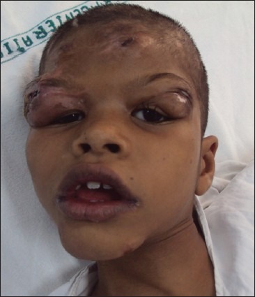
A 9-year-old boy weighing 20 kg, diagnosed case of PKAN, admitted at our institute with a history of right eye lid oedema with maggots for debridement. He had a history of fall 7 days back and sustained injury over the right eyeball from a wooden stick. The child also had a history of intermittent abnormal flexor posturing and spasm of both the upper limbs for last 7 years, which became persistent and fixed during last 2 years. He also reported of frequent falls due to progressively worsening spasm and was unable to perform any definitive motor function. His developmental milestones were delayed with slurred speech. He was able to communicate only with his parents. His younger sibling was also having similar illness. He had a history of generalised tonic-clonic seizures for the past 3 years and was on syrups of sodium valproate and lorazepam. Since admission to our institute, he was receiving intravenous (IV) antibiotics and eye drops.
Physical examination revealed cognitive dysfunction, facial dysmorphism, rigidity and involuntary movements of upper limbs. Airway examination showed microstomia, micrognathia, retrognathia, Modified Mallampatti class II, with normal thyromental distance and neck movements [Figure 1]. Magnetic resonance imaging (MRI) brain revealed specific pattern of hyperintensity within hypointensity of anteromedial globus pallidus with “eye-of-the-tiger” sign. Rest of the investigations, including blood chemistry were within normal limits.
Figure 1.

Child with panthothenate kinase associated neurodegeneration presenting with right eyelid oedema and maggots
High-risk consent was obtained from the child's parents and a bed was arranged in the paediatric intensive care for post-operative ventilation if required. On the day of surgery, oral antiepileptics and IV ranitidine 20 mg and routine antibiotics were administered as IV cannula was in situ. A difficult airway cart including supraglottic devices, video-laryngoscope, and fibreoptic bronchoscope (FOB) were kept ready to manage the anticipated difficult airway. Injections of midazolam and sodium valproate were loaded to treat convulsions. The child's father was allowed to stay in the operating room (OR) till induction. Standard monitoring including electrocardiogram, pulse oximetry (SpO2), and non-invasive blood pressure (NIBP) were attached. Before induction, his vital signs recorded were a heart rate of 116 beats/min, NIBP 86/38 mm Hg, respiratory rate 26/min and SpO298%. IV fentanyl 2 mcg/kg was administered, and the child was pre-oxygenated with 100% oxygen (6 L/min) for 3 minutes. For induction, IV propofol was administered in titrated doses (total of 35 mg) and anaesthesia was then gradually deepened with halothane (1to 3% in 100% O2). After ensuring adequate depth of anaesthesia (loss of eyelash reflex and relaxed jaw), check video-laryngoscopy was done to assess the upper airway patency. Glottic view seen was Cormack and Lehane's (CL) grade IIIa . ProSeal laryngeal mask airway (PLMA) of size 2.0 was inserted in the first attempt and suction catheter was passed through the gastric channel. As adequate tidal volume was achieved with manual ventilation, assisted pressure support ventilation (pressure support ventilation with apnoea backup ventilation using the Drager Primus® anaesthesia machine) was started and anaesthesia was maintained with isoflurane (1.0 to-1.2 MAC) in O2: Air (FiO20.5). Topical procaine anaesthetic drops (0.5%) were instilled in the operating eye and 200 mg IV paracetamol were administered for intra-operative analgesia. Ringer lactate was administered for fluid management. Intra-operative period was uneventful. Antiemetic prophylaxis (2 mg of ondansetron) was given 10 min before the end of surgery. On the completion of surgery, isoflurane was turned off and 100% O2 was administered. When adequate tidal volume and regular respiration were achieved, the PLMA was removed after gastric and oropharyngeal suctioning. Child was turned left lateral and was observed in OR for next 10 minutes, before shifting to post-anaesthesia care unit (PACU). Oxygen supplementation was continued through face mask. His father was allowed to stay in PACU. There was no episode of seizure, muscle spasm, vomiting or airway obstruction noticed postoperatively. After 2 h of stay in PACU, child was shifted to ward.
DISCUSSION
Panthothenate kinase associated neurodegeneration is a rare, autosomal recessive, neurodegenerative disorder characterised by the accumulation of iron in the basal ganglia of the human brain. The responsible gene (PANK 2) for the disease has been localised in the short arm of chromosome no. 20, which is required for the phosphorylation of pantothenic acid in the formation of coenzyme A.[2,5] Defective phosphorylation results in under-utilisation of cystine in the body. This excessive cystine causes chelation of iron resulting in excessive free radical generation, lipid peroxidation, axonal dystrophy, and apoptotic cell death resulting in neuraxonal degeneration.[6] The onset of disease is usually in the first or second decade of life; however, it can occur at any age. It may be familial or sporadic, and the average survival time after the diagnosis is about 10 to-15 years.
Characteristic neurological features include progressive rigidity (oromandibular rigidity, trismus, dysphagia), involuntary movements (chorea, athetosis), seizures, cognitive dysfunctions, visual impairment (optic atrophy, retinitis pigmentosa), and difficulties in articulation, swallowing, and chewing.[2,3,4,7] These signs and symptoms influence the pre-anaesthetic preparation of the patient, as well as intra-operative and post-operative anaesthesia management.[2]
Hinkelbein et al.[2] reviewed the case reports concerning the anaesthetic management of patients with PKAN and found successful and safe use of anaesthetic drugs like opioids (fentanyl, remifentanil), inhalational agents (nitrous oxide, halothane, sevoflurane, isoflurane), induction agents (propofol, thiopentone), and muscle relaxants (succinylcholine, vecuronium, pancuronium). In most of these case reports, airway was secured using endotracheal intubation.[4,7,8] Owing to immobility, hyperkalaemic cardiac arrest induced by succinylcholine is always a possibility. There are several reports of autopsies revealing muscle wasting secondary to poor nutrition and diffuse axonal changes in the brain which may involve upper motor neuron lesions to an unpredictable extent, thereby increasing the possibility of hyperkalaemic cardiac arrest.[8] Intrathecal baclofen to relieve spasticity and dexmedetomidine during MRI were also used in these patients.[9,10] Stereotactic pallidotomy and thalamotomy have been attempted for dystonia with good results.[4,7,11]
Presence of involuntary movements (chorea, athetosis), rigidity and seizures interfere with the placement of IV cannula, arterial line, positioning for regional anaesthesia and awake intubation. Involuntary movements may not completely disappear with the induction of anaesthesia and reappear on emergence from anaesthesia. Attempts for awake intubation techniques like FOB and tracheostomy under local anaesthesia can intensify these involuntary movements.[4,7]
The child reported here was mentally disabled, uncooperative, and had signs of difficult airway with a progressive neurological disorder. Although awake FOB is the gold standard for difficult airway management, it is unsuitable for such patient. GA is always needed for the definitive procedures in mentally retarded patients. This child was also having risk of aspiration and endotracheal intubation is considered to be a gold standard for securing the airway in these patients. However, successful airway management using PLMA® in a patient with difficult airway and at increased risk of gastric aspiration has been reported in the literature.[12] Supraglottic airway devices have advantages in ophthalmic surgery as they cause minimal changes in haemodynamics, intraocular pressure, minimal trauma to the airway and decrease intubation and extubation response.[13,14] We chose PLMA® as it allows the passage of suction catheter through the gastric tube and protects against gastric aspiration. We also administered antiaspiration and antiemetic prophylaxis. Video-laryngoscopy was done to assess CL grading to be prepared for intubation in the emergent situation. Keegan et al.[4] reported reintubation and mechanical ventilation in the post-operative period due to the dynamic upper airway obstruction and acute respiratory failure in an 11-year-old girl with PKAN for stereotactic thalamotomy. Emergency tracheostomy during induction and re-intubation in the post-operative period may be needed. Hence, all the necessary preparations should be done and made available to secure the airway in such scenarios.[4,8]
We avoided neuromuscular blockade as ophthalmic surgery did not need muscular paralysis, and lungs were ventilated with assisted pressure support ventilation with EtCO2 monitoring. Sevoflurane was also avoided during induction as seizure-like movements of the extremities have been observed during induction of anaesthesia with sevoflurane. Sevoflurane is known to produce epileptiform activity in EEG that weakly portends seizure activity.[15] There are several case reports of sevoflurane provoking seizure like activity, particularly in children and in conjunction with high concentrations and hypocapnia.[15] Therefore, propofol was used for induction of anaesthesia using its antiepileptic properties to our benefit. The child did not have any abnormal movement during anaesthesia and surgery. Multimodal analgesia was used for pain relief. He had no respiratory depression or upper airway obstruction post-operatively.
CONCLUSION
We could successfully manage the child with PKAN and difficult airway using PLMA® without muscle relaxants for ophthalmic surgery. No case has been reported in the literature using ProSeal LMA for airway management in a child with PKAN. Balanced anaesthesia and careful monitoring are key to a safe outcome.
Footnotes
Source of Support: Nil
Conflict of Interest: None declared.
REFERENCES
- 1.Hallervorden J, Spatz H. Eigenartige Erkrankung im extrapyramidalen System mit besonderer Beteiligung des Globus pallidus und der Substantia nigra: Ein Beitrag zu den Beziehungen zwischen diesen beiden Zentren. Z Ges Neurol Psychiatric. 1922;79:254–302. [Google Scholar]
- 2.Hinkelbein J, Kalenka A, Alb M. Anesthesia for patients with panthothenate-kinase-associated neurodegeneration (Hallervorden-Spatz disease) - A literature review. Acta Neuropsychiatr. 2006;18:168–72. doi: 10.1111/j.1601-5215.2006.00144.x. [DOI] [PubMed] [Google Scholar]
- 3.Kapoor S, Hortnagel K, Gogia S, Paul R, Malhotra V, Prakash A. Pantothenate kinase associated neurodegeneration (Hallervorden-Spatz syndrome) Indian J Pediatr. 2005;72:261–3. [PubMed] [Google Scholar]
- 4.Keegan MT, Flick RP, Matsumoto JY, Davis DH, Lanier WL. Anesthetic management for two-stage computer-assisted, stereotactic thalamotomy in a child with Hallervorden-Spatz Disease. J Neurosurg Anesthesiol. 2000;12:107–11. doi: 10.1097/00008506-200004000-00006. [DOI] [PubMed] [Google Scholar]
- 5.Hayflick SJ, Westaway SK, Levinson B, Zhou B, Johnson MA, Ching KH, et al. Genetic, clinical, and radiographic delineation of Hallervorden-Spatz syndrome. N Engl J Med. 2003;348:33–40. doi: 10.1056/NEJMoa020817. [DOI] [PubMed] [Google Scholar]
- 6.Rock CO, Calder RB, Karim MA, Jackowski S. Pantothenate kinase regulation of the intracellular concentration of coenzyme A. J Biol Chem. 2000;275:1377–83. doi: 10.1074/jbc.275.2.1377. [DOI] [PubMed] [Google Scholar]
- 7.Balas I, Kovacs N, Hollody K. Staged bilateral stereotactic pallidothalamotomy for life-threatening dystonia in a child with Hallervorden-Spatz disease. Mov Disord. 2006;21:82–5. doi: 10.1002/mds.20655. [DOI] [PubMed] [Google Scholar]
- 8.Roy RC, McLain S, Wise A, Shaffner LD. Anesthetic management of a patient with Hallervorden-Spatz disease. Anesthesiology. 1983;58:382–4. doi: 10.1097/00000542-198304000-00017. [DOI] [PubMed] [Google Scholar]
- 9.Madhusudhana Rao B, Radhakrishnan M. Dexmedetomidine for a patient with Hallervorden-Spatz syndrome during magnetic resonance imaging: A case report. J Anesth. 2013;27:963–4. doi: 10.1007/s00540-013-1652-2. [DOI] [PubMed] [Google Scholar]
- 10.Lee C, Chu Y, Chuang C, Chen C, Tsou M, Chan K. Intrathecal baclofen facilitated postanesthetic tracheal extubation in a dystonic patient associated with neurodegeneration of brain iron accumulation (Hallervorden-Spatz Disease) Neurosci Med. 2011;2:351–4. [Google Scholar]
- 11.Justesen CR, Penn RD, Kroin JS, Egel RT. Stereotactic pallidotomy in a child with Hallervorden-Spatz disease. Case report. J Neurosurg. 1999;90:551–4. doi: 10.3171/jns.1999.90.3.0551. [DOI] [PubMed] [Google Scholar]
- 12.Twigg SJ, Cook TM. Anaesthesia in an adult with Rubenstein-Taybi syndrome using the ProSeal laryngeal mask airway. Br J Anaesth. 2002;89:786–7. [PubMed] [Google Scholar]
- 13.Gulati M, Mohta M, Ahuja S, Gupta VP. Comparison of laryngeal mask airway with tracheal tube for ophthalmic surgery in paediatric patients. Anaesth Intensive Care. 2004;32:383–9. doi: 10.1177/0310057X0403200314. [DOI] [PubMed] [Google Scholar]
- 14.Ates Y, Alanoglu Z, Uysalel A. Use of the laryngeal mask airway during ophthalmic surgery results in stable circulation and few complications: A prospective audit. Acta Anaesthesiol Scand. 1998;42:1180–3. doi: 10.1111/j.1399-6576.1998.tb05273.x. [DOI] [PubMed] [Google Scholar]
- 15.Perks A, Cheema S, Mohanraj R. Anaesthesia and epilepsy. Br J Anaesth. 2012;108:562–71. doi: 10.1093/bja/aes027. [DOI] [PubMed] [Google Scholar]


