

Abstract
Eczema herpeticum is a rare and potentially life-threatening viral infection. We present the case of a 54-year-old man who presented to the emergency eye clinic with a dendritic ulcer and a facial rash. An initial diagnosis of herpes zoster ophthalmicus was suspected. On follow-up, the rash had begun to rapidly disseminate and was no longer respecting dermatome boundaries. A diagnosis of eczema herpeticum was made and appropriate treatment started. This case highlights the importance of a comprehensive history and close inspection of skin lesions in patients with herpetic eye disease.
Background
Eczema herpeticum (also known as Kaposi's varicelliform eruption) is a rare and potentially life-threatening viral cutaneous infection. It is most commonly caused by herpes simplex virus type 1 (HSV-1), which becomes superimposed on patients whose skin barrier has been disrupted (typically by pre-existing atopic dermatitis).1 The condition presents as clusters of punctated vesicopustules with associated cutaneous pain. Once HSV has broken the skin barrier, it can rapidly disseminate and potentially involve multiple organs.2
In severe cases where systemic spread has occurred, admission to hospital for intravenous aciclovir and intensive supportive treatment may be required.
Case presentation
A 54-year-old man with a background of eczema and asthma presented to the emergency eye clinic with a 2-day history of worsening pain, photophobia and discharge from his left eye. Examination revealed a visual acuity of 6/36 in his left eye (baseline 6/12) and a 3×3 mm dendritic infiltrate on the cornea. An initial diagnosis of herpes simplex keratitis was made, although the presence of mild pruritus and erythema in the distribution of the ophthalmic division of the trigeminal nerve (V1), led to suspicion of herpes zoster ophthalmicus. The patient was given a course of oral aciclovir 800 mg five times a day, guttae aciclovir 3% five times a day, and guttae ciprofloxacin 0.3% four times a day, with a plan to review after 3 days.
At follow-up with a different clinician it was noted that while the corneal dendritic ulcer had largely resolved with visual acuity improving to 6/12, the facial rash had developed into an erythematous vesicopustular rash with cutaneous dendrites (figure 1), which had spread across the midline and did not respect the boundaries of the V1 dermatome (figure 2). Upper and lower lids on the left side were swollen, although there was a full range of extraocular movements with no proptosis and no relative afferent pupillary defect. A diagnosis of eczema herpeticum was suspected, and a detailed history revealed the patient had a current exacerbation of his eczema.
Figure 1.
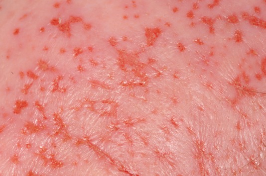
Photograph of frontal area of head, showing multiple dendritic/geographic lesions.
Figure 2.
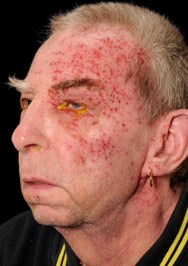
Photograph showing distribution of eruption. Note lesions crossing the midline and not confined to the V1 dermatome.
An urgent review with dermatology was arranged, and eczema herpeticum was confirmed.
Investigations
In this case, the clinical diagnosis did not require investigations. If an underlying immunocompromise is suspected, further blood tests and investigations can be undertaken as clinically appropriate. If required, confirmation of the diagnosis can be made with HSV cultures, HSV PCR, or using a Tzanck smear, where multinucleated giant cells are found in HSV, herpes zoster and cytomegalovirus infections.
Differential diagnosis
The differential diagnosis for a vesicopustular rash can be divided into non-infectious and infectious causes. Non-infectious causes include pustular psoriasis, drug hypersensitivity, vasculitis and bullous lupus erythematosus. Infectious causes include eczema herpeticum, herpes zoster, hand, foot and mouth disease, impetigo and cellulitis.3
In this case, the history and association with a corneal ulcer makes eczema herpeticum the most likely diagnosis.
Treatment
Once the diagnosis had been confirmed, a course of oral aciclovir 800 mg five times a day was extended for a further 10 days. The patient was given a fucidin cream for the wet areas of the rash around his ears, polymyxin B and bacitracin cream for the facial rash, and a betamethasone and fucidic acid cream for the eczema flare up on his legs.
As the patient was systemically well, admission for intravenous aciclovir was not felt to be appropriate on this occasion.
Outcome and follow-up
The patient declined any further follow-up with dermatology, but at his ophthalmology follow-up a further 3 days later, it was noted that the rash had started to resolve and there was no remaining corneal epithelial defect. The patient continued to report improving symptoms and a week later the rash had completely resolved.
Discussion
Eczema herpeticum is a disseminated superinfection of the skin, usually by HSV-1 or HSV-2, on a pre-existing epithelial disruption. The pattern of HSV infection is of particular interest at the moment. Traditionally, HSV-1 was predominantly the cause of oral lesions and HSV-2 the cause of genital lesions. Interestingly, the epidemiology of both subtypes has changed over the past few decades. HSV-1 is now responsible for around 80% of oral infections and 20% of genital lesions. The reverse applies to HSV-2, which causes genital lesions in 80% of cases and oral lesions in 20%.4 This change is primarily believed to be due to a change in sexual practice including increased orogenital contact.
While, as in this case, skin disruption is most commonly caused by atopic dermatitis, other cases in the literature describe eczema herpeticum following burns,5 pemphigus vulgaris and pityriasis rubra pilaris.6
Eczema herpeticum is a rare condition despite the fact that atopic dermatitis is a common skin problem and that around 60% of adults are HSV seropositive.7 Recent studies have shown that the subset of patients with atopic dermatitis who are prone to eczema herpeticum (ADEH+) have a distinct phenotype; they have more severe atopic skin disease, are more likely to have a food allergy and asthma, and are more prone to cutaneous skin infections.2 7 Research into cell-mediated immunity defects in the ADEH+ group of patients has found an association with the human leucocyte antigen B7 allele and that expression of interferon (IFN)-γ was lower in CD8+ T cells and monocytes when exposed to HSV.8 An in vitro study has found that regulators of type I and type III IFNs, which are important in fighting HSV infections, are downregulated.9
Our patient presented with a dendritic ulcer on the cornea and an erythematous rash on the face, and a diagnosis of herpes zoster ophthalmicus was suspected initially. This highlights the importance of a comprehensive history and close inspection of the skin lesions. It is unusual for herpes zoster ophthalmicus to form dendrite or geographic patterns within the cutaneous tissues. This patient's atopic diathesis makes eczema herpeticum more likely following a herpes simplex keratitis infection. The potential for serious sequalae to develop means this condition needs to be followed closely.
Learning points.
Eczema herpeticum should be considered in a patient presenting with herpetic eye disease and rash.
Owing to the nature of herpes simplex and its propensity to spread in epithelium, there are typically skin dendrites/geographic patterns and these should allow rapid diagnosis.
Multidisciplinary involvement is advised, with the dermatologists assessing whether a patient warrants admission for intravenous antivirals.
Footnotes
Contributors: AS arranged for clinical imaging. AS and TY-Z contributed to writing the report. MM was the consultant in charge.
Competing interests: None.
Patient consent: Obtained.
Provenance and peer review: Not commissioned; externally peer reviewed.
References
- 1.Olson J, Robles D, Kirby P et al. Kaposi varicelliform eruption (eczema herpeticum). Dermatol Online J 2008;14:2. [PubMed] [Google Scholar]
- 2.Leung D. Why is eczema herpeticum unexpectedly rare? Antiviral Res 2013;98:153–7. 10.1016/j.antiviral.2013.02.010 [DOI] [PMC free article] [PubMed] [Google Scholar]
- 3.Mackool B, Goverman J, Nazarian R. A 43 year-old woman with fever and a generalised rash. N Engl J Med 2012;366:1825–34. 10.1056/NEJMcpc1111572 [DOI] [PubMed] [Google Scholar]
- 4.Mell HK. Management of oral and genital herpes in the emergency department. Emerg Med Clin North Am 2008;26:457–73. 10.1016/j.emc.2008.02.001 [DOI] [PubMed] [Google Scholar]
- 5.Bartralot R, Garcia-Patos V, Rodriguez-Cano L et al. Kaposi's varicelliform eruption in a patient with healing second degree burns. Clin Exp Dermatol 1996;21:127–30. 10.1111/j.1365-2230.1996.tb00035.x [DOI] [PubMed] [Google Scholar]
- 6.Cavalie M, Giacchero D, Cardot-Leccia N et al. Kaposi's varicelliform eruption in a patient with pityriasis rubra pilaris (pityriasis rubra pilaris herpeticum). J Eur Acad Dermatol Venereol 2013;27:1585–6. 10.1111/jdv.12120 [DOI] [PubMed] [Google Scholar]
- 7.Beck L, Boguniewicz M, Hata T et al. Phenotype of atopic dermatitis subjects with a history of eczema herpeticum. J Allergy Clin Immunol 2009;124:260–9. 10.1016/j.jaci.2009.05.020 [DOI] [PMC free article] [PubMed] [Google Scholar]
- 8.Mathias R, Weinberg A, Bogunewicz M et al. Atopic dermatitis complicated by eczema herpeticum is associated with HLA B7 and reduced interferon-γ-producing CD8+ T cells. Br J Dermatol 2013;169:700–3. 10.1111/bjd.12382 [DOI] [PMC free article] [PubMed] [Google Scholar]
- 9.Bin L, Edwards M, Helser R et al. Identification of novel gene signatures in patients with atopic dermatitis complicated by eczema herpeticum. J Allergy Clin Immunol 2014;134:848–55. 10.1016/j.jaci.2014.07.018 [DOI] [PMC free article] [PubMed] [Google Scholar]