Abstract
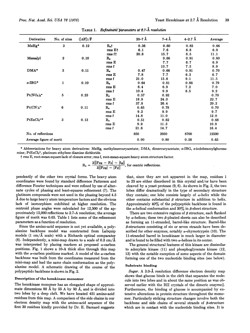
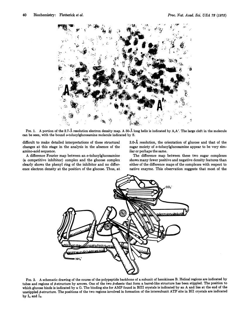
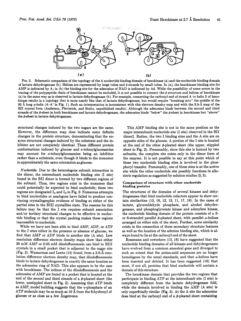
From a 2.7-A resolution electron density map we have built a model of the polypeptide backbone of a monomer of yeast hexokinase B (EC 2.7.1.1). This map was obtained from a third crystal form of hexokinase, called BIII, which exhibits space group P212121 and which contains only one monomer per asymmetric unit. The 51,000 molecular weight monomer has an elongated shape (80 A by 55 A by 50 A) and is divided into two lobes by a deep central cleft. The polypeptide chain is folded into three structural domains, one of which is predominantly alpha-helical and two of which each contain a beta-pleated sheet flanked by alpha-helices. Both glucose and AMP bind to these crystals and produce significant alterations in the protein structure. Glucose binds in the deep cleft, as was observed previously in the BII crystal of the dimeric enzyme. AMP, however, binds to a site that is different from the major intersubunit ATP binding site observed in the crystalline dimer. The AMP is found near one of the beta-pleated sheets. From our current interpretation of this electron density map we conclude that neither of the two nucleotide binding regions has the same structure as has been observed for the nucleotide binding regions of the dehydrogenases, adenylate kinase, and phosphoglycerate kinase, although some similarities exist.
Full text
PDF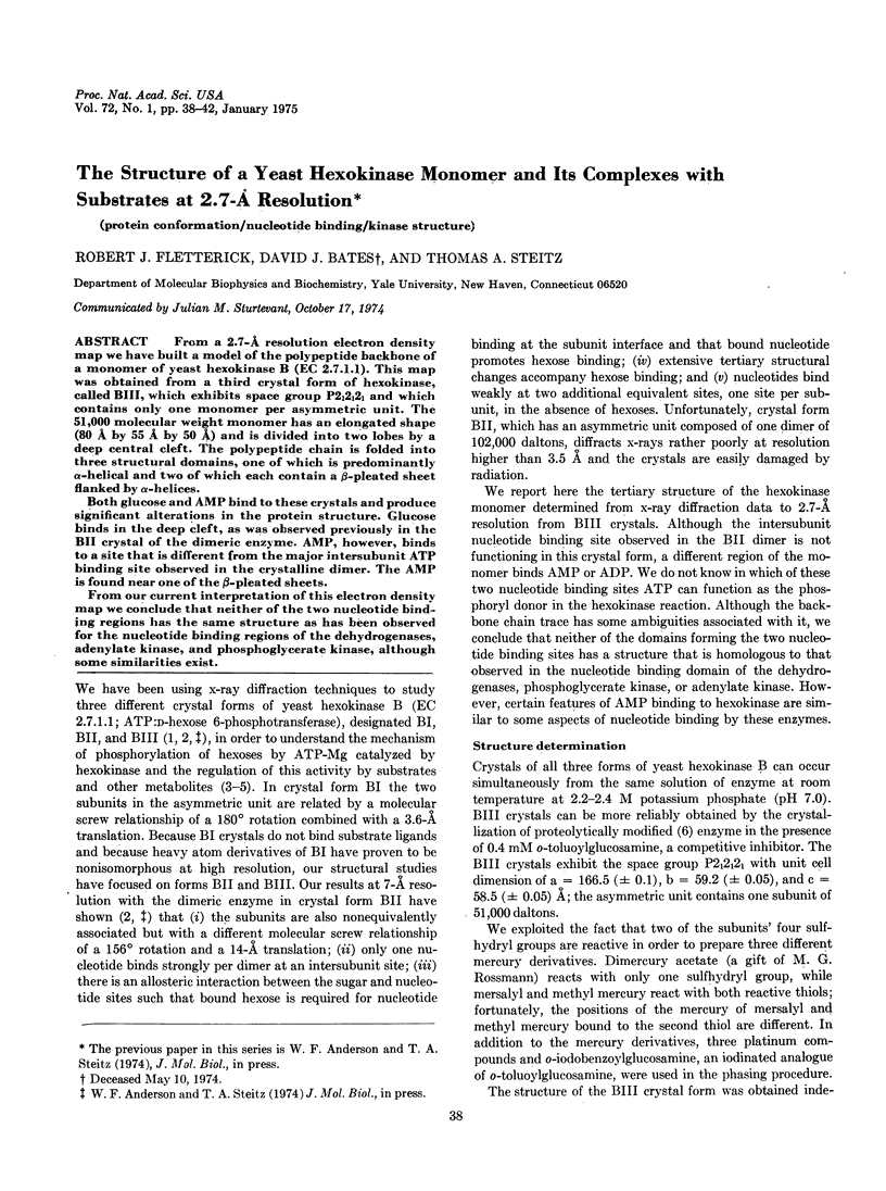
Images in this article
Selected References
These references are in PubMed. This may not be the complete list of references from this article.
- Adams M. J., Haas D. J., Jeffery B. A., McPherson A., Jr, Mermall H. L., Rossmann M. G., Schevitz R. W., Wonacott A. J. Low resolution study of crystalline L-lactate dehydrogenase. J Mol Biol. 1969 Apr;41(2):159–188. doi: 10.1016/0022-2836(69)90383-0. [DOI] [PubMed] [Google Scholar]
- Ainslie G. R., Jr, Shill J. P., Neet K. E. Transients and cooperativity. A slow transition model for relating transients and cooperative kinetics of enzymes. J Biol Chem. 1972 Nov 10;247(21):7088–7096. [PubMed] [Google Scholar]
- Anderson W. F., Fletterick R. J., Steitz T. A. Structure of yeast hexokinase. 3. Low resolution structure of a second crystal form showing a different quaternary structure, heterologous interaction of subunits and substrate binding. J Mol Biol. 1974 Jun 25;86(2):261–269. doi: 10.1016/0022-2836(74)90017-5. [DOI] [PubMed] [Google Scholar]
- Birktoft J. J., Blow D. M. Structure of crystalline -chymotrypsin. V. The atomic structure of tosyl- -chymotrypsin at 2 A resolution. J Mol Biol. 1972 Jul 21;68(2):187–240. doi: 10.1016/0022-2836(72)90210-0. [DOI] [PubMed] [Google Scholar]
- Blake C. C., Evans P. R. Structure of horse muscle phosphoglycerate kinase. Some results on the chain conformation, substrate binding and evolution of the molecule from a 3 angstrom Fourier map. J Mol Biol. 1974 Apr 25;84(4):585–601. doi: 10.1016/0022-2836(74)90118-1. [DOI] [PubMed] [Google Scholar]
- Bryant T. N., Watson H. C., Wendell P. L. Structure of yeast phosphoglycerate kinase. Nature. 1974 Jan 4;247(5435):14–17. doi: 10.1038/247014a0. [DOI] [PubMed] [Google Scholar]
- Brändén C. I., Eklund H., Nordström B., Boiwe T., Söderlund G., Zeppezauer E., Ohlsson I., Akeson A. Structure of liver alcohol dehydrogenase at 2.9-angstrom resolution. Proc Natl Acad Sci U S A. 1973 Aug;70(8):2439–2442. doi: 10.1073/pnas.70.8.2439. [DOI] [PMC free article] [PubMed] [Google Scholar]
- Kosow D. P., Rose I. A. Activators of yeast hexokinase. J Biol Chem. 1971 Apr 25;246(8):2618–2625. [PubMed] [Google Scholar]
- Lazarus N. R., Derechin M., Barnard E. A. Yeast hexokinase. 3. Sulfhydryl groups and protein dissociation. Biochemistry. 1968 Jun;7(6):2390–2400. doi: 10.1021/bi00846a049. [DOI] [PubMed] [Google Scholar]
- Richards F. M. The matching of physical models to three-dimensional electron-density maps: a simple optical device. J Mol Biol. 1968 Oct 14;37(1):225–230. doi: 10.1016/0022-2836(68)90085-5. [DOI] [PubMed] [Google Scholar]
- Rossmann M. G., Moras D., Olsen K. W. Chemical and biological evolution of nucleotide-binding protein. Nature. 1974 Jul 19;250(463):194–199. doi: 10.1038/250194a0. [DOI] [PubMed] [Google Scholar]
- Schmidt J. J., Colowick S. P. Identification of a peptide sequence involved in association of subunits of yeast hexokinases. Arch Biochem Biophys. 1973 Oct;158(2):471–477. doi: 10.1016/0003-9861(73)90538-9. [DOI] [PubMed] [Google Scholar]
- Schulz G. E., Elzinga M., Marx F., Schrimer R. H. Three dimensional structure of adenyl kinase. Nature. 1974 Jul 12;250(462):120–123. doi: 10.1038/250120a0. [DOI] [PubMed] [Google Scholar]
- Schulz G. E., Schirmer R. H. Topological comparison of adenyl kinase with other proteins. Nature. 1974 Jul 12;250(462):142–144. doi: 10.1038/250142a0. [DOI] [PubMed] [Google Scholar]
- Steitz T. A., Fletterick R. J., Hwang K. J. Structure of yeast hexokinase. II. A 6 angstrom resolution electron density map showing molecular shape and heterologous interaction of subunits. J Mol Biol. 1973 Aug 15;78(3):551–561. doi: 10.1016/0022-2836(73)90475-0. [DOI] [PubMed] [Google Scholar]
- Wassarman P. M., Lentz P. J., Jr The interaction of tetra-iodofluorescein with dogfish muscle lactate dehydrogenase: a chemical and x-ray crystallographic study. J Mol Biol. 1971 Sep 28;60(3):509–522. doi: 10.1016/0022-2836(71)90185-9. [DOI] [PubMed] [Google Scholar]


